0312
Characterizing microstructural patterns within the cortico-striato-thalamo-cortical circuit in Parkinson’s disease1Department of Medical imaging center, Clinical Medical College, Yangzhou University, Yangzhou, China, 2MR Research China, GE Healthcare, Beijing, China, 3Clinical Medical College, Yangzhou University, Yangzhou, China
Synopsis
Keywords: Parkinson's Disease, Diffusion/other diffusion imaging techniques
Motivation: Parkinson’s disease (PD) pathologically involves regional impairments and network disturbances. However, its microstructural abnormalities remain to be further elucidated via an appropriate neuroimaging approach.
Goal(s): We thus aimed to investigate the microstructural patterns of PD as mapped by diffusion kurtosis imaging (DKI).
Approach: The intergroup difference and classification performance of global microstructural complexity were analyzed, respectively. The network disruptions were explored in terms of structural connectivity, network covariance and modular connectivity.
Results: Our findings indicated that PD encountered globally impaired microstructural complexity, disturbed structural connectivity between basal ganglia and cortices, aberrant network covariance within the striatum and thalamus and altered modular connectivity.
Impact: These findings verified the potential clinical application of DKI for the exploration of microstructural patterns in PD, contributing complementary imaging features that offer insights into the neurodegenerative process.
Introduction
Converging evidence from magnetic resonance imaging studies has highlighted the hypothesis that Parkinson’s disease (PD) can be conceptualized as a disconnection syndrome, featuring prominent disruptions of the cortico-striato-thalamo-cortical (CSTC) circuit at the levels of structure, function and metabolism1, 2. Given that structural networks are the anatomical substrates of brain function across a range of spatial scales, structural disconnections are assumed to be indicative of pathological status3, 4. As an alternative to diffusion tensor imaging, diffusion kurtosis imaging (DKI) was introduced to PD, revealing impaired microstructures in the basal ganglia (BG), which are identified as the hub of CSTC circuit5.Nevertheless, whether DKI is capable of tracing the microstructural abnormalities of functional cortices and elucidating the pattern of disconnection in PD remain to be verified. We thus, hypothesized that 1) patients with PD suffered from global microstructural impairments in gray matters, which could be sensitively captured by DKI with favorable classification performance, and 2) the microstructure alterations in PD were synchronized between sub-regions of BG and functionally defined cortical areas, presenting as network perturbations within the CSTC circuit centered on the BG.
Materials and Methods
Subjects76 (45 male and 31 female) patients with PD, and 80 (45 male and 35 female) matched healthy controls (HCs) were recruited. The Unified Parkinson’s Disease Rating Scale part III and Hoehn-Yahr scale were scored for disease severity and stage of PD.
MRI experiment
MRI experiments were performed using a 3.0-tesla MRI scanner (Discovery MR750, GE, USA) with an 8-channel phased array head coil. DKI data were obtained as follows: TR: 5800 ms; TE: 100 ms; FOV: 240 × 240 mm2; matrix size: 100 × 100; voxel size: 2.4 × 2.4 × 2.4 mm3; b-values (0, 1250 and 2500 s/mm2); diffusion gradient directions: 30; number of slices: 35; and total scan time: 5 minutes and 30 seconds.
Data analysis
DKI data were preprocessed by DKE software and MATLAB. The bilateral striatum (putamen, caudate and pallidum) and thalamus were selected as regions of interest (ROIs). A seed-based correlation analysis and calculations of the winner-take-all (WTA) map and network within the CSTC circuit were performed using BCCT toolkit. For the definition of seed ROIs, the bilateral cerebral cortices were grouped into seven modules based on the Yeo atlas6.
Statistical analysis
The DKI data in each group were compared globally using two-sample t tests. The significance threshold was set as an uncorrected cluster-defining threshold of p < 0.001 and a familywise error-corrected significance of p < 0.05. Classification performance was analyzed using support vector machine strategy. Permutation tests were utilized to analyze the intergroup differences in structural connectivity, WTA map and the network matrix that were statistically significant at p < 0.05.
Results
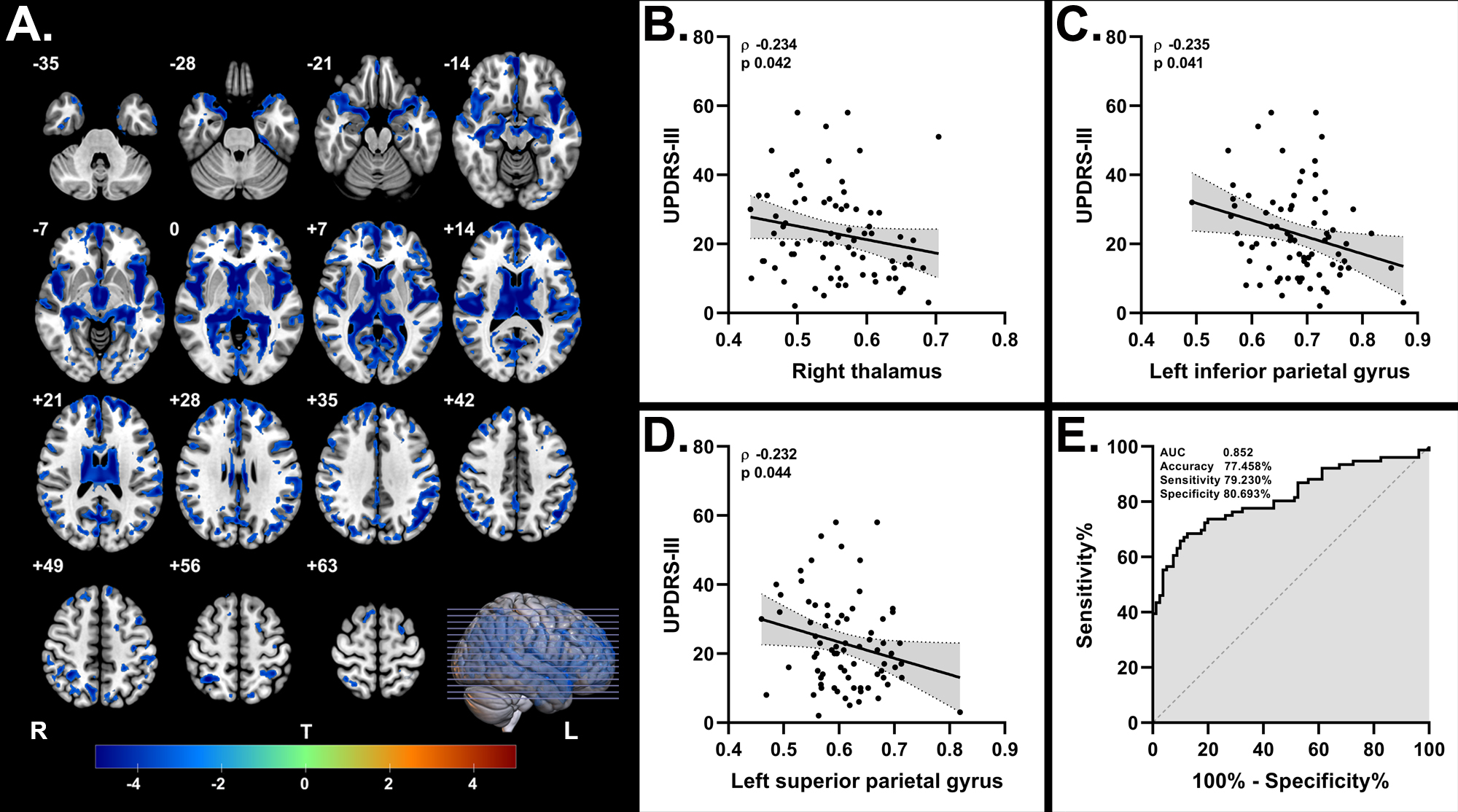
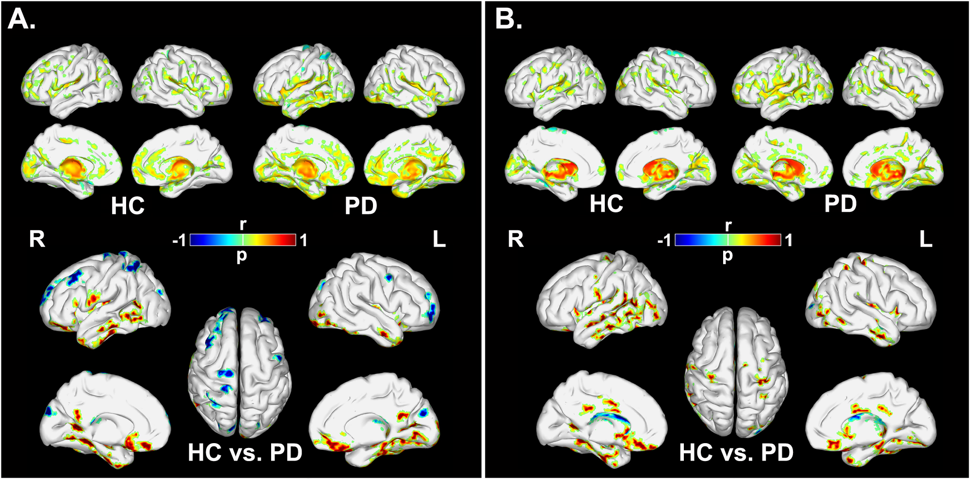
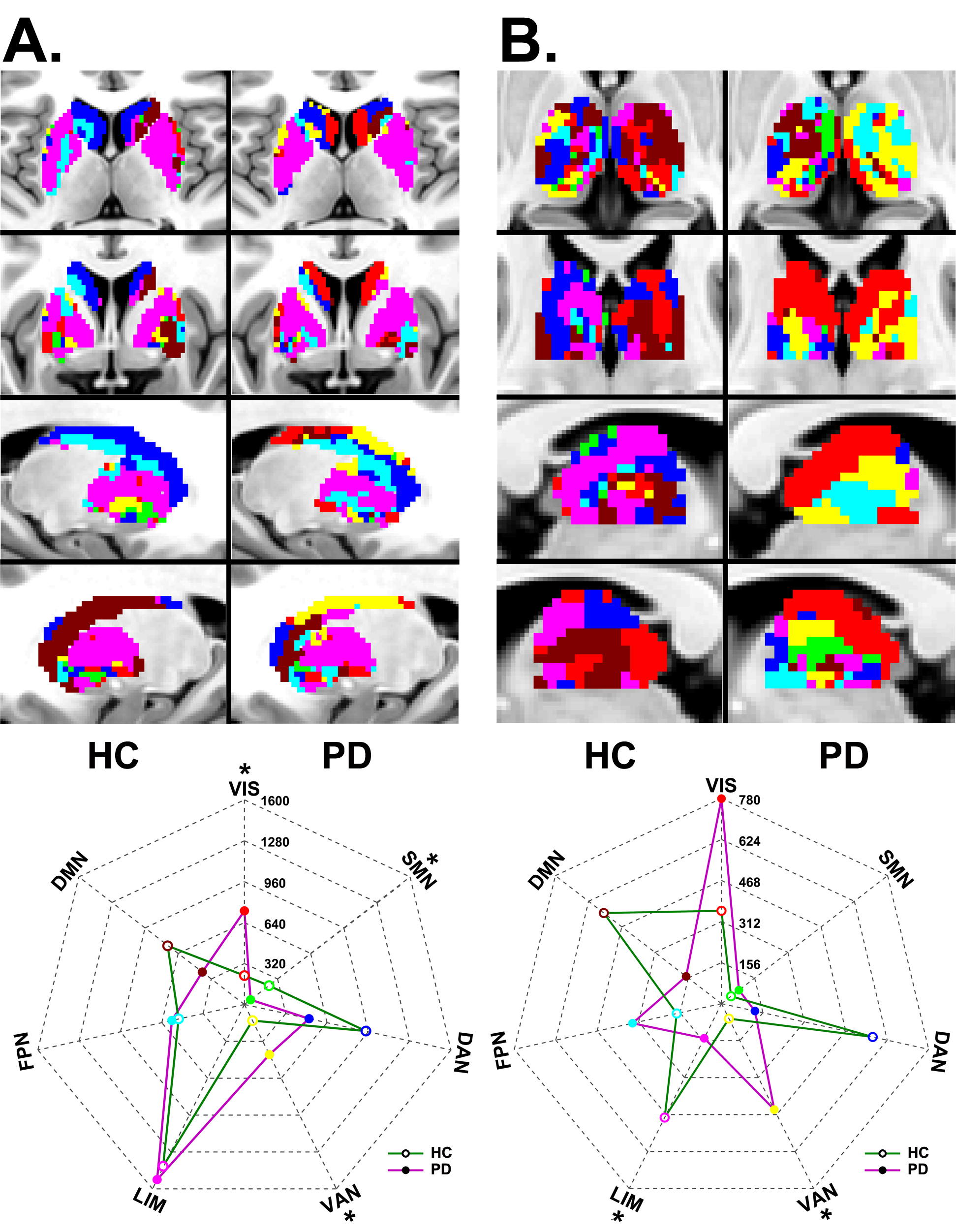
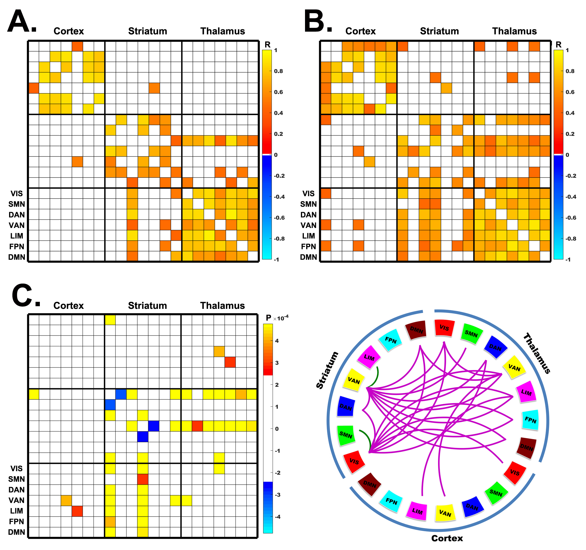
Patients with PD exhibited global microstructural impairments (frontal, parietal, occipital and temporal cortices) that served as an efficient diagnostic indicator, with an area under the curve of 0.817 (Fig. 1). Disrupted structural connections between the striatum and cortices as well as between the thalamus and cortices were widely distributed in PD group (Fig. 2). Aberrant covariance of the striatocortical circuitry and thalamocortical circuitry was observed in PD patients (Fig. 3), who also showed disrupted modular connectivity within the striatum and thalamus as well as across structures of the cortex, striatum and thalamus (Fig. 4).Discussion and conclusion
This study investigated the cortical alteration and network imbalance within the CSTC circuit from the perspective of microstructures in PD. We discovered that patients with PD suffered from microstructural injuries throughout the cortex (frontal, parietal, occipital and temporal) beyond those in BG regions, and the corresponding imaging features were beneficial to the clinical differentiation of PD patients from healthy controls with an AUC of 0.817. The interactions in the CSTC circuit in PD were microstructurally disturbed, presenting extensive disconnection between BG regions and functional cortices as well as aberrant modular connectivity that was mainly relevant to the VIS, SMN, VAN and LIM modules. These findings thus highlighted the potential of DKI for the diagnosis of PD and identified the microstructural pattern of neurodegeneration within the CSTC circuit.In conclusion, our DKI findings indicated that patients with PD encountered microstructural patterns of neurodegeneration, including globally impaired microstructural complexity, disturbed structural connectivity between BG subregions and cortices, aberrant network covariance within the striatum and thalamus and altered modular connectivity within the CSTC circuit. This study verified that DKI may be a promising tool for the comprehensive exploration of microstructural abnormalities and contributed novel evidence toward an understanding of pathological degradation in PD.
Acknowledgements
References
1. Zeng W, Fan W, Kong X, Liu X, Liu L, Cao Z, Zhang X, Yang X, Cheng C, Wu Y, Xu Y, Cao X. Altered intra- and inter-network connectivity in drug-naive patients with early parkinson's disease. Frontiers in aging neuroscience. 2022;14:783634.
2. Shang S, Wu J, Zhang H, Chen H, Cao Z, Chen YC, Yin X. Motor asymmetry related cerebral perfusion patterns in parkinson's disease: An arterial spin labeling study. Human brain mapping. 2021;42:298-309.
3. Dai Z, Lin Q, Li T, Wang X, Yuan H, Yu X, He Y, Wang H. Disrupted structural and functional brain networks in alzheimer's disease. Neurobiology of aging. 2019;75:71-82.
4. Gu Z, Jamison KW, Sabuncu MR, Kuceyeski A. Heritability and interindividual variability of regional structure-function coupling. Nature communications. 2021;12:4894.
5. Bingbing G, Yujing Z, Yanwei M, Chunbo D, Weiwei W, Shiyun T, Yangyingqiu L, Jin S, Qingwei S, Ailian L, Lizhi X. Diffusion kurtosis imaging of microstructural changes in gray matter nucleus in parkinson disease. Frontiers in neurology. 2020;11:252.
6. Yeo BT, Krienen FM, Sepulcre J, Sabuncu MR, Lashkari D, Hollinshead M, Roffman JL, Smoller JW, Zollei L, Polimeni JR, Fischl B, Liu H, Buckner RL. The organization of the human cerebral cortex estimated by intrinsic functional connectivity. Journal of neurophysiology. 2011;106:1125-1165.
Figures