0311
Serial neuromelanin MRI detects progression of depigmentation over 6 months in early Parkinson’s1School of Medicine, Mental Health & Clinical Neurosciences, University of Nottingham, Nottingham, United Kingdom, 2Sir Peter Mansfield Imaging Centre, University of Nottingham, Nottingham, United Kingdom, 3National Institute for Health Research, Nottingham Biomedical Research Centre, Nottingham, United Kingdom, 4Neurology, Queen’s medial Centre, Nottingham, United Kingdom, 5Neurology, Queen's medical Centre, Nottingham, United Kingdom
Synopsis
Keywords: Parkinson's Disease, Parkinson's Disease, Neuromelanin MRI
Motivation: The potential of neuromelanin-MRI in tracking disease progression is underexplored.
Goal(s): We investigated the detectability of changes of contrast in substructures of the substantia nigra over 6 months.
Approach: We compared changes of contrast between healthy controls and subjects with early PD.
Results: Depigmentation progresses over 6 months in early PD, but not in healthy controls, advancing the use of neuromelanin-MRI as a potential marker of PD.
Impact: Our study addresses the knowledge gap of neuromelanin-MRI as a potential progression marker of early PD, which will be particularly helpful in the context of future disease-modifying trials.
Introduction
Dopamine imaging effectively quantifies striatal dopaminergic deficits, but faces challenges in tracking disease progression. Neuromelanin-sensitive MRI (NM-MRI) is a promising novel approach sensitive to the neuromelanin-iron complex in the substantia nigra [1-2]. While its diagnostic value in Parkinson’s disease (PD) has been established in case-control studies (ie.[3-4]), its potential as a progression biomarker remains underexplored [5-6] especially in the early disease stage when disease modifying treatment is expected to be most effective. Previous findings have shown that dorsolateral substantia nigra (dlSN), where nigrosome-1 is located, could be the earliest region affected by PD, but with much longer follow-up span. Hence, in this study, we investigated the detectability of neuromelanin-related contrast loss in dlSN at as early as 6 months follow-up. We hypothesize that the depigmentation progresses over 6 months in early PD, but not in healthy controls (HC).Methods
85 early stage PD (62 males; disease duration <4 years; average 1 year; age: 66.8 / 9.96) and 53 age- and sex- matched HC (40 males; age: 68.0 / 8.85) with the complete set of baseline (BL) and follow-up (FL) NM-MRI from InsIghtPD study (Trial registration: https://classic.clinicaltrials.gov/ct2/show/NCT05631158) was used for this analysis. The BL-FU intervals in the HC and PD group are 189±32 days and 201±30 days, respectively.The NM-MRIs were acquired on a 3T GE Discovery MR750 scanner using Fast Spin Echo (flip angle=150, TE=min full, TR=900, Echoes=1, ETL=3, Multi-phase=3, FOV=18.0, slice_thickness=2.0, spacing=0.2, slice_number=12).
We processed the structural T1w and NM-MRI sequence using a pipeline developed in-house (https://github.com/SPMIC-UoN/parkinsons_pipeline). This pipeline brings all NM-MRI images into MNI152 space for further analysis. All data passed both pre- and post-pipeline Quality Control.
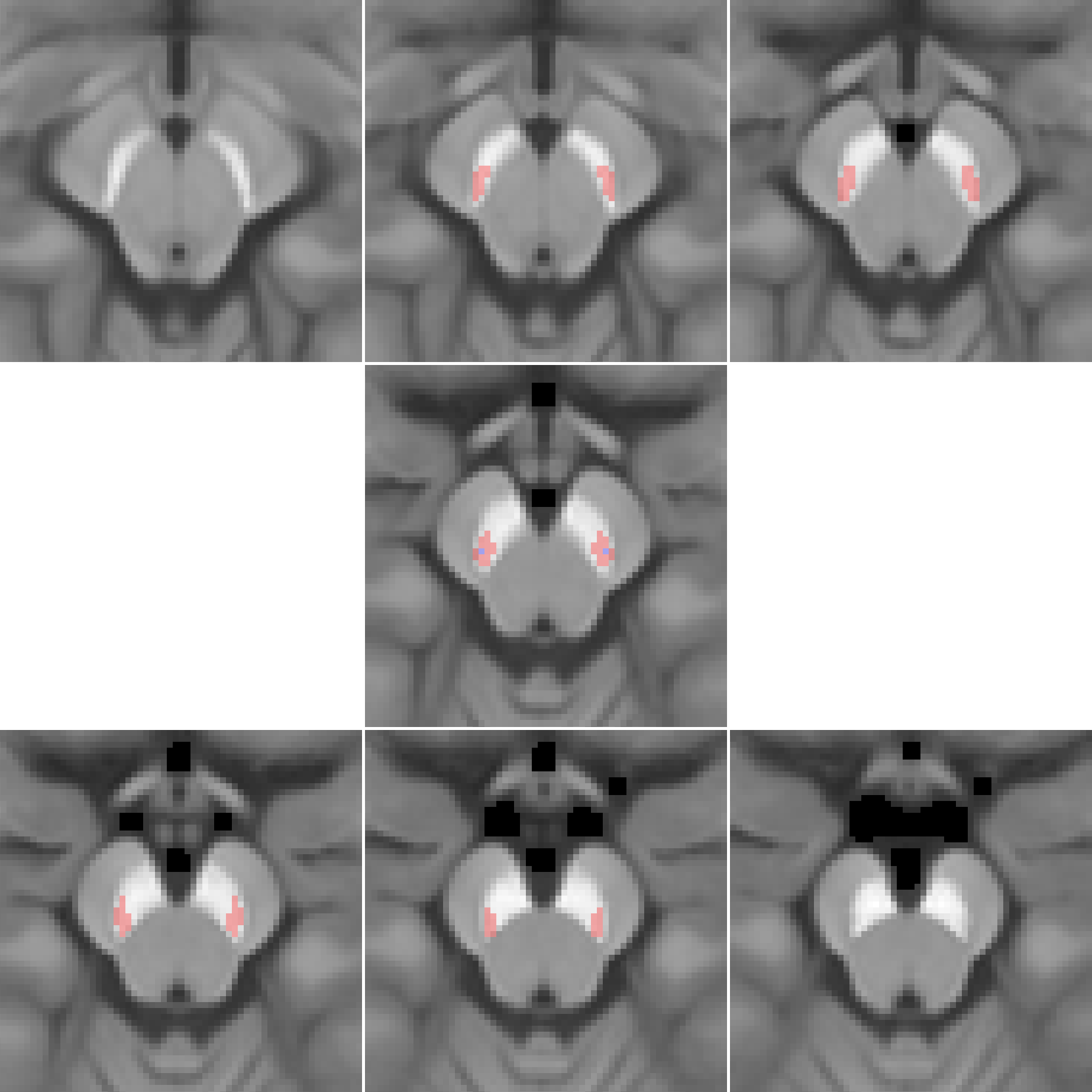
We established the ROIs of dlSN using anatomical a priori knowledge and a region-growing method. Starting from the two MNI152 coordinates representing the centroids of the mass for the bilateral nigrosome-1 [7], we grew these seeds into a sub-nigral areas (referred as “hot spot”) containing the voxels that shows the most depigmentation around the time of diagnosis in PD. The full analysis plan has been prepublished (DOI:10.17639/nott.7347)
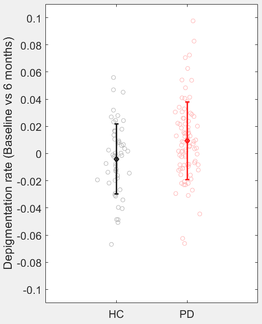
The dorsolateral nigral pigmentation index (dlNNMC) was first measured at each timepoint in both groups as dlNNMC=(μ_SN-μ_BG)/μ_BG, where μ_SN corresponds to the mean intensity value over in the SN and μ_BG corresponds to the mean intensity value over a delineated background area within the cerebral peduncles. The annual depigmentation rate was calculated using dlNNMC: 365*(dlNNMC _bl-dlNNMC _fu)/Ndays, where Ndays is the number of days between BL and 6 months.
The annual change of neuromelanin metrics estimated from the baseline versus 6 months difference for HC and PD was compared with zero, separately using one-tailed single group t-test. To compare the changes between HC and PD, an unpaired t-test at each time point was used. P-value <0.05 was considered as the threshold for statistical significance.
Results
Using the dlSN region (Figure 1) we found that the calculated annual depigmentation rate in controls was not significantly different from zero (P=0.2703), whilst the result in PD was significant (P=0.0028) in keeping with contrast loss reflecting depigmentation. Additionally, there was a significant difference between PD and HC (P=0.0059) (Figure 2).Discussion
To our best knowledge, this is the first NM-MRI study measuring pigmentation change in relevant substructures of pigmented SN over such a short follow-up span. In this study, we focused on the depigmentation rate of dlSN to best reflect changes in early clinical PD (Braak&Braak stage 3) and revealed that depigmentation can be detected at 6 months FU in PD. One previous study with 2 years follow-up have demonstrated 10% and 3% annual percentage change in SN volume and contrast ratio [8]. Our approach for calculating the depigmentation rate is different from theirs and thus difficult to make a direct comparison. However, we demonstrated that our robust template-based analysis pipeline of NM-MRI at 3T can detect minimal changes (~1%) in early PD over 6 months. In addition, we found as expected a near-zero depigmentation rate in HC which significantly differed from PD. The age range of the healthy controls corresponds to the period of minimal pigmentation change in agreement with our previous study of the physiological changes in healthy ageing. The findings are part of an on-going study with further planned analyses to study depigmentation over longer intervals in order to establish NM-MRI study as a clinically useful progression marker in Parkinson’s disease.Conclusion
We report NM contrast loss at 6 month in early PD, suggesting that the NM-MRI at 3T can detect subtle depigmentation in the absence of overt clinical deterioration, highlighting its potential as adjunct disease progression markerAcknowledgements
We thank Weston Brain Institute for funding this project NIHR Nottingham Biomedical Research Centre grant for funding Stefan PszczolkowskiReferences
1. M. Sasaki et al., ‘Neuromelanin magnetic resonance imaging of locus ceruleus and substantia nigra in Parkinson’s disease’, NeuroReport, vol. 17, no. 11, pp. 1215–1218, Jul. 2006, doi: 10.1097/01.wnr.0000227984.84927.a7.
2. P. Trujillo et al., ‘Contrast mechanisms associated with neuromelanin-MRI’, Magn. Reson. Med., vol. 78, no. 5, pp. 1790–1800, 2017, doi: 10.1002/mrm.26584. K. Ogisu et al., ‘3D neuromelanin-sensitive magnetic resonance imaging with semi-automated volume measurement of the substantia nigra pars compacta for diagnosis of Parkinson’s disease’, Neuroradiology, vol. 55, no. 6, pp. 719–724, Jun. 2013, doi: 10.1007/s00234-013-1171-8.
3. D. E. Huddleston, J. Langley, J. Sedlacik, K. Boelmans, S. A. Factor, and X. P. Hu, ‘In vivo detection of lateral–ventral tier nigral degeneration in Parkinson’s disease’, Hum. Brain Mapp., vol. 38, no. 5, pp. 2627–2634, 2017, doi: 10.1002/hbm.23547.
4. G. Castellanos et al., ‘Automated neuromelanin imaging as a diagnostic biomarker for Parkinson’s Disease’, Mov. Disord., vol. 30, no. 7, pp. 945–952, Jun. 2015, doi: 10.1002/mds.26201.
5. S. T. Schwarz, Y. Xing, P. Tomar, N. Bajaj, and D. P. Auer, ‘In vivo assessment of brainstem depigmentation in Parkinson Disease: Potential as a severity marker for multicenter studies’, Radiology, vol. 283, no. 3, pp. 789–798, Jun. 2017, doi: 10/gf3qnn.
6. N. Pavese, ‘Is neuromelanin the imaging biomarker for the early diagnosis of Parkinson’s disease that we were looking for?’, Parkinsonism Relat. Disord., vol. 58, pp. 1–2, Jan. 2019, doi: 10.1016/j.parkreldis.2018.12.013.
7. J. Langley, D. E. Huddleston, B. Crosson, D. D. Song, S. A. Factor, and X. Hu, ‘Multimodal assessment of nigrosomal degeneration in Parkinson’s disease’, Parkinsonism Relat. Disord., vol. 80, pp. 102–107, Nov. 2020, doi: 10.1016/j.parkreldis.2020.09.021.
8: Gaurav R, Yahia-Cherif L, Pyatigorskaya N, et al. Longitudinal Changes in Neuromelanin MRI Signal in Parkinson's Disease: A Progression Marker. Mov Disord. 2021;36(7):1592-1602. doi:10.1002/mds.28531
Figures