0302
Acute impact of soccer ball heading on brain tissue electrical conductivity1Neuroscience Research Australia, Sydney, Australia, 2School of Health Sciences and Social Work, Griffith University, Gold Coast, Australia, 3Queensland Academy of Sport, Queensland, Australia, 4School of Psychology, Faculty of Science, The University of Sydney, Sydney, Australia, 5School of Medical Sciences (Neuroscience), Faculty of Medicine and Health, The University of Sydney, Sydney, Australia, 6Menzies Health Institute Queensland, Griffith University, Gold Coast, Australia, 7Lambert Initiative for Cannabinoid Therapeutics, The University of Sydney, Sydney, Australia, 8School of Psychology, The University of New South Wales, Sydney, Australia
Synopsis
Keywords: Traumatic Brain Injury, Brain
Motivation: The effects of sub-concussive head impacts are not well understood. New biomarkers are needed to detect sub-concussion.
Goal(s): Our goal was to investigate the acute effects of sub-concussive impacts using MREPT.
Approach: Fourteen soccer players were scanned with MREPT in two separate sessions after performing the task of either heading or kicking soccer balls for 20 minutes.
Results: Electrical conductivity measured in multiple brain regions such as inferior fronto-occipital fasciculus after the heading session was significantly less than that measured following kicking, indicating that MREPT could be a useful tool for detecting sub-concussive injury.
Impact: The finding that heading soccer balls for a short period can cause significant acute decreases in brain electrical conductivity suggests that this activity may have detrimental short term effects on brain function.
Introduction
Sub-concussive head impacts are impacts to the head that cause sudden brain movement without neurological symptoms. People who play certain sports such as football, soccer and boxing are more prone to sub-concussive head impacts. Although difficult to diagnose sub-concussion clinically, evidence shows a variety of potential effects on sub-concussion, such as negative short-term effects on memory following boxing sparring sessions1. A significant relationship was found between head impact exposure and change of fractional anisotropy in the left inferior fronto-occipital fasciculus tract, resulting from a single season of youth football2, and a recent systematic review of contact sport found decreased NAA and creatine in MRS studies3. Neuroimaging biomarkers are needed to better understand the effects of sub-concussion and its diagnosis. Electric properties of tissue including conductivity and permittivity can provide auxiliary information for disease diagnosis and monitoring. Magnetic resonance electrical properties tomography (MREPT) is emerging as a noninvasive imaging method for mapping tissue electrical properties4-7. Here, we aimed to investigate the acute effects of soccer heading using MREPT.Methods
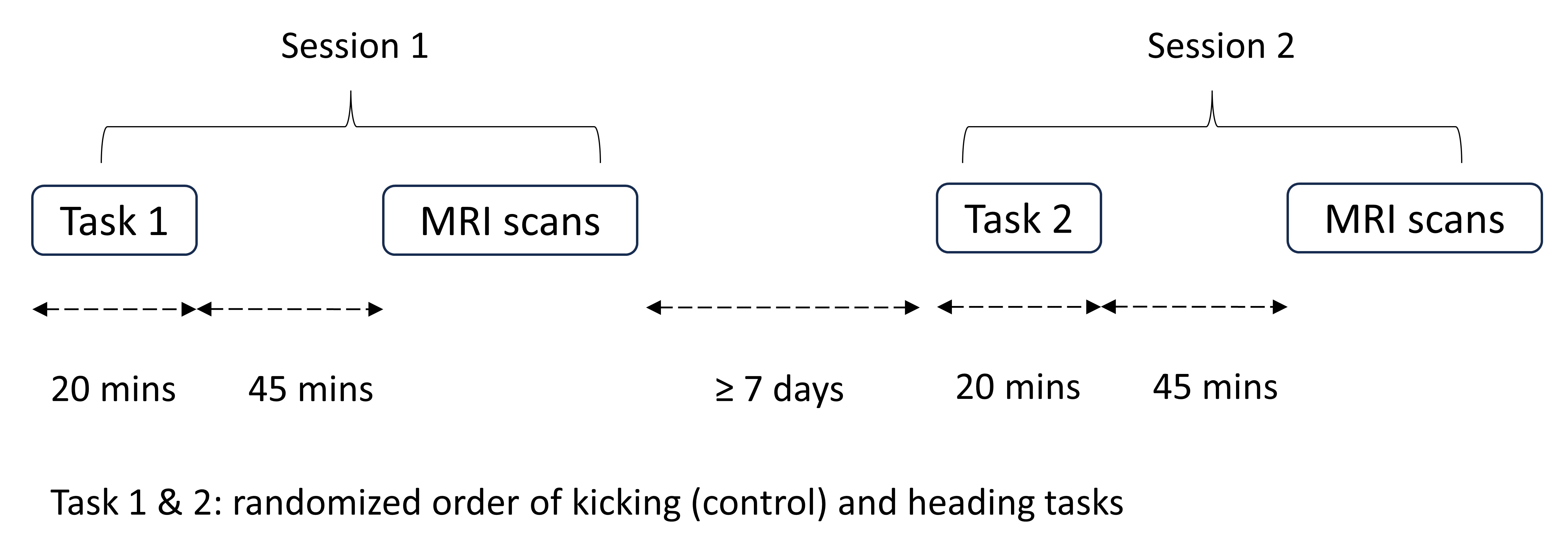
Data acquisitionThis work was approved by the University of Sydney human research ethics committee (HREC 2021/515), and informed consent was obtained from all participants. Fourteen male soccer players (25.4 ± 4.8 years old; thirteen right-handed, one left-handed) were scanned at 3T (Ingenia CX, Philips Healthcare, Best, The Netherlands) on two occasions (separated by ≥ 7 days). Participants performed either a heading task involving 20 headers in 20 mins (launched at 35 km/h over 12m) or a kicking task (control). Figure 1 shows the experimental timeline of the two sessions involving heading & kicking tasks separately. 45 mins after each task, the participants were scanned using balanced fast field echo (bFFE: TR/TE = 2.52/1.26 ms, nonselective RF pulses, flip angle 25°, resolution 1 mm isotropic, sagittal slices) and T1-weighted turbo field echo (TFE: TR/TE = 7.06/3.25 ms, resolution 0.75×0.75×0.9 mm3, sagittal slices, FOV 320×320 mm2).
Data analysis
In this study, phase-based MREPT was adopted as $$$\sigma=\triangledown^{2}\phi_{\pm}/2\mu_{0}\omega$$$ where σ denotes conductivity (unit: S/m), μ0=4π×10-7 H/m is the magnetic permeability of free space, ω denotes angular Larmor frequency, $$$\triangledown^{2}$$$ denotes Laplacian operation, and $$$\phi_{\pm}$$$ denotes transceive phase.
T1-weighted TFE images were co-registered and segmented into white matter, gray matter and CSF using FSL to alleviate boundary artifacts in the calculation. Within each tissue type, an average parabolic phase fitting method was used to reduce artifacts amplified in the Laplacian8,9, and the second derivatives of the fitted phase were taken to calculate conductivity.
The conductivity maps of each participant from both sessions were normalized into MNI space (voxel size 2 mm isotropic) using SPM 12, and the differential conductivity map of each participant was obtained by subtracting the normalized conductivity map of heading session from that of kicking session. The differential conductivity maps of fourteen participants were analyzed using one-sample t test to determine if the conductivity values of these two sessions were significantly different. FDR correction was applied to p values, and significance level α=0.05. The minimum cluster size was 3.
Results
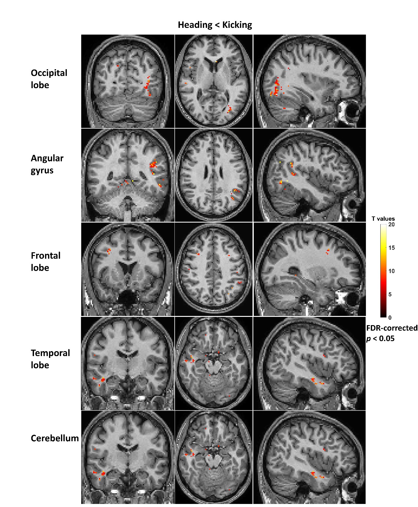
Figure 2 show T value maps (FDR-corrected, p<0.05) of conductivity difference between the kicking session and heading sessions from fourteen participants. Conductivity following the heading session was significantly smaller than that after the kicking session in the left inferior and middle occipital lobe, including inferior fronto-occipital fasciculus (IFOF) and inferior longitudinal fasciculus (ILF), and angular gyrus, frontal lobe, temporal lobe and cerebellum. There were no significant brain regions of increased conductivity following the heading session.Discussion and conclusion
In summary, the soccer heading task led to significantly lower conductivity values in multiple brain regions, especially in the left inferior fronto-occipital fasciculus and inferior longitudinal fasciculus. Areas in the left parietal lobe have previously been reported as potentially affected by heading2,11,12. MREPT may be useful to investigate sub-concussive impacts on brain structures and functions, as a potential modality to aid diagnosis and monitoring of sub-concussive/concussive/brain injury noninvasively, though a large-scale study is warranted.Acknowledgements
The authors acknowledge the support of Dr. Iain Ball and Mr. Brendan Moran. The authors acknowledge the facilities and scientific and technical assistance of the National Imaging Facility, a National Collaborative Research Infrastructure Strategy (NCRIS) capability, at Neuroscience Research Australia and UNSW. This work is a sub component of a larger study driven by Danielle McCartney and Nathan Delang.References
1. Di Virgilio TG, Ietswaart M, Wilson L, et al. Understanding the consequences of repetitive subconcussive head impacts in sport: brain changes and dampened motor control are seen after boxing practice. Front Hum Neurosci. 2019;13:294.
2. Bahrami N, Sharma D, Rosenthal S, et al. Subconcussive head impact exposure and white matter tract changes over a single season of youth football. Radiology. 2016;281(3):919-26.
3. Delang N, Irwin C, Peek AL, et al. The effect of contact/collision sport participation without concussion on neurometabolites: A systematic review and meta-analysis of magnetic resonance spectroscopy studies. J Neurochem. 2023;00:1-18.
4. Haacke EM, Petropoulos LS, Nilges EW, et al. Extracting of conductivity and permittivity using magnetic resonance imaging. Phys. Med. Biol. 1991;36(6):723-734.
5. Wen H. Noninvasive quantitative mapping of conductivity and dielectric properties distributions using RF wave propagation effects in high-field MRI. Physics of medical imaging international society for optics and photonics. 2003; 5030:471-478.
6. Katscher U, Voigt T, Findeklee C, et al. Determination of electric conductivity and local SAR via B1 mapping. IEEE T. Med. Imaging. 2009;28(9):1365-1374.
7. Voigt T, Katscher U, Doessel O. Quantitative conductivity and permittivity imaging of the human brain using electric properties tomography. Magn. Reson. Med. 2011;66(2):456-466.
8. Katscher U, Djamshidi K, Voigt T, et al. Estimation of breast tumor conductivity using parabolic phase fitting. 20th Annual Meeting of Internal Society of Magnetic Resonance in Medicine. 2012;3482.
9. Cao J, Ball I, Humburg P, et al. Repeatability of brain phase-based magnetic resonance electric properties tomography methods and effect of compressed SENSE and RF shimming. Phys Eng Sci Med. 2023;46(2):753-66.
10. Di Virgilio TG, Hunter A, Wilson L, et al. Evidence for acute electrophysiological and cognitive changes following routine soccer heading. EBioMedicine. 2016;13:66-71.
11. Lipton ML, Kim N, Zimmerman ME, et al. Soccer heading is associated with white matter microstructural and cognitive abnormalities. Radiology. 2013;268(3):850-7.
12. Oliveira TG, Ifrah C, Fleysher R, et al. Soccer heading and concussion are not associated with reduced brain volume or cortical thickness. PLoS One. 2020;15(8):e0235609.
Figures