0301
Association of white matter brain diffusivity properties with football exposure in former professional American-style football players1Athinoula A Martinos Center for Medical Imaging, Massachusetts General Hospital, Charlestown, MA, United States, 2Harvard T. H. Chan School of Public Health, Boston, MA, United States, 3Neuroendocrine Unit, Massachusetts General Hospital, Boston, MA, United States, 4Department of Physical Medicine and Rehabilitation, Spaulding Rehabilitation Hospital, Charlestown, MA, United States, 5Football Players Health Study, Harvard Medical School, Boston, MA, United States, 6Department of Radiology, Massachusetts General Hospital, Boston, MA, United States, 7Cardiology Division, Massachusetts General Hospital, Boston, MA, United States
Synopsis
Keywords: Traumatic Brain Injury, Traumatic brain injury
Motivation: The possible long-term effects of repetitive head impacts experienced by American-style professional football players are poorly understood. White matter injury is a known sequela of head trauma. Peak-width skeletonized mean diffusivity measurements have been associated with cerebrovascular disease.
Goal(s): Our goal is to evaluate the association of peak-width skeletonized diffusion values with football exposure.
Approach: We measured peak-width skeletonized diffusion values in 103 retired professional football players who underwent multi-shell diffusion imaging.
Results: Age, hypertension, body-mass index, concussion signs and symptom history score, total years of non-professional play, and episodes of loss of consciousness were significantly associated with peak-width skeletonized diffusion values.
Impact: Measured peak-width skeletonized diffusion values in white matter may provide an improved understanding of the association between football exposure and later-in-life brain microstructural integrity.
Introduction
The possible long-term effects of repetitive head impacts experienced by former professional American-style football (ASF) players are poorly understood. White matter (WM) injury is a known sequela of head trauma. Peak-width skeletonized (PS) mean diffusivity (PSMD) values in WM have been associated with cognitive dysfunction.1 Although well-studied in patients with small vessel disease1 and vascular cognitive dementia2, there has been no investigation of PSMD in former professional ASF players. Our study aimed to investigate the association of PSMD with football exposure in former professional players. We also examined changes in PS values in diffusion kurtosis and neurite orientation dispersion and density images.3Methods
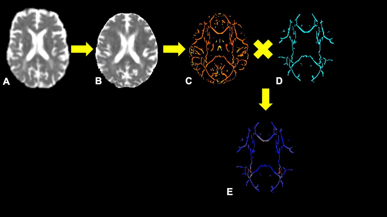
Former professional American football players who had played since 1960 and were less than 60 were enrolled.4 Participants underwent 3T MRI scans acquired using a 20-channel head and neck coil. Multiple shell diffusion imaging was acquired using 30 directions with b-value=1000 s/mm2, and 2500 s/mm2 (2x2x2 mm3), and 10 b-value=0 s/mm2 images acquired using blipped simultaneous multi-slice5 echo planar imaging. Orientation dispersion (OD),6 intracellular volume fraction (ICVF),6 isotropic volume fraction (ISOVF),6 mean kurtosis (MK),7 axial kurtosis (AK), radial kurtosis (RK),7 mean diffusivity (MD),7 axial diffusivity (AD),7 radial diffusivity (RD)7 and fractional anisotropy (FA)7 were calculated. Peak-width skeletonized values were calculated for each participant1 (Figure 1). In brief, FA images were skeletonized using the tract-based spatial statistics (TBSS) toolkit.8 MD were projected onto the skeleton using the FA-derived parameters. PSMD was defined as the difference between the 5th and 95th percentiles of the values in the projected image after further masking using a template skeleton mask.9 Analyses were repeated for AD, RD, FA, OD, ICVF, ISOVF, MK, AK, and RK.Football exposure variables included the number of seasons played professionally, the number of years played at the non-professional level (pre-high school, high school, and college), the number of episodes of loss of consciousness (LOC), and a concussion signs and symptoms history score (CSS)10. The CSS score was calculated by summing the self-reported frequency of the following ten features after a football-related injury: headache, nausea, dizziness, LOC episodes, memory problems, disorientation, confusion, seizure, visual problems, or feeling unsteady. Main field position was dichotomized into linemen (offensive and defensive linemen and linebacker) versus all other positions. A self-reported history of using performance-enhancing drugs during playing years was recorded.
Univariable linear regression was used to assess the association between PS values and football exposure variables. Age, race, co-morbidities (i.e., hypertension, diabetes mellitus, hyperlipidemia, body mass index [BMI]), and alcohol and tobacco use were taken into consideration. Statistical analyses were performed using JMP 16.2.0. P-values <0.5 were considered statistically significant.
Results
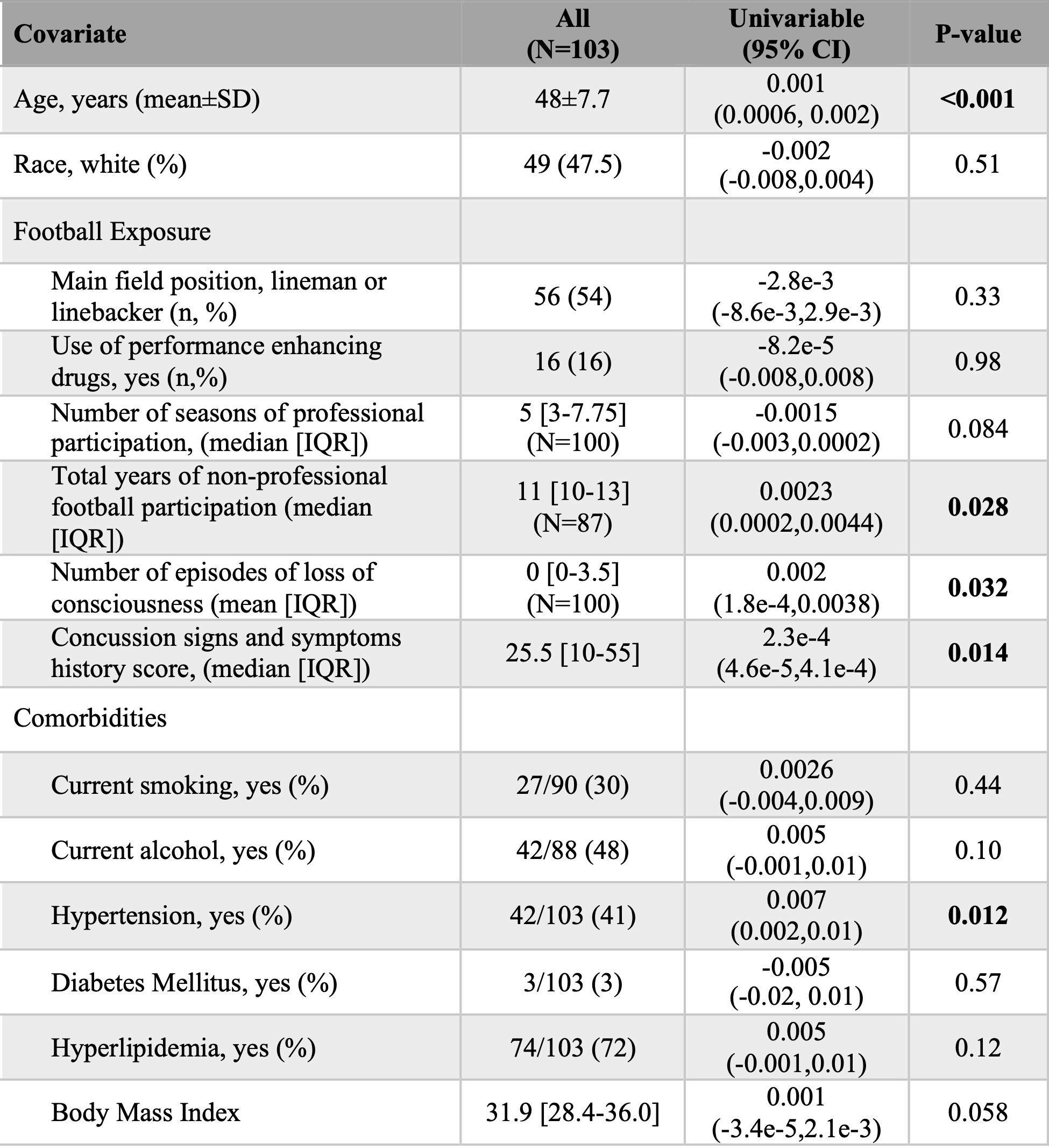
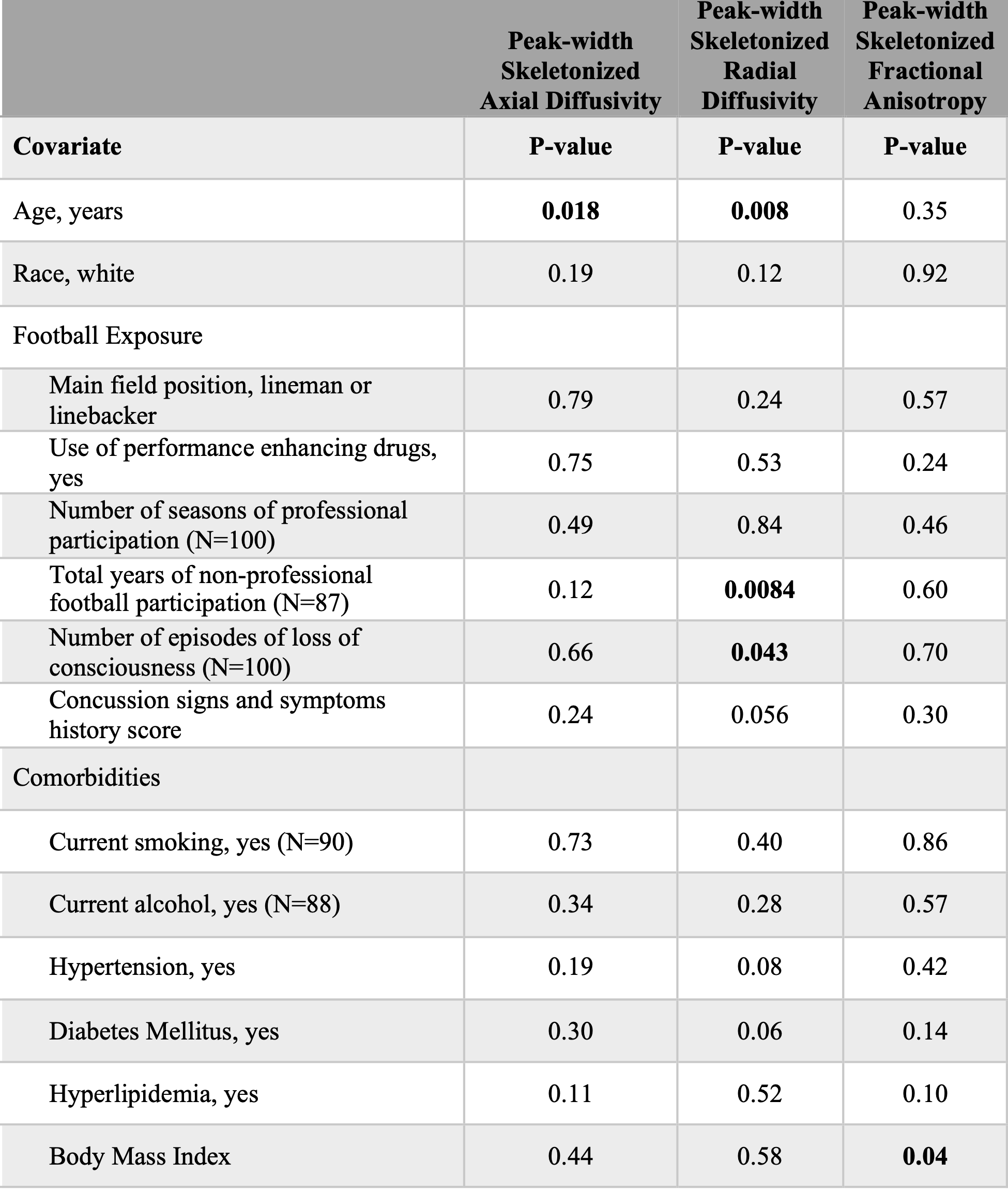
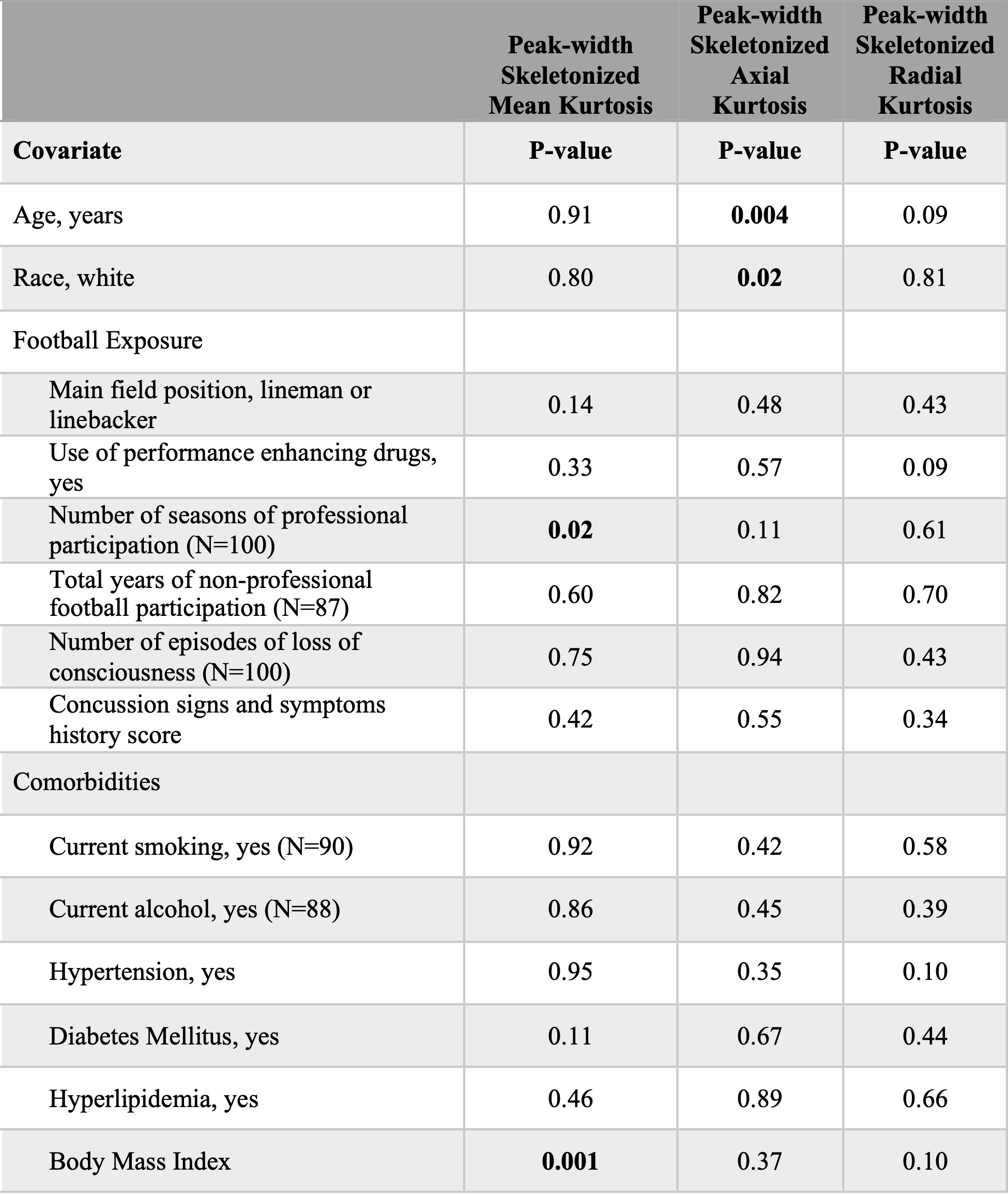
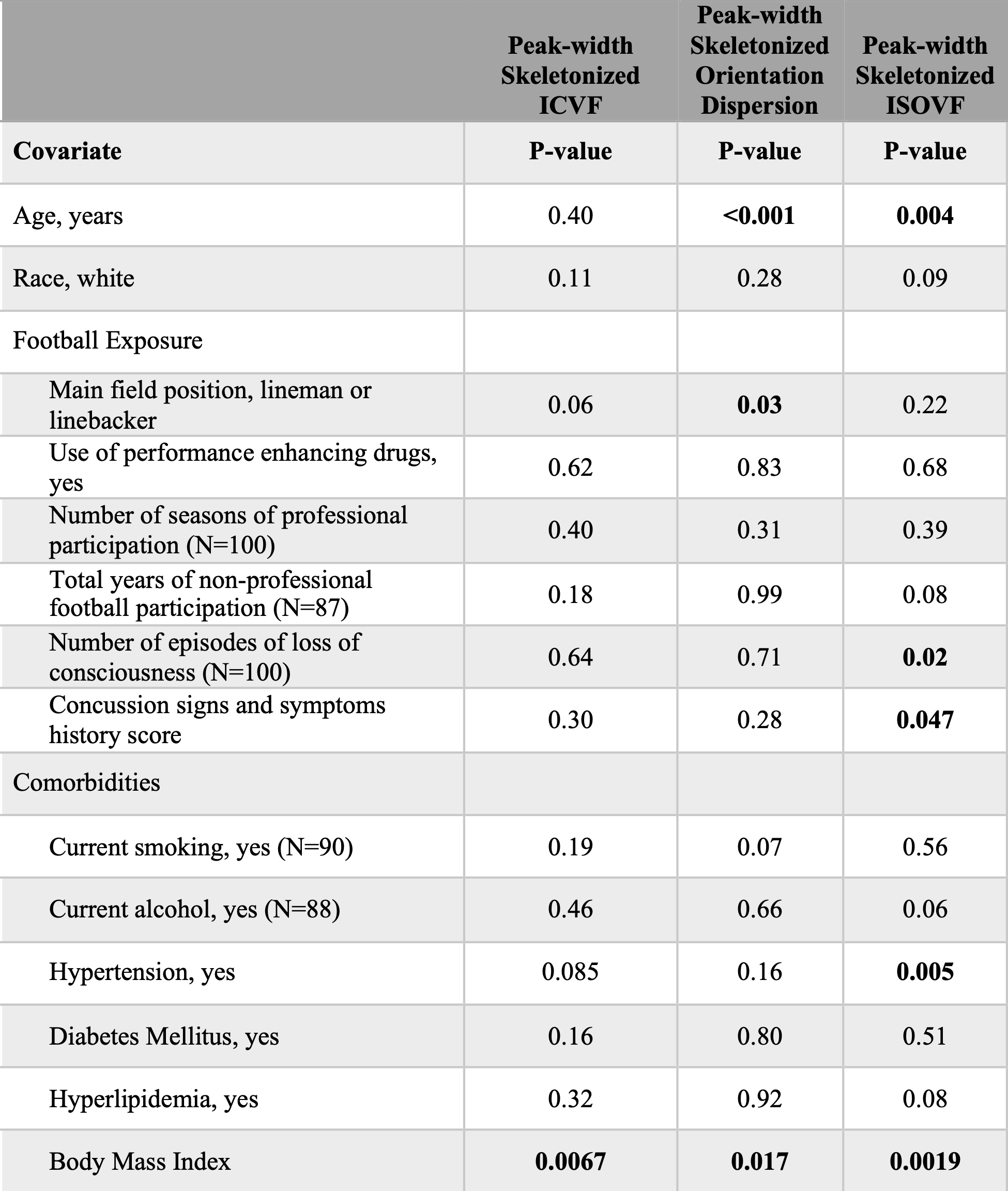
103 former professional ASF players were evaluated. Summaries of demographics and football exposures are provided in Figure 2, and univariable analyses for predictors of PSMD. Age and hypertension were significant predictors of increased PSMD. The CSS score, the number of LOC episodes, and the number of years of non-professional play were significant univariable predictors of increased PSMD. After adjusting for age and hypertension, CSS (P=0.012), the total number of years of non-professional play (P=0.018) remained a significant predictor of increased PSMD, but not the number of LOC episodes (P=0.11).Repeating the analysis for AD, RD, and FA (Figure 3) showed that only PSRD was associated with football exposure (years of non-professional play and the number of LOC episodes). Like PSMD, when adjusting for age, only years of non-professional play remained significant (P=0.0066) but not LOC (P=0.077). Results for kurtosis metrics (PSMK, PSAK, PSRK) (Figure 4) showed only the number of professional seasons associated with PSMK, which remained significant (P=0.03) even after adjusting for BMI. Results for NODDI metrics (PSICVF, PSOD, PSISOVF) (Figure 5) found that only the main position played was associated with PSOD, but this was no longer significant (P=0.23) after adjusting for age and BMI. PSISOVF was found to be associated with CSS and the number of LOC episodes. CSS remained significant after adjusting for age, hypertension, and BMI (P=0.046) but not the number of LOC episodes (P=0.056).
Discussion
For former American football players, the number of years of non-professional football play was significantly associated with greater PSMD and PSRD, even after adjusting for comorbidities. We also found that PSMK was inversely associated with the number of professional seasons played. Previous studies have found decreases in MK after TBI11 and associations between MK and head impact exposure in high school football players.12 Future studies with larger sample sizes are needed to understand better the association between football exposure and later-in-life brain microstructure, as measured by PSMD, PSRD, PSMK, and PSISOVF.Acknowledgements
We thank Drs. Andre van der Kouwe and John Kirsch for providing pulse sequences that were used in this study and assistance in protocol optimization. We also thank the Harvard Football Players Health Study Navigators, Athinoula A Martinos Center Radiology Technologists, and Kiran Dhakal, PhD for their assistance in data acquisition. We thank Philip Ayres, Bir Kafle, Dean Marengi Jr., and Heather DiGregorio for their assistance in data analysis.References
1. Baykara E, Gesierich B, Adam R, Tuladhar AM, Biesbroek JM, Koek HL, Ropele S, Jouvent E, Alzheimer's Disease Neuroimaging I, Chabriat H, et al. A novel imaging marker for small vessel disease based on skeletonization of white matter tracts and diffusion histograms. Ann Neurol. 2016;80:581-592
2. Zanon Zotin MC, Yilmaz P, Sveikata L, Schoemaker D, van Veluw SJ, Etherton MR, Charidimou A, Greenberg SM, Duering M, Viswanathan A. Peak width of skeletonized mean diffusivity: A neuroimaging marker for white matter injury. Radiology. 2023;306:e212780
3. Zhang H, Schneider T, Wheeler-Kingshott CA, Alexander DC. NODDI: Practical in vivo neurite orientation dispersion and density imaging of the human brain. Neuroimage. 2012;61:1000-1016
4. Zafonte R, Pascual-Leone A, Baggish A, Weisskopf MG, Taylor HA, Connor A, Baker J, Cohan S, Valdivia C, Courtney TK, et al. The football players' health study at Harvard University: Design and objectives. Am J Ind Med. 2019;62:643-654
5. Setsompop K, Gagoski BA, Polimeni JR, Witzel T, Wedeen VJ, Wald LL. Blipped-controlled aliasing in parallel imaging for simultaneous multislice echo planar imaging with reduced g-factor penalty. Magn Reson Med. 2012;67:1210-1224
6. Daducci A, Canales-Rodriguez EJ, Zhang H, Dyrby TB, Alexander DC, Thiran JP. Accelerated microstructure imaging via convex optimization (AMICO) from diffusion MRI data. Neuroimage. 2015;105:32-44
7. Tabesh A, Jensen JH, Ardekani BA, Helpern JA. Estimation of tensors and tensor-derived measures in diffusional kurtosis imaging. Magn Reson Med. 2011;65:823-836
8. Smith SM, Jenkinson M, Johansen-Berg H, Rueckert D, Nichols TE, Mackay CE, Watkins KE, Ciccarelli O, Cader MZ, Matthews PM, et al. Tract-based spatial statistics: Voxelwise analysis of multi-subject diffusion data. Neuroimage. 2006;31:1487-1505
9. PSMD marker. 2021;2023
10. Grashow R, Weisskopf MG, Miller KK, Nathan DM, Zafonte R, Speizer FE, Courtney TK, Baggish A, Taylor HA, Pascual-Leone A, et al. Association of concussion symptoms with testosterone levels and erectile dysfunction in former professional US-style football players. JAMA Neurol. 2019;76:1428-1438
11. Stokum JA, Sours C, Zhuo J, Kane R, Shanmuganathan K, Gullapalli RP. A longitudinal evaluation of diffusion kurtosis imaging in patients with mild traumatic brain injury. Brain Inj. 2015;29:47-57
12. Davenport EM, Apkarian K, Whitlow CT, Urban JE, Jensen JH, Szuch E, Espeland MA, Jung Y, Rosenbaum DA, Gioia GA, et al. Abnormalities in diffusional kurtosis metrics related to head impact exposure in a season of high school varsity football. J Neurotrauma. 2016;33:2133-2146
Figures