0295
Ventilation and Gas Exchange Delay maps in Pre-clinical Thoracic Insufficiency Syndrome Models using Dynamic Hyperpolarized-Xenon MRI1University of Pennsylvania, Philadelphia, PA, United States, 2Children’s Hospital of Philadelphia, Philadelphia, PA, United States, 3Boston Children’s Hospital, Boston, MA, United States
Synopsis
Keywords: Hyperpolarized MR (Gas), MSK, Hyperpolarized MR (Gas), Dynamic Imaging, Pediatric, Signal Modeling, Skeletal, Preclinical, Lung, Flow, Analysis/Processing
Motivation: The pressing need to develop and evaluate novel treatments of thoracic insufficiency syndrome (TIS), a condition with significant implications for compromised pulmonary growth and function.
Goal(s): To gain deeper insights into the altered lung function associated with TIS, ultimately improving patient care and outcomes.
Approach: Dynamic hyperpolarized-xenon-129 MRI was used to assess ventilation and gas exchange dynamics in pre-clinical models of TIS.
Results: Significant ventilation and gas exchange delays were found in the TIS animals compared to the healthy ones.
Impact: Our findings hold the promise to improve TIS assessment and treatment monitoring by providing crucial insights into altered lung function. This study offers a valuable tool for clinicians/researchers, fostering improved patient care and stimulating further TIS management and intervention research.
Introduction
Thoracic insufficiency syndrome (TIS) can result from various genetic and developmental factors and leads to compromised pulmonary growth and function1,2. Left untreated, TIS often progresses to restrictive lung disease, elevating the risk of pulmonary hypertension and chronic respiratory failure, and thereby increasing mortality3. Conventional evaluations of treatment effectiveness for TIS rely solely on pulmonary function tests, which are challenging to obtain pre-intervention and offer only global assessments of lung function. Here, we propose a novel analytical approach assessing TIS development and progression in pre-clinical models using dynamic hyperpolarized xenon-129 MRI.Methods
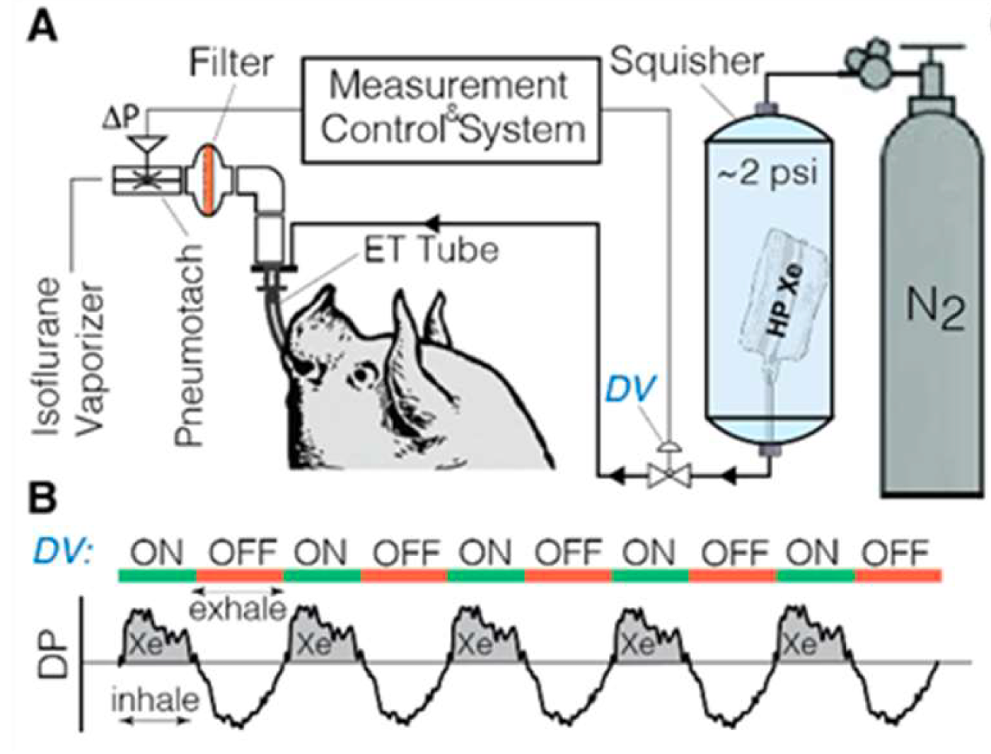
We imaged two 5-month-old Yucatan miniature pigs, one healthy control and one with right-sided (T3-9) rib tethering performed at 6 weeks-old (TIS model), as well as four New-Zealand white rabbits: one healthy and three with right-sided rib tethering. All images were acquired in prone position on 1.5T Siemens scanner during spontaneous breathing. Xemed prototype system was used to polarize 87% enriched xenon-129. Pigs were imaged using an 8-channel xenon-129 coil (Stark Contrast), while rabbits were imaged with a custom birdcage designed coil (Stark Contrast). All animal studies were approved by the Institutional Animal Care and Use Committee of the University of Pennsylvania.As depicted in Figure 1, polarized xenon bags were placed under ~2psi pressure inside a custom-built plastic container (squisher) directly connected to the endotracheal (ET) tube blocked by a pneumatic diaphragm valve (DV) that is normally closed. Inhale/exhale breathing curves were collected from the pneumotach to actuate the DV at each breath, delivering a small dose of polarized-xenon (rabbits = 6mL/kg, total of 750mL; pigs = 10mL/kg, total of 2L).
3D-spiral interleaves were acquired continuously over approximately three minutes. To quantify both ventilation and gas exchange, the frequencies of each RF excitation pulse were alternated between the gas phase (GP, 0 ppm) and dissolved phase (DP, 200 ppm) resonances with TR/TE = 7.63/0.62ms and GP/DP flip angles of 4°/30°. Images were reconstructed onto 80×80x80 grids with FOV of 150mm3 isotropic for pigs and 120mm3 for rabbits. Phase-binning was used to retrospectively bin each interleave into 16 phases of a representative breathing cycle. Symmetric image normalization with cross correlation metric in the ANTs toolbox was used to co-register all frames to the end-inhale frame to allow voxel-wise analysis. The 16 points of the GP signal representing an average breath were fit to piecewise sigmoid functions as
$$S(t) = \begin{cases} \frac{TV}{1 + e^{-(t - T_I)/\tau_I}} + \text{FRC} & \text{if } t < t_p \\ \frac{TV}{1 + e^{-(t - T_E)/\tau_E}} + \text{FRC} & \text{if } t \geq t_p \end{cases}$$
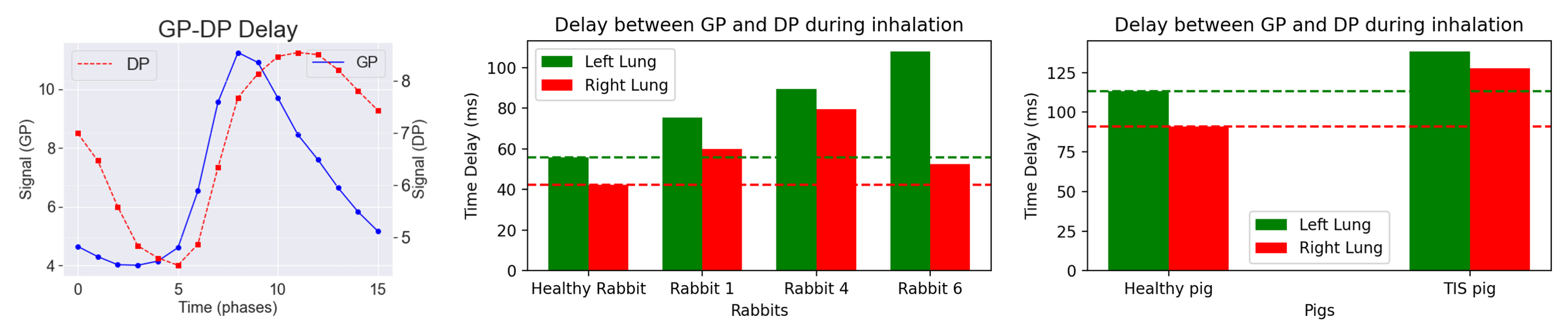
where TV, FRC, $$$T_{I/E}$$$, $$$\tau_{I/E}$$$, and $$$t_p$$$ represent the tidal volume, functional residual capacity, infliction point at inhalation/exhalation, inhalation/exhalation time constant, and the switching timepoint between inhalation and exhalation, respectively. The timepoints on each sigmoid curve at which the signal is 50% of the total amplitude were defined as arrival time (inhalation) and departure time (exhalation). Fractional ventilation was calculated as TV/(TV+FRC). Finally, the time delay between GP and DP (GP-DP delay) was measured as the arrival time of DP signal minus the arrival time of GP signal at each voxel.
Results/Discussion
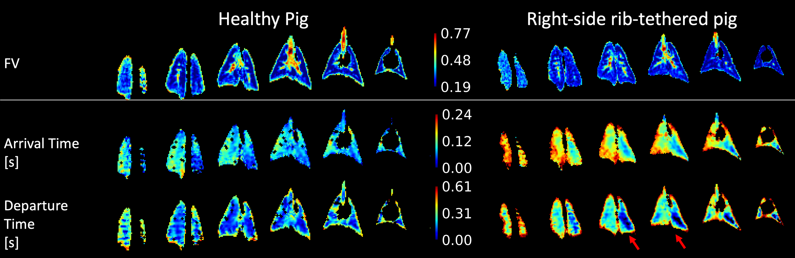
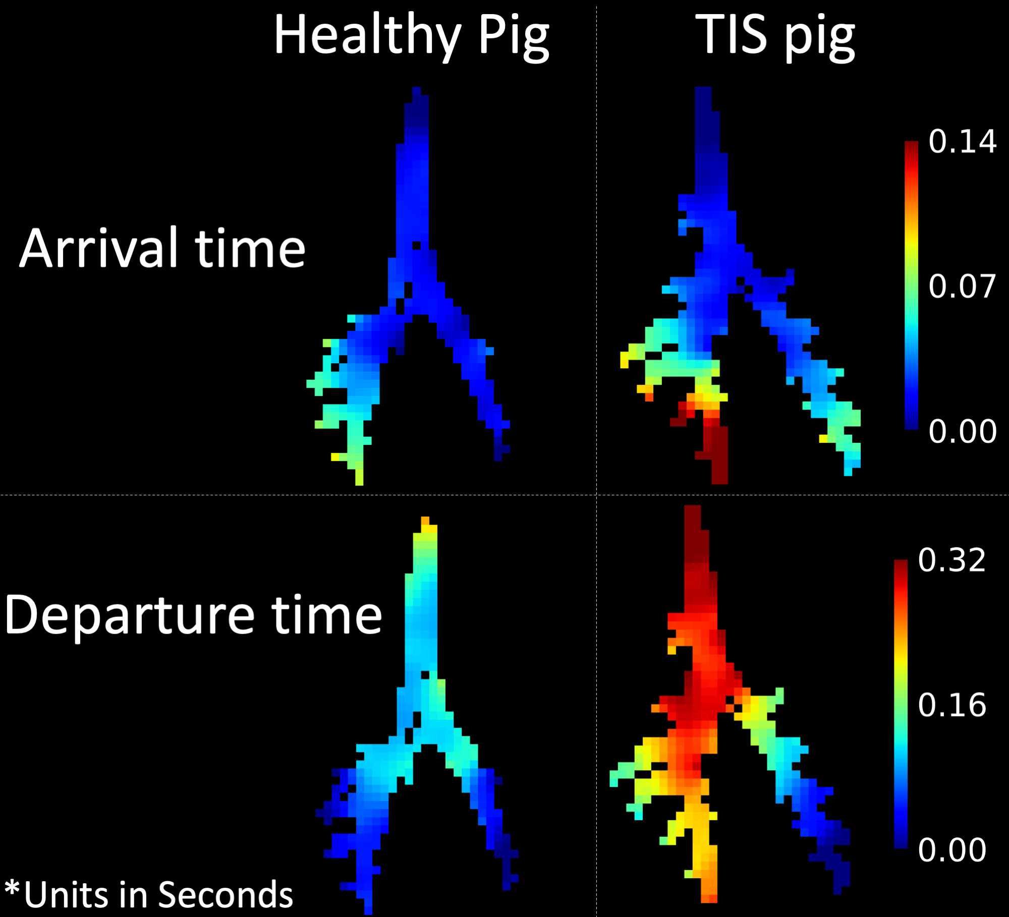
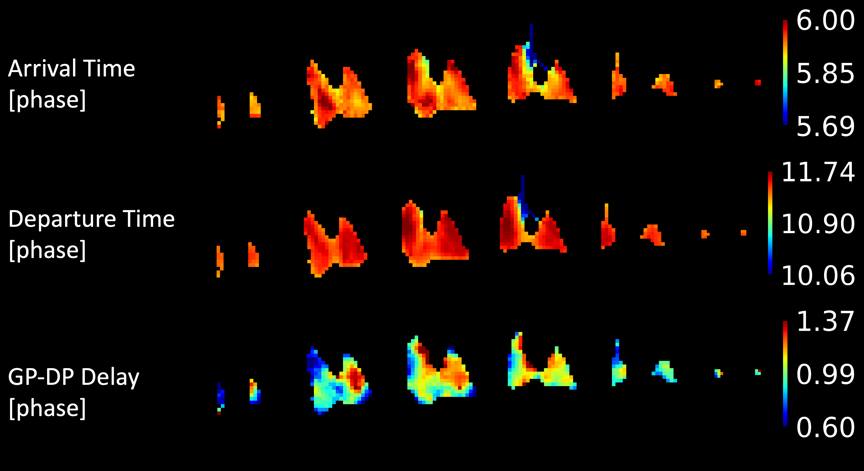
Figure 2 shows arrival/departure times and FV maps for both TIS and healthy pigs. While FV differences between pigs are minimal, arrival and departure time measurements reveal significant delays during both inhalation and exhalation in the tethered lung compared to the contralateral lung. Figure 3 highlights the airways, illustrating differences between the left and right bronchi in the TIS pig. In figure 4, increased delay between GP and DP can be seen in TIS animals compared to healthies. These delays were higher in the left lung compared to right lung for all animals. Moreover, observed GP-DP delays were longer in pigs compared to rabbits, suggests that they are species- or size-dependent. Figure 5 shows the arrival/departure times as well as the GP-DP delay maps for a TIS rabbit. Delayed xenon arrival was observed in the posterior of the right tethered lung compared to the left lung. Moreover, increased GP-DP delays were observed in the left lung compared to the right tethered lung. GP-DP delay could increase as a result of alveolar wall-thickening, airway restrictions, or differences in breathing rate or inhale/exhale ratio; however, different breathing patterns would have less effect on the voxel level analysis shown in figure 5, making it less likely to be the cause in this case.Conclusion
Hyperpolarized xenon-129 dynamic-MRI reveals altered ventilation and gas exchange dynamics resulting from TIS, potentially offering an improved tool for diagnosis and treatment monitoring, as well as additional insight into the altered lung function associated with this disease.Acknowledgements
This work was funded by the US National Institute of Health and the Wyss-Campbell Center for Thoracic Insufficiency at Children's Hospital of Philadelphia .References
- Karol, L. A. et al. Pulmonary function following early thoracic fusion in non-neuromuscular scoliosis. J Bone Joint Surg Am 90, 1272-1281 (2008). https://doi.org:10.2106/jbjs.G.00184
- Tsiligiannis, T. & Grivas, T. Pulmonary function in children with idiopathic scoliosis. Scoliosis 7, 7 (2012). https://doi.org:10.1186/1748-7161-7-7
- Pehrsson, K., Larsson, S., Oden, A. & Nachemson, A. Long-term follow-up of patients with untreated scoliosis. A study of mortality, causes of death, and symptoms. Spine (Phila Pa 1976) 17, 1091-1096 (1992). https://doi.org:10.1097/00007632-199209000-00014
Figures