0293
Comparison of diagnostic image quality of a 24-channel pediatric screen-printed coil to commercially available coils in clinical body MR exams1Stanford University Department of Radiology, Palo Alto, CA, United States
Synopsis
Keywords: Body, Pediatric
Motivation: To evaluate diagnostic image quality of novel 24-channel screen-printed flexible coil array in pediatric body MR at 3T
Goal(s): Whether the diagnostic image quality of novel 24-channel screen-printed flexible coil array in pediatric body MR at 3T is comparable to commercially available coils
Approach: Retrospective case-control study with pediatric patients who underwent clinical body MR exams using either screen-printed coils or commercial coils. A single radiologist reviewed image quality using anonymized randomized image stacks.
Results: A flexible screen-printed pediatric MRI receive coil yielded similar diagnostic image quality to commercial coils on body MRI exams performed in the clinical setting.
Impact: Screen-printed pediatric MRI receive coil yielded comparable diagnostic image quality to commercial coils on body MRI exams performed in the clinical setting on 3T scanners while offering the advantages of comfort, flexibility, and ease of use.
Introduction
MRI is the preferred modality for imaging a wide array of pediatric conditions given its lack of ionizing radiation. However, pediatric patients’ smaller size and limited ability to understand and follow instructions have often led to decreased signal-to-noise ratio (SNR), and one of the contributing factors is the use of large and relatively inflexible coils. Such coils are heavy and are often placed at a distance to the anatomy of interest, reducing SNR. They also generally have suboptimal geometry for pediatric applications, resulting in reduced acceleration capability. Finally, they can cause discomfort and physical as well as emotional distress in the patient and caregiver. Novel screen-printed MRI coils allow for a closer fit to the patient and, given they may be printed on flexible substrates such as fabric, have been shown to be preferred over commercially available coils by patients, parents, and caregivers (1-8). This project aims to evaluate the comparative image quality of a 24-channel screen-printed flexible coil array dedicated to pediatric body MRI at 3T versus conventional coils routinely used for pediatric imaging.Methods
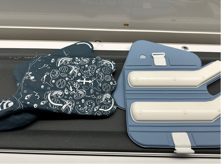
This retrospective study was done with IRB approval and waived consent/assent. Twenty (N=20) pediatric patients underwent MR of the heart (n=8), chest (n=3), and abdomen and pelvis (n=9) using screen-printed flexible receive coils on one of three 3T scanners (GE MR 750, SIGNA PET/MR, SIGNA Premier, GE Healthcare, Waukesha, WI) using a 24-channel phased-array coil (InkSpace Imaging, Pleasanton, CA). A case-control study was performed, with 1:2 matching of cases to control subjects based on age, body part, sex, and scanner type. Control patients were scanned with the standard coil in the clinical workflow, which included a 32-channel coil or 16 channel flex coils (GE Healthcare, Waukesha, WI). Anonymized, randomized and scrollable image stacks were provided to a radiologist (ABS, 9 years of experience interpreting MRI studies) who classified each exam by using a Likert scale (1=non-diagnostic; 2=substantial deficits in image quality impacting clinical utility; 3= deficits in image quality not impacting clinical utility; 4=no deficits in image quality; and 5=excellent image quality) on three scoring categories: apparent signal-to-noise ratio, image blurring, and overall image quality. All reviews were conducted on two sequences from each exam: a coronal single-shot T2 sequence and an axial T1-weighted postcontrast sequence for MRI of the abdomen and pelvis; a coronal respiratory triggered T2 weighted 3D FSE sequence and a 3D SPGR with 2-point Dixon fat suppression for MRI of the chest, and an axial T1 weighted postcontrast sequence and an axial ungated contrast-enhanced MRA sequence for MR of the heart. Wilcoxon signed rank test was used to compare the image quality scores between the control and test cases, with Bonferroni-Holm method applied to correct for multiple comparison categories.Results
Twenty study participants were evaluated with the screen-printed coil (age range, 2 days to 15 years old; 7 male and 13 female subjects). The radiologist determined that all 20 cases are diagnostically acceptable, with an average overall image quality score of 3.85 ± 0.75, compared to 3.75 ± 0.90 for the standard coils (P = 0.810). None of the study participants needed to repeat the exam with a different coil. Exams performed with the screen-printed coils yielded mean score for apparent SNR of 3.75 ± 0.74 compared to 3.73 ± 0.94 for standard coils, and a mean score for blurring of 3.98 ± 0.92 compared to 3.96 ± 0.82 for standard coils. Differences in apparent SNR and blurring scores were not statistically significant (P = 0.920 and 0.841, respectively).Discussion
We showed that clinical body MRI exams performed with novel screen-printed coils on 3T scanners yielded comparable diagnostic image quality to those performed with widely available commercial coils in pediatric patients. Given their ease of use by technologists, superior comfort, and preference by parents, caregivers and technologists over conventional adult coils, screen-printed coils show promise in increasing the utilization and improving the user experience of MRI for pediatric body imaging. Limitations of this study include only one reader, a small number of patients, and lack of an imaging atlas to guide scoring. Additionally, due to the practical considerations of scan time and requirement for general anesthesia for some of the study participants, all but one subject was scanned using either the new screen-printed coil or the conventional commercial coil. No direct comparisons of the coils were made in the same subject during the same exam.Conclusion
A flexible screen-printed pediatric MRI receive coil yielded similar diagnostic image quality to standard coils on body MRI exams performed in the clinical setting.Acknowledgements
No acknowledgement found.References
1. Zhang T, Grafendorfer T, Cheng JY, et al. A semiflexible 64-channel receive-only phased array for pediatric body MRI at 3T. Magn Reson Med 2016;76(3):1015–1021.
2. Keil B, Alagappan V, Mareyam A, et al. Size-optimized 32-channel brain arrays for 3 T pediatric imaging. Magn Reson Med 2011;66(6):1777–1787
3. Lopez Rios N, Foias A, Lodygensky G, Dehaes M, Cohen-Adad J. Size-adaptable 13-channel receive array for brain MRI in human neonates at 3 T.
4. Corea JR, Flynn AM, Lechêne B, et al. Screen-printed flexible MRI receive coils. Nat Commun 2016;7(1):10839.
5. Corea JR, Lechene PB, Lustig M, Arias AC. Materials and methods for higher performance screen-printed flexible MRI receive coils. Magn Reson Med 2017;78(2): 775–783.
6. Corea J, Lechene BP, Grafendorfer T, Robb F, Arias AC, Lustig M. Printed receive coil arrays with high SNR [abstr]. In: Proceedings of the Twenty-Fourth Meeting of the International Society for Magnetic Resonance in Medicine. Berkeley, Calif: International Society for Magnetic Resonance in Medicine, 2016; 2146.
7. Winkler SA, Corea J, Lechene B, et al. First clinical pilot study using screen-printed flexible MRI receive coils for pediatric applications [abstr]. In: Proceedings of the Twenty-Sixth Meeting of the International Society for Magnetic Resonance in Medicine. Berkeley, Calif: International Society for Magnetic Resonance in Medicine, 2018.
8. Simone Angela Winkler, Joseph Corea, Balthazar Lechêne, Kendall O’Brien, John Ross Bonanni, Akshay Chaudhari, Marcus Alley, Valentina Taviani, Thomas Grafendorfer, Fraser Robb, Greig Scott, John Pauly, Michael Lustig, Ana Claudia Arias, and Shreyas Vasanawala. Evaluation of a Flexible 12-Channel Screen-printed Pediatric MRI Coil. Radiology 2019 291:1, 180-185
Figures