0290
Accelerated Free-Breathing Liver Fat/R2* Quantification in Pediatrics Using XD-GRASP Multi-Echo Stack-of-Radial MRI: A Retrospective Study1Radiological Sciences, University of California Los Angeles, Los Angeles, CA, United States, 2Biomedical Engineering, University of Memphis, Memphis, TN, United States, 3MR Application Predevelopment, Siemens Healthineers AG, Erlangen, Germany, 4Radiology, Cincinnati Children's Hospital Medical Center, Cincinnati, OH, United States, 5Diagnostic Imaging, St. Jude Children’s Research Hospital, Memphis, TN, United States, 6MR R&D Collaborations, Siemens Medical Solutions USA, Inc, Los Angeles, CA, United States, 7MR R&D Collaborations, Siemens Medical Solutions USA, Inc, Austin, TX, United States
Synopsis
Keywords: Body, Liver, Fat and iron quantification
Motivation: Free-breathing liver fat and iron quantification is of growing interest in pediatric patients. However, motion compensation is necessary for quantification accuracy, resulting in prolonged acquisition times.
Goal(s): This study was to retrospectively evaluate a newly developed technique with XD-GRASP reconstruction in pediatric patients.
Approach: Data with oversampled k-space radial views were undersampled to fewer radial views. All data were reconstructed and compared between a self-gating method and the proposed XD-GRASP method.
Results: When applied to data with undersampled radial views, the proposed XD-GRASP method reduced image artifacts and improved PDFF and R2* results compared to the self-gating method.
Impact: The proposed multi-echo stack-of-radial MRI method using motion-resolved reconstruction and multi-dimensional regularization may allow accelerated free-breathing liver PDFF and R2* mapping in pediatric patients.
INTRODUCTION
There is a growing interest to evaluate and quantify liver fat and iron in children1. Although breath-hold three-dimensional (3D) multi-echo Cartesian gradient-echo imaging is a well-validated method for liver fat and iron quantification in adults2-4, it is challenging to implement in children who have difficulty with breath-holding. Alternatively, 3D stack-of-radial imaging can provide accurate liver proton density fat fraction (PDFF) and R2* quantification in pediatric patients using free-breathing acquisitions5-10, with self-gating to correct possible measured R2* biases due to respiratory motion8-10. However, after self-gating, only a subset of radial views is used for reconstruction, causing possible residual undersampling artifacts. This often necessitates prolonged acquisition of additional radial views8-10. A recent study proposed to address this limitation using a modified extra-dimensional golden-angle radial sparse parallel (XD-GRASP) approach, and validated this technique in adult subjects11.The purpose of this study was to perform a retrospective evaluation of the free-breathing stack-of-radial liver PDFF and R2* quantification technique with XD-GRASP reconstruction11 in pediatric patients, using the oversampled data as a reference.
METHODS
Approval from the local institution review board was obtained. Raw data of free-breathing stack-of-radial acquisitions from a previous study10 were retrospectively used for our analysis. MRI performed at 1.5T (MAGNETOM AvantoFit, Siemens Healthineers, Erlangen, Germany) for the evaluation of liver iron overload in 21 clinical pediatric subjects without global fat/water swapping were included in this study (15 females, 14.7±4.2 yrs, BMI: 24.8±6.4 kg/m2). The acquisition parameters are in Table 1.All scans were acquired with 700 radial views (oversampled with a sampling factor of 2.0). The raw data were undersampled to 352 and 176 radial views (sampling factors 1.0 and 0.5 respectively). All data were reconstructed with a self-gating method applied to accept data acquired near end-expiration with a 40% acceptance rate (termed as “stable40”)8 and the proposed XD-GRASP method11 for comparison. Processing steps included k-space shift correction6,7,12, self-gating8,9, multi-dimensional regularization11 and PDFF/R2* quantification4.
For the multi-dimensional regularization11, 4 motion states were resolved by self-gating. The following cost function was minimized: 0.5||Ax-y||22+||Wx||1, where A is an operator collectively applying relevant processing steps such as coil sensitivity map multiplication and non-uniform Fourier transformation; x is the image to be reconstructed; y is the acquired k-space data; ||x||2 is the Euclidean l2-norm, and ||x||1 is the l1-norm. W is the redundant Haar wavelet transformation applied in the in-plane spatial and motion-state dimensions whose low- and high-pass filters are separately scaled empirically11.
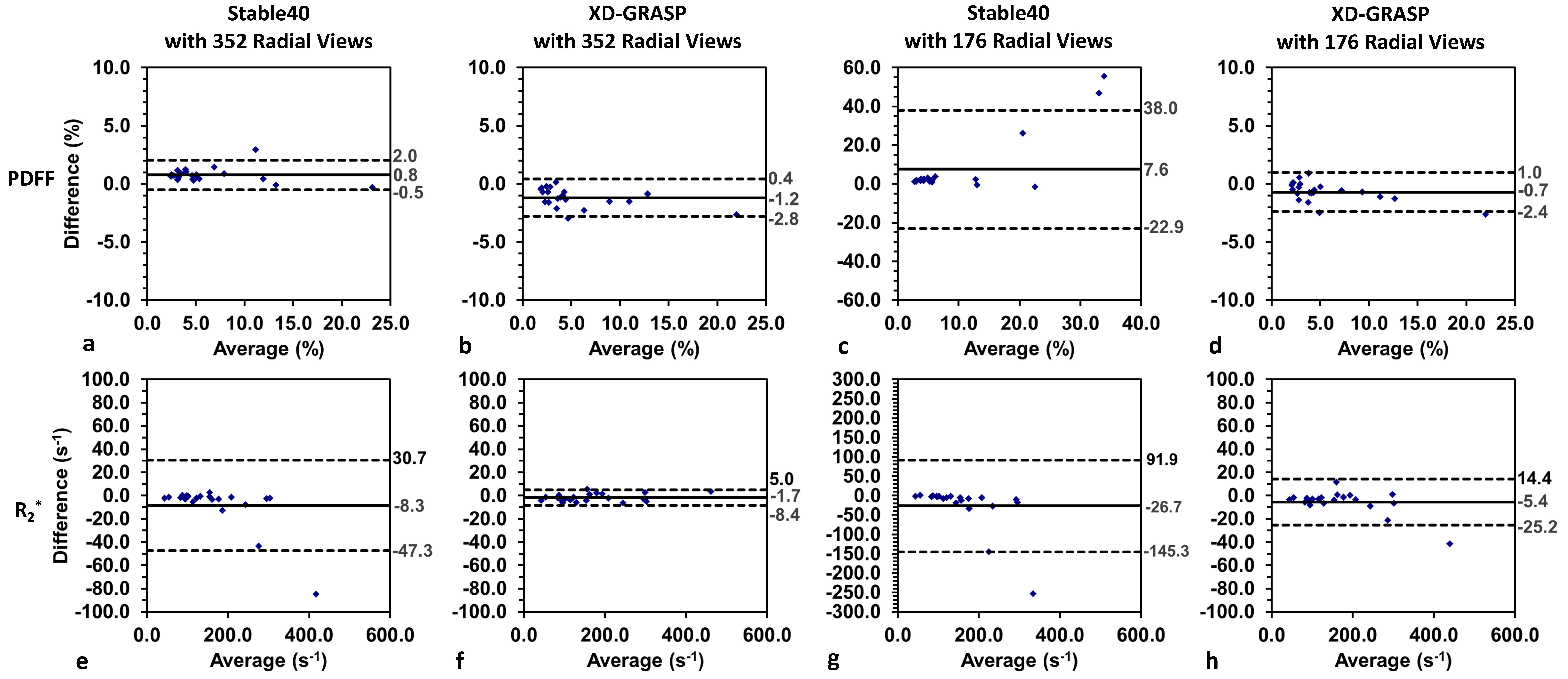
The same whole-liver regions of interest (ROIs) on a mid-level slice for all subjects from the previous study10 were used. PDFF and R2* values were measured as mean ± standard deviation (SD). Using the results of the stable40 method on data with 700 radial views, Bland-Altman analysis was performed to determine mean differences (MD) and limits of agreement (LoA = MD ± 1.96×SD) of different methods, reported as [MD; lower LoA, upper LoA].
RESULTS & DISCUSSION
Representative self-gating signal curves from one subject with different radial views are shown in Figure 1. Example echo images, PDFF and R2* maps of two subjects reconstructed with the stable40 and XD-GRASP methods using data with 352 and 176 views (sampling factor = 1.0 and 0.5 respectively) are shown in Figures 2 and 3. Reduced artifacts on images and maps using the proposed XD-GRASP method were observed.Bland-Altman plots comparing the reference PDFF and R2* results from the stable40 method on the data with 700 views to those of the stable40 and XD-GRASP methods with 352 and 176 views are shown in Figure 4. MD and LoA for the proposed XD-GRASP method using 176 views were [-5.4;-25.2,14.4] s-1 for R2* and [-0.7;-2.4,1.0]% for PDFF, which had lower biases than the stable40 method using 352 radial views: [-8.3;-47.3,30.7] s-1 for R2* and [0.8;-0.5,2.0]% for PDFF.
Compared to actual acquisition times of 144-417 seconds in the original protocols with 700 views10, the sampling factor of 0.5 (i.e., an acceleration factor of 4) was feasible with the proposed XD-GRASP method, leading to an estimated/calculated acquisition time of 47-137 seconds, i.e., 67% time saving (Table 1).
CONCLUSION
A multi-echo stack-of-radial MRI method using motion-resolved reconstruction and multi-dimensional regularization for free-breathing liver PDFF and R2* quantification was evaluated retrospectively in pediatric patients. Using reference results from oversampled k-space data, our proposed method demonstrated reduced artifacts and improved PDFF/R2* quantification accuracy on the undersampled k-space data compared to a self-gating method. In this study, an acceleration factor of 4 and an approximate acquisition time saving of 97-280 seconds (67% shorter) were achieved. This proposed method may allow accelerated free-breathing liver PDFF and R2* mapping in pediatric subjects.Acknowledgements
No acknowledgement found.References
1. Barshop NJ, Sirlin CB, Schwimmer JB, Lavine JE. Epidemiology, pathogenesis and potential treatments of paediatric non-alcoholic fatty liver disease. Aliment Pharmacol Ther. 2008;28:13-24.
2. Yu H, Shimakawa A, McKenzie CA, Brodsky E, Brittain JH, Reeder SB. Multiecho water‐fat separation and simultaneous R2* estimation with multifrequency fat spectrum modeling. Magn Reson Med. 2008;60:1122-1134.
3. Hernando D, Levin YS, Sirlin CB, Reeder SB. Quantification of liver iron with MRI: state of the art and remaining challenges. J Magn Reson Imaging. 2014;40:1003-1021.
4. Zhong X, Nickel MD, Kannengiesser SAR, Dale BM, Kiefer B, Bashir MR. Liver fat quantification using a multi‐step adaptive fitting approach with multi‐echo GRE imaging. Magn Reson Med. 2014;72:1353-1365.
5. Peters DC, Derbyshire JA, McVeigh ER. Centering the projection reconstruction trajectory: reducing gradient delay errors. Magn Reson Med. 2003;50:1-6.
6. Armstrong T, Dregely I, Stemmer A, et al. Free-breathing liver fat quantification using a multiecho 3D stack-of-radial technique. Magn Reson Med. 2018;79:370-382.
7. Armstrong T, Ly KV, Murthy S, et al. Free-breathing quantification of hepatic fat in healthy children and children with nonalcoholic fatty liver disease using a multi-echo 3-D stack-of-radial MRI technique. Pediatr Radiol. 2018;48:941-953.
8. Zhong X, Armstrong T, Nickel MD, et al. Effect of respiratory motion on free‐breathing 3D stack‐of‐radial liver R2* relaxometry and improved quantification accuracy using self-gating. Magn Reson Med 2020;83:1964-1978.
9. Zhong X, Hu HH, Armstrong T, Li X, Lee YH, Tsao TC, Nickel MD, Kannengiesser SAR, Dale BM, Deshpande V, Kiefer B, Wu HH. Free-breathing volumetric liver R2* and proton density fat fraction quantification in pediatric patients using stack-of-radial MRI with self-gating motion compensation. J Magn Reson Imaging 2021;53:118-129.
10. Rohani SC, Morin CE, Zhong X, Kannengiesser S, Shrestha U, Goode C, Holtrop J, Khan A, Loeffler RB, Hankins JS, Hillenbrand CM, Tipirneni-Sajja A. Hepatic iron quantification using a free-breathing 3D radial gradient echo technique and validation with a 2D biopsy-calibrated R2* relaxometry method. J Magn Reson Imaging 2022;55:1407-1416.
11. Zhong X, Nickel MD, Kannengiesser SAR, Dale BM, Wu HH, Deshpande V. Improved free-breathing volumetric liver fat and R2* quantification using 3D stack-of-radial GRE Dixon MRI and XD-GRASP reconstruction. In Proceedings of the 31st Annual Meeting of ISMRM, Toronto, Canada, 2023. p. 1217.
12. Zhong X, Armstrong T, Gao C, Nickel MD, Han F, Dale BM, Li X, Kafali SG, Hu P, Wu HH, Deshpande V. Accelerated k-space shift calibration for free-breathing stack-of-radial MRI quantification of liver fat and R2*. Magn Reson Med 2022;87:281-291.
Figures
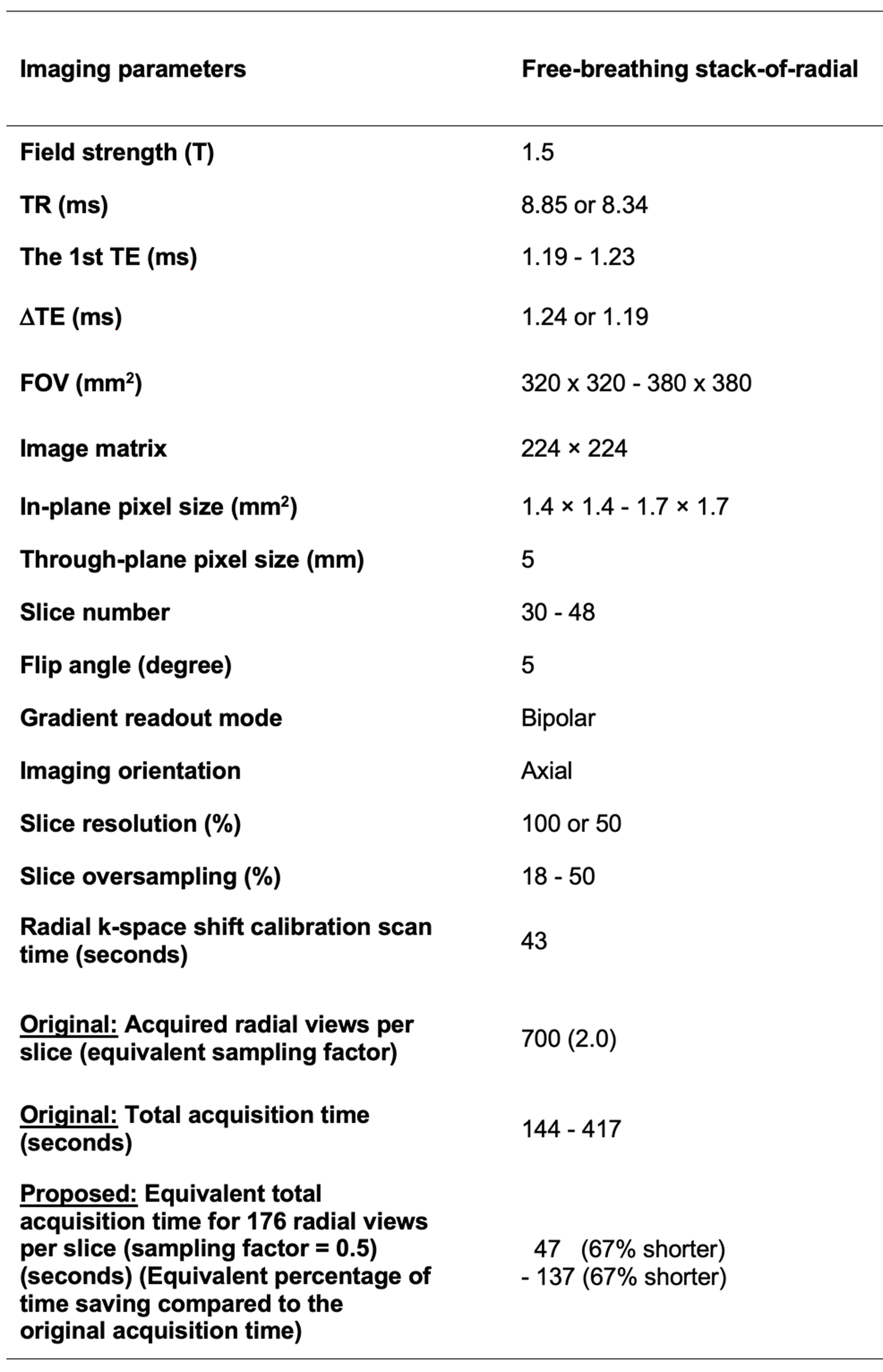
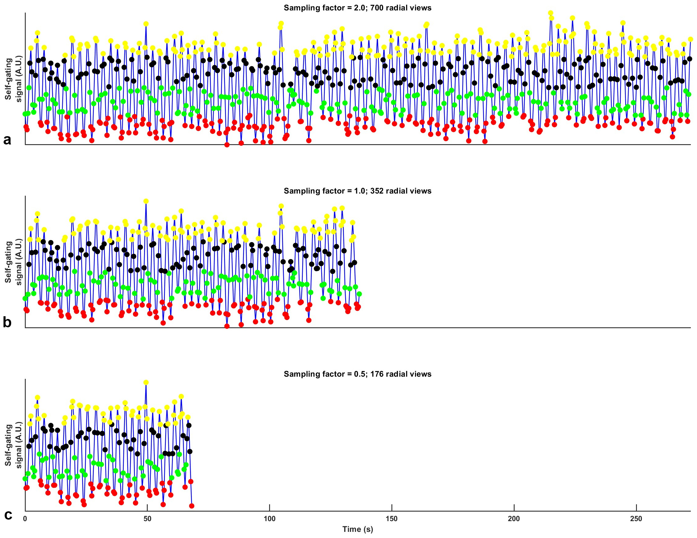
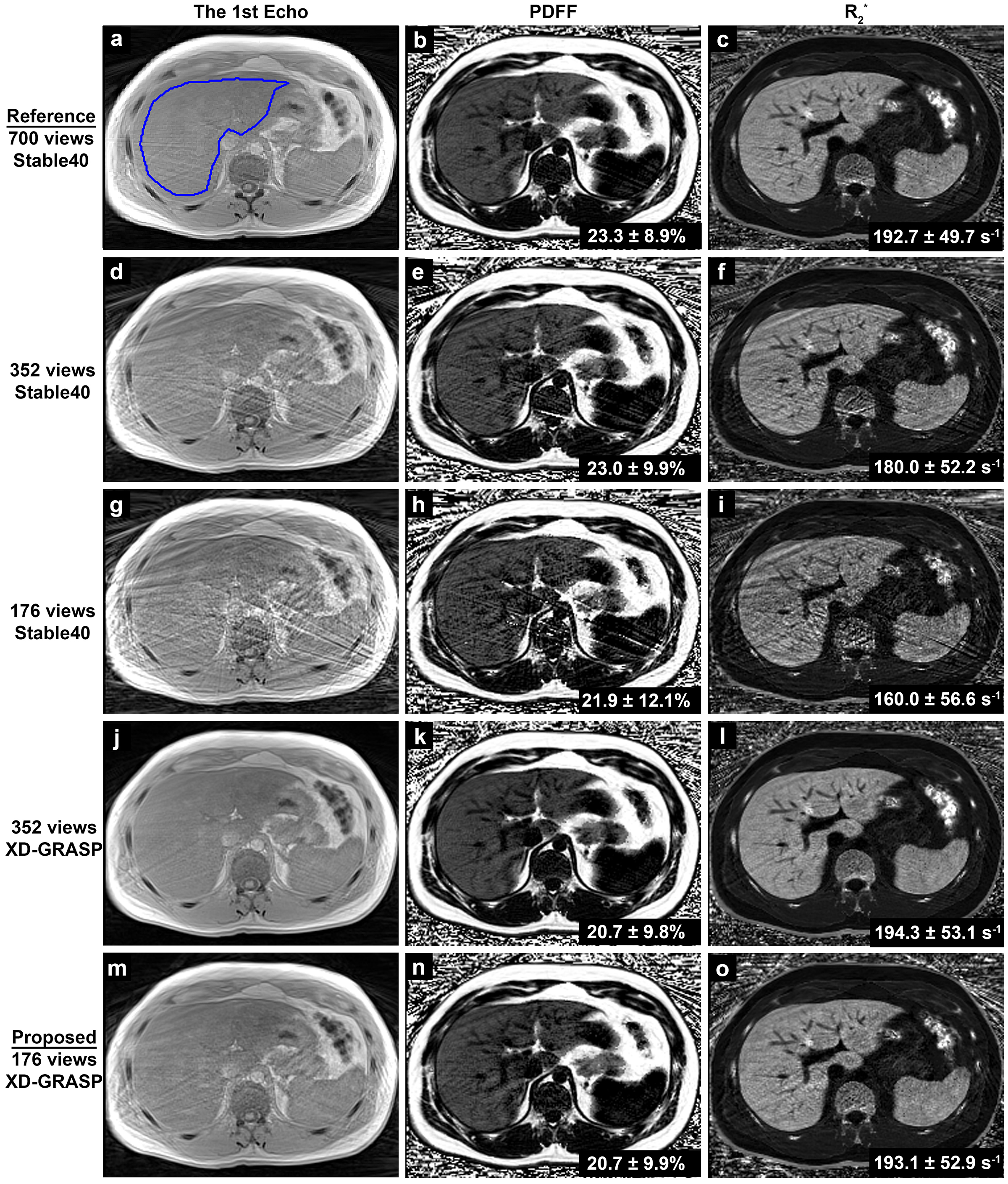
Figure 2 Example echo images, PDFF and R2* maps of one subject reconstructed with the methods of stable40 (a-i) and XD-GRASP (j-o) using data with 700, 352 and 176 views. The reference results are from the stable40 method on the data with 700 views (a-c). All methods show results at the first motion state (the end expiration state). The images and maps are cropped for better visualization. The PDFF and R2* values within the whole-liver contour (indicated by the blue outline in a) are reported as mean ± SD.
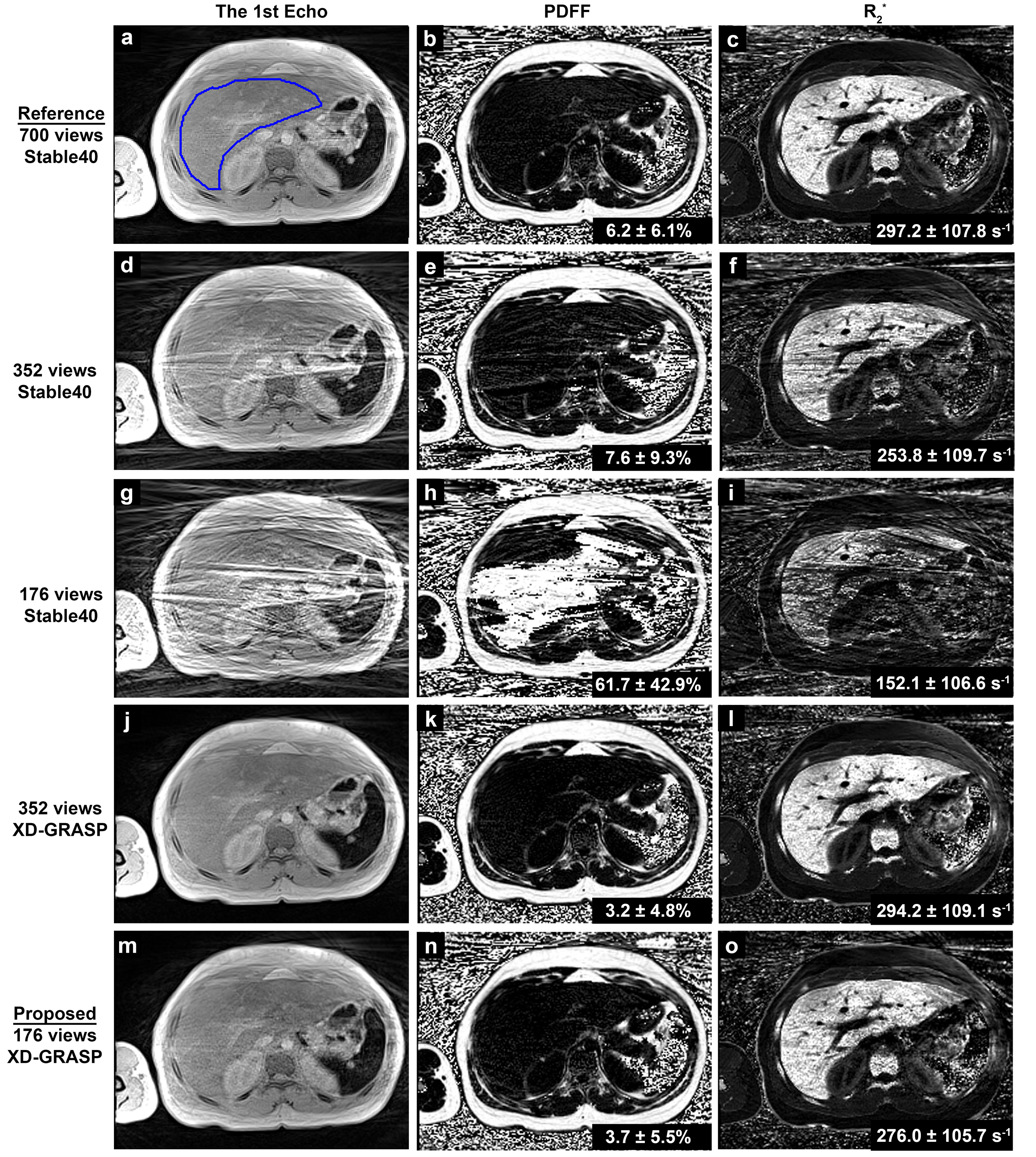
Figure 3 Example echo images, PDFF and R2* maps of a different subject reconstructed with the methods of stable40 (a-i) and XD-GRASP (j-o) using data with 700, 352 and 176 views. The undersampling artifacts are pronounced on the echo images of the stable40 method using the data with 176 views (g), accompanied by local fat/water swapping problems on the PDFF map (h) and aberrations on the R2* map (i), leading to erroneous PDFF and R2* values.