0277
Fast 3D Neuro T2-FLAIR with Learned Sampling and fully 3D Model Based Deep learning1Department of Medical Physics, University of Wisconsin-Madison, Madison, WI, United States, 2Department of Medicine, University of Wisconsin-Madison, Madison, WI, United States, 3Department of Radiology, University of Wisconsin-Madison, Madison, WI, United States
Synopsis
Keywords: White Matter, Brain
Motivation: T2-FLAIR is an essential contrast for clinical neuroimaging. However, the inherently long scan time limits its application in screening.
Goal(s): We aim to accelerate 3D T2-FLAIR scan while maintaining sufficient image quality.
Approach: We developed a framework that simultaneously learns a sampling pattern and a fully 3D deep learning reconstruction neural network. This allows exploiting the optimization space in both sampling and reconstruction.
Results: Learned sampling pattern with MoDL reconstruction trained with added Gaussian noise was able to provide high quality T2-FLAIR scan with 1x1x1.6mm resolution in 1 min 39s.
Impact: This work confirms the feasibility of a short 3D T2-FLAIR scan, provides insights for optimization strategies, and could lead to clinical implementation.
Introduction
T2 weighted fluid attenuated inversion recovery (T2-FLAIR) imaging plays an essential role in detection of white matter lesions and has become a routine scan in clinical protocols for stroke1,2, multiple sclerosis3, tumors 4, etc. While 3D T2-FLAIR scans can be acquired in clinical scan times leveraging variable flip angle, long echo train length (ETL) 5, and parallel imaging, high resolution protocols require longer scan times. This limits the utility of 3D T2-FLAIR for neurologic screening applications and in cases of motion. Further, achieving sufficient resolution for lesion segmentation is challenging in clinical settings. While techniques have been proposed to generally accelerate 3D T2-FLAIR including Wave-CAIPI6 and compressed sensing, these techniques are generally limited by parallel imaging factors. Here, we aim to shorten 3D T2-FLAIR scan for clinical screening purposes while maintaining sufficient image quality using learned sampling and deep learning (DL) image reconstruction. Previously, we developed a framework to achieve high quality fast 4D Flow imaging 7 from retrospectively undersampled ground truth images. In this work, we investigated simultaneously optimized sampling and a fully 3D model based deep learning (3D-MoDL) 8 reconstruction network to enable accelerated T2-FLAIR imaging.Methods
Training data: A total of 16 cases of T2-FLAIR CUBE data were used for training and validation. The data was acquired on 3T scanners (Signa Premier, GE Healthcare) with a 48-ch head coil, resolution 1x1x1.6mm3, matrix size 256x256x130, parallel acceleration 2x2. ARC (GE Healthcare) and an L1-Wavelet reconstruction 9 were used to obtain pseudo ground truth images for training. Coil sensitivity maps were estimated with JSENSE 10 and compressed to 12 channels using principal component analysis.Optimization: As is illustrated in Figure 1, the sampling along readout is uniform while the PE locations are not confined to a Cartesian grid, rather, they are trained together with a 3D-MoDL to optimize the image quality with respect to ground truth images, evaluated using normalized mean squared error (NMSE). Density compensation factors are learned simultaneously. At each iteration, we sample the ground truth images with the current sampling coordinates using differentiable 3D non-uniform Fourier transform (NUFFT), coil sensitivity maps, and subsequently reconstruct with MoDL. The CNN regularizer consists of convolutional layers and residual connections. The NUFFT was implemented by bindings to torchkbnufft and PyTorch 11–13, which allowed for gradient tracking of the coordinates and Adam 14 optimizer for learning. It has been observed that adding noise to the ground truths can improve the generalization performance 15. We trained our framework with complex zero-means Gaussian noise added to the ground truths, with 𝝈 estimated from the edge of its kspace data, as well as with no noise added and no learnable sampling pattern for comparisons. The sampling pattern was initialized with the same Poisson disc for all three scenarios.
Image acquisition and analysis: Prospective scans (n=6) were performed with 3D T2 FLAIR sequences. Details on the scan parameters are shown in Figure 4(a). We acquired accelerated scans with Poisson disc sampling and the two learned sampling patterns trained with and without Gaussian noise. The accelerated scans were reconstructed with its corresponding 3D-MoDL. A reference scan was reconstructed with L1-Wavelet and regarded as the ground truth image for NMSE calculations.
Results
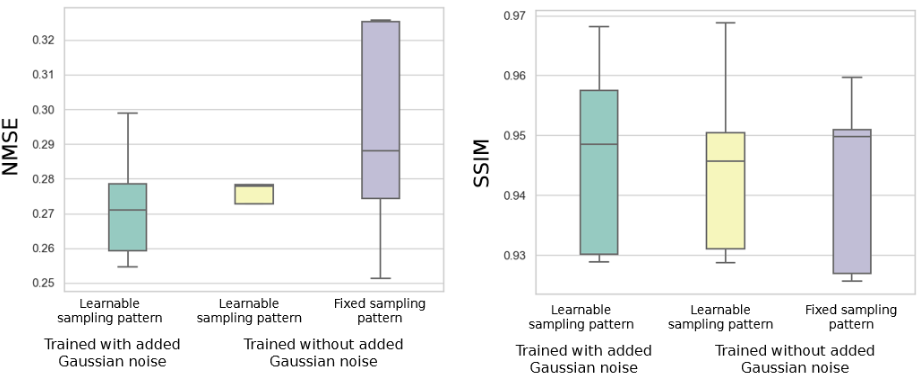
We optimized sampling patterns with 2704 phase encodes, which is approximately 9.2x accelerated (Figure 2). The number of training epochs was fixed, and the training and evaluation loss kept decreasing for all three optimization strategies (Figure 3(a)). However, as shown in Figure 3(b), the NMSE and SSIM of an additional test case deteriorated after some epochs for training without added Gaussian noise. This indicates overfitting, and adding noise reduces this behavior. Axial views of the test case are shown in Figure 3(c). Images from prospective scans are illustrated in Figure 4(b). Overall, training with a learnable sampling pattern and added Gaussian noise yielded the best image, while the fixed Poisson disc sampling tends to have the most residual aliasing (red arrows). Figure 5 shows NMSE and SSIM of the prospective scans from the three different optimization strategies.Discussion and Conclusions
In this work, we demonstrated the feasibility of a high quality brain T2-FLAIR scan in 1min39s by optimizing the sampling pattern and 3D DL reconstruction simultaneously. We confirmed that adding noise to the ground truth is a valid approach for improving generalization ability. Without learnable sampling, the fixed Poisson disc sampling with DL reconstruction provides images with more structured noise, yet NMSE is not sensitive to such artifacts (Figure 3(c), Figure 4(b)). Reader studies could be conducted in the future to better evaluate the quality of differently optimized frameworks.Acknowledgements
We gratefully acknowledge funding support from NIH grants R01AG075788 and R21NS125094 and research support from GE Healthcare.References
1. Noguchi K, Ogawa T, Inugami A, et al. Acute subarachnoid hemorrhage: MR imaging with fluid-attenuated inversion recovery pulse sequences. Radiology. 1995;196(3):773-777. doi:10.1148/radiology.196.3.7644642
2. Leiva-Salinas C, Wintermark M. Imaging of Ischemic Stroke. Neuroimaging Clin N Am. 2010;20(4):455-468. doi:10.1016/j.nic.2010.07.002
3. Tan IL, Pouwels PJW, van Schijndel RA, Adèr HJ, Manoliu RA, Barkhof F. Isotropic 3D fast FLAIR imaging of the brain in multiple sclerosis patients: initial experience. Eur Radiol. 2002;12(3):559-567. doi:10.1007/s00330-001-1170-8
4. Ellingson BM, Bendszus M, Boxerman J, et al. Editor’s choice: Consensus recommendations for a standardized Brain Tumor Imaging Protocol in clinical trials. Neuro-Oncology. 2015;17(9):1188. doi:10.1093/neuonc/nov095
5. Busse RF, Hariharan H, Vu A, Brittain JH. Fast spin echo sequences with very long echo trains: design of variable refocusing flip angle schedules and generation of clinical T2 contrast. Magn Reson Med. 2006;55(5):1030-1037. doi:10.1002/mrm.20863
6. Polak D, Cauley S, Huang SY, et al. Highly-accelerated volumetric brain examination using optimized wave-CAIPI encoding. J Magn Reson Imaging. 2019;50(3):961-974. doi:10.1002/jmri.26678
7. Tang C, Rivera-Rivera Leonardo, Eisenmenger L, Johnson KM. Machine Learned Wave Encoded Neurovascular 4D Flow. In: ISMRM Annual Meeting &Exhibition; 2023, Program #0705.
8. Aggarwal HK, Mani MP, Jacob M. MoDL: Model-Based Deep Learning Architecture for Inverse Problems. IEEE Transactions on Medical Imaging. 2019;38(2):394-405. doi:10.1109/TMI.2018.2865356
9. Lustig M, Donoho D, Pauly JM. Sparse MRI: The application of compressed sensing for rapid MR imaging. Magn Reson Med. 2007;58(6):1182-1195. doi:10.1002/mrm.21391
10. Ying L, Sheng J. Joint image reconstruction and sensitivity estimation in SENSE (JSENSE). Magn Reson Med. 2007;57(6):1196-1202. doi:10.1002/mrm.21245
11. Muckley MJ, Stern R, Murrell T, Knoll F. TorchKbNufft: A High-Level, Hardware-Agnostic Non-Uniform Fast Fourier Transform. In: ; 2020.
12. Gossard A. Bindings-NUFFT-pytorch. Published online December 14, 2021. Accessed November 9, 2022. https://github.com/albangossard/Bindings-NUFFT-pytorch
13. Paszke A, Gross S, Massa F, et al. PyTorch: An Imperative Style, High-Performance Deep Learning Library. Advances in Neural Information Processing Systems. 2019;32:8026-8037.
14. Kingma DP, Ba J. Adam: A Method for Stochastic Optimization. arXiv:14126980 [cs]. Published online January 29, 2017. Accessed April 9, 2022. http://arxiv.org/abs/1412.6980
15. Bishop CM. Training with Noise is Equivalent to Tikhonov Regularization. Neural Computation. 1995;7(1):108-116. doi:10.1162/neco.1995.7.1.108
Figures
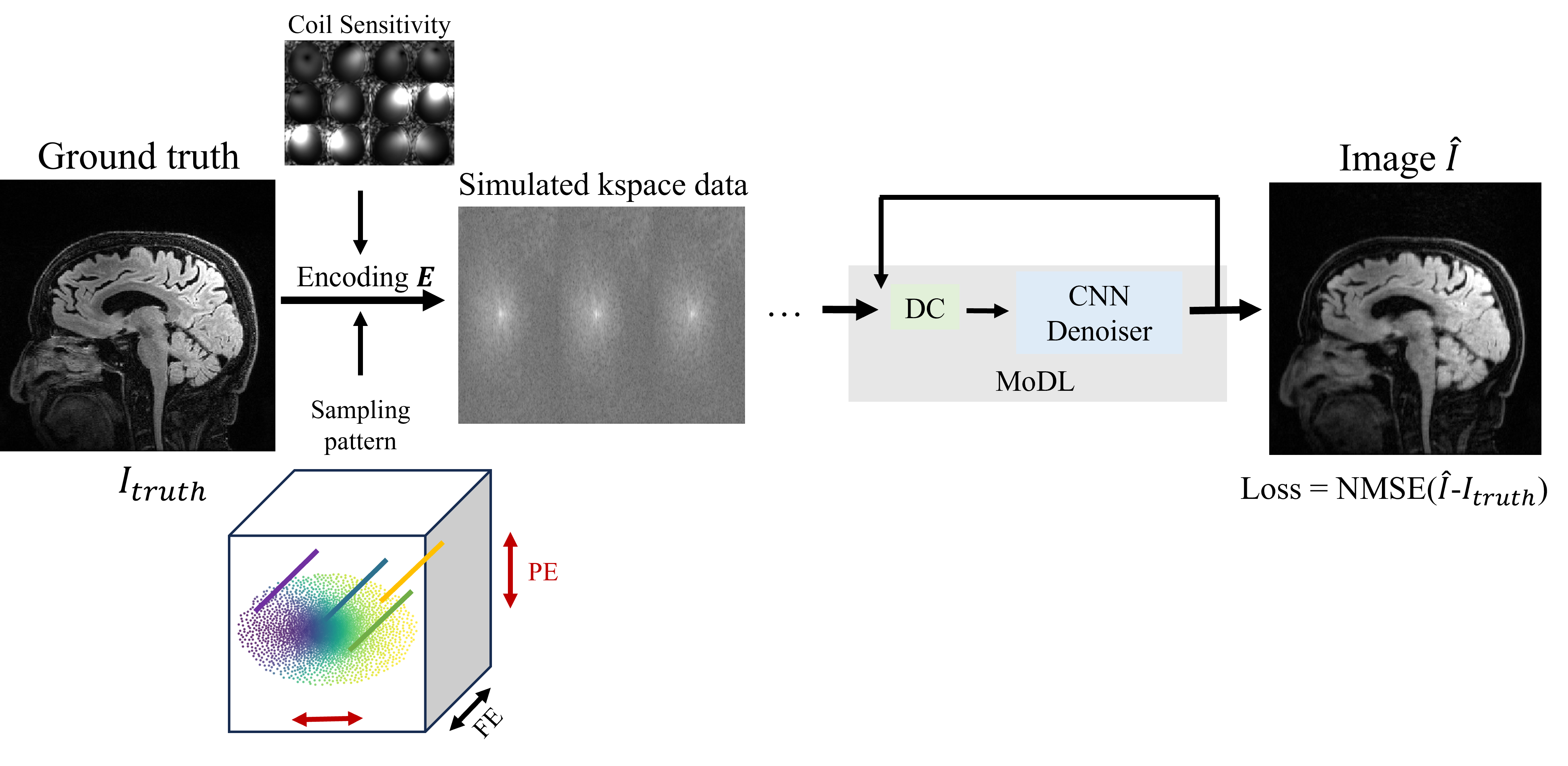
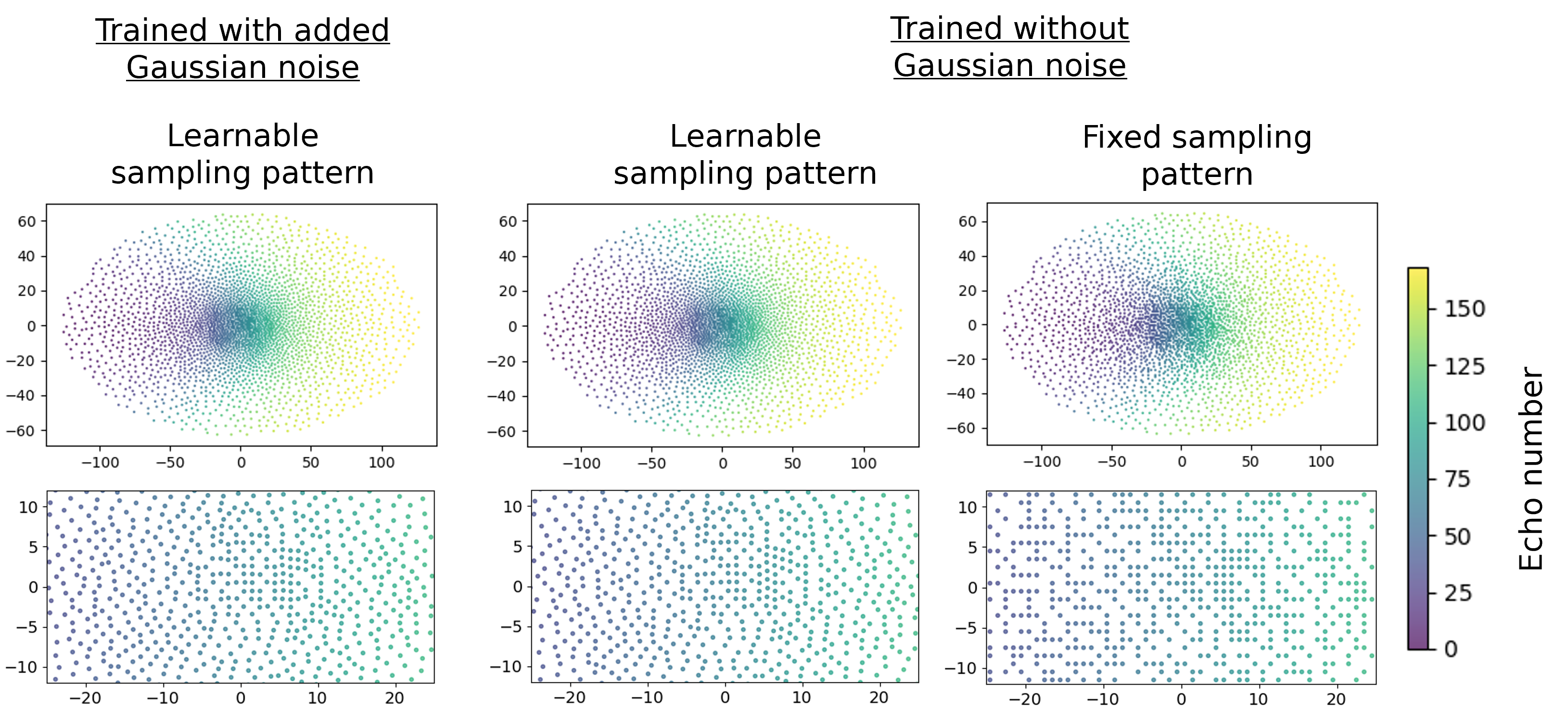
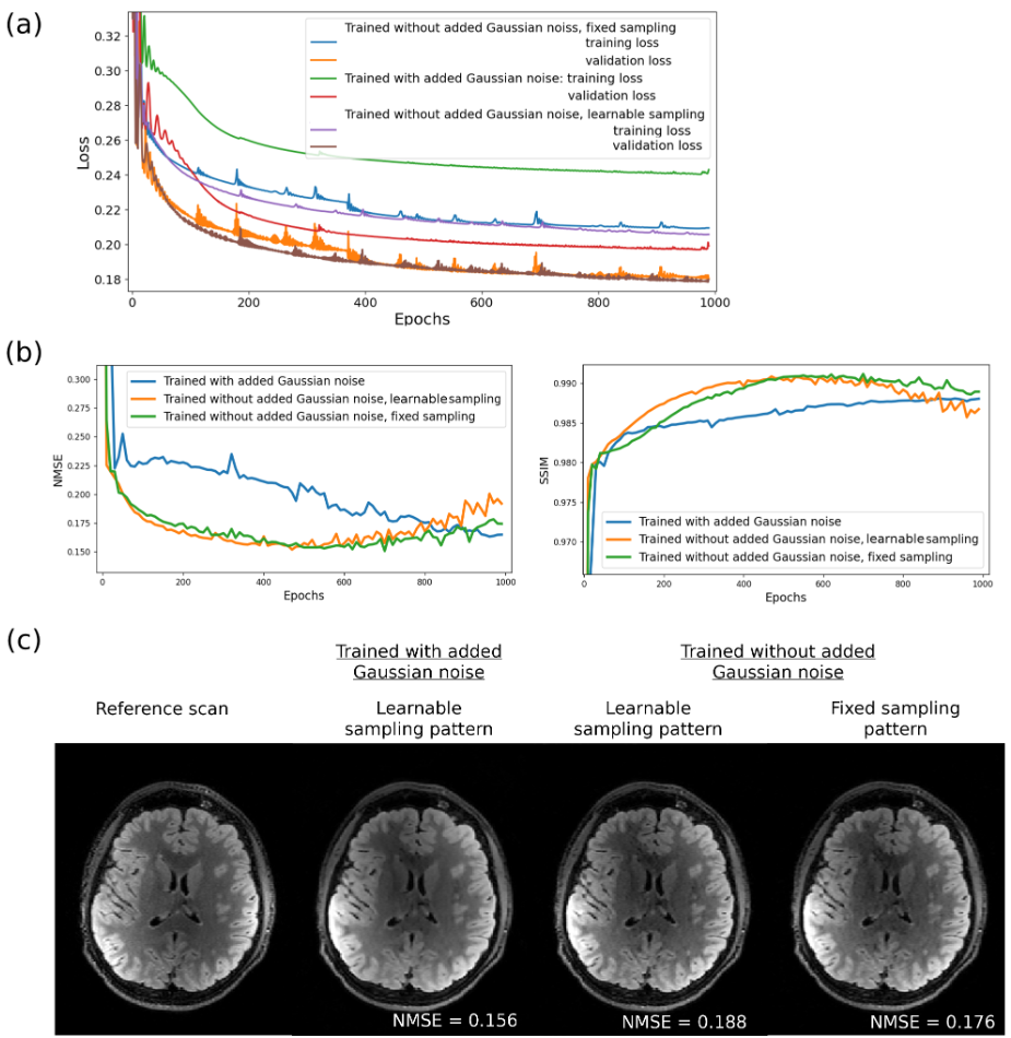
Figure 3. (a) Training and validation loss for the three different optimization strategies.
(b) NMSE and SSIM of a test case at saved framework states.
(c) Axial view of the reference and the retrospectively undersampled test case at the end of optimization.
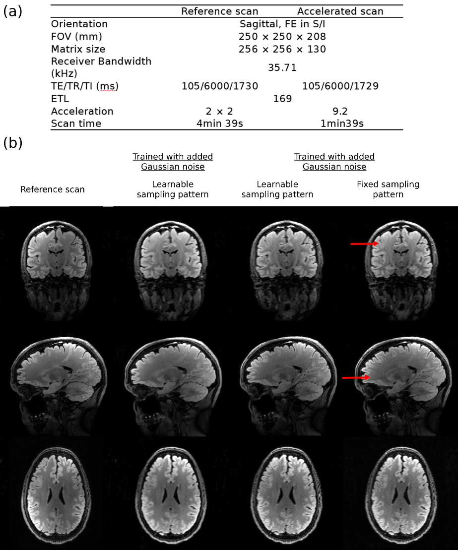
Figure 4. (a) Details on parameters for the prospective volunteer scans.
(b) Coronal, sagittal and axial views of the accelerated and reference volunteer scan.