0265
Advancements in Prostate Luminal Water Imaging: Radial Turbo Spin-Echo Acquisition and Spatial Regularization1University of Southern California, Los Angeles, CA, United States, 2Siemens Healthineers, Los Angeles, CA, United States
Synopsis
Keywords: Prostate, Cancer
Motivation: Non-invasive and efficient diagnostic methods for prostate cancer are urgently needed to enhance patient experience and improve diagnostic accuracy.
Goal(s): To develop and validate a novel radial turbo spin-echo (rTSE) sequence for luminal water imaging (LWI) that reduces MRI scan times while maintaining image quality.
Approach: Employed a radial k-space trajectory for the rTSE sequence, optimized on volunteers, followed by paired comparison with the MESE sequence. Implemented spatial regularization to stabilize the T2 decay curve fitting.
Results: The rTSE sequence halved scan times without compromising image quality or diagnostic precision. Spatial regularization significantly improved the homogeneity and smoothness of LWF, demonstrating better outcomes.
Impact: The rTSE sequence enhances prostate MRI by cutting scan times and discomfort without losing diagnostic accuracy. Spatial regularization refines tissue analysis for earlier cancer detection and reduces noise for clearer luminal water fraction maps.
Introduction
MRI stands at the forefront of non-invasive diagnosis of prostate cancer, with luminal water imaging (LWI) emerging as a promising method for differentiating cancerous prostate tissue based on luminal water fractions1,2. We introduce two significant refinements to LWI: a novel radial turbo spin-echo (rTSE) sequence to considerably shorten scan duration and a spatial regularization approach to enhance the smoothness constraint in T2 decay curve fitting to reduce noise in resultant fraction map.Methods
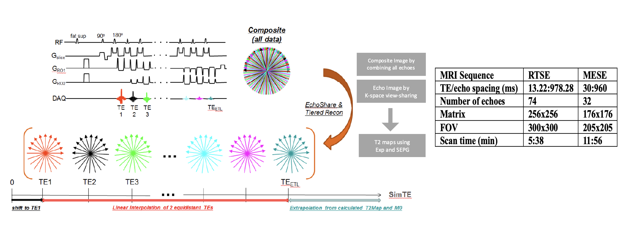
Technical Development: The rTSE sequence was meticulously designed to achieve a balance between efficiency and the reliability of prostate LWI (Fig. 1). By employing a radial k-space trajectory and sliding-window view-sharing reconstruction, the sequence circumvents the susceptibility to motion artifacts and accelerates acquisition3. Based on preliminary parameter optimization on a few healthy volunteers, we implemented a <6 min long rTSE protocol. In addition, to address the ill-posed nature of the T2 decay curve fitting process, we implemented a spatial regularization scheme. This approach accounts for the spatial correlation between adjacent voxels, augmenting the stability of the optimization problem and mitigating the influence of noise. The mathematical model was designed to achieve a homogenized smoothing effect across the prostate imaging plane, ensuring the LWF values' consistency and reliability. Alternating direction method of multipliers (ADMM) is used to solve the convex optimization problem in the T2 decay fitting considering all pixels collectively at the same time rather than independently within the fitting process. This promises greater mathematical stability of the LWF estimation, enhancing the homogeneity and smoothness of LWF map.Experiments and Data Analysis: Four healthy volunteers and three patients who had been scheduled for a clinical MRI study for highly suspicious prostate cancer were prospectively recruited after informed consent. In healthy volunteers, rTSE and multi-echo spin-echo (MESE, a conventional LWI sequence) scans were acquired consecutively for paired comparison, and a second rTSE scan was also acquired for scan-rescan analysis. In patients, rTSE and MESE were acquired in addition to the routine protocol.
Results
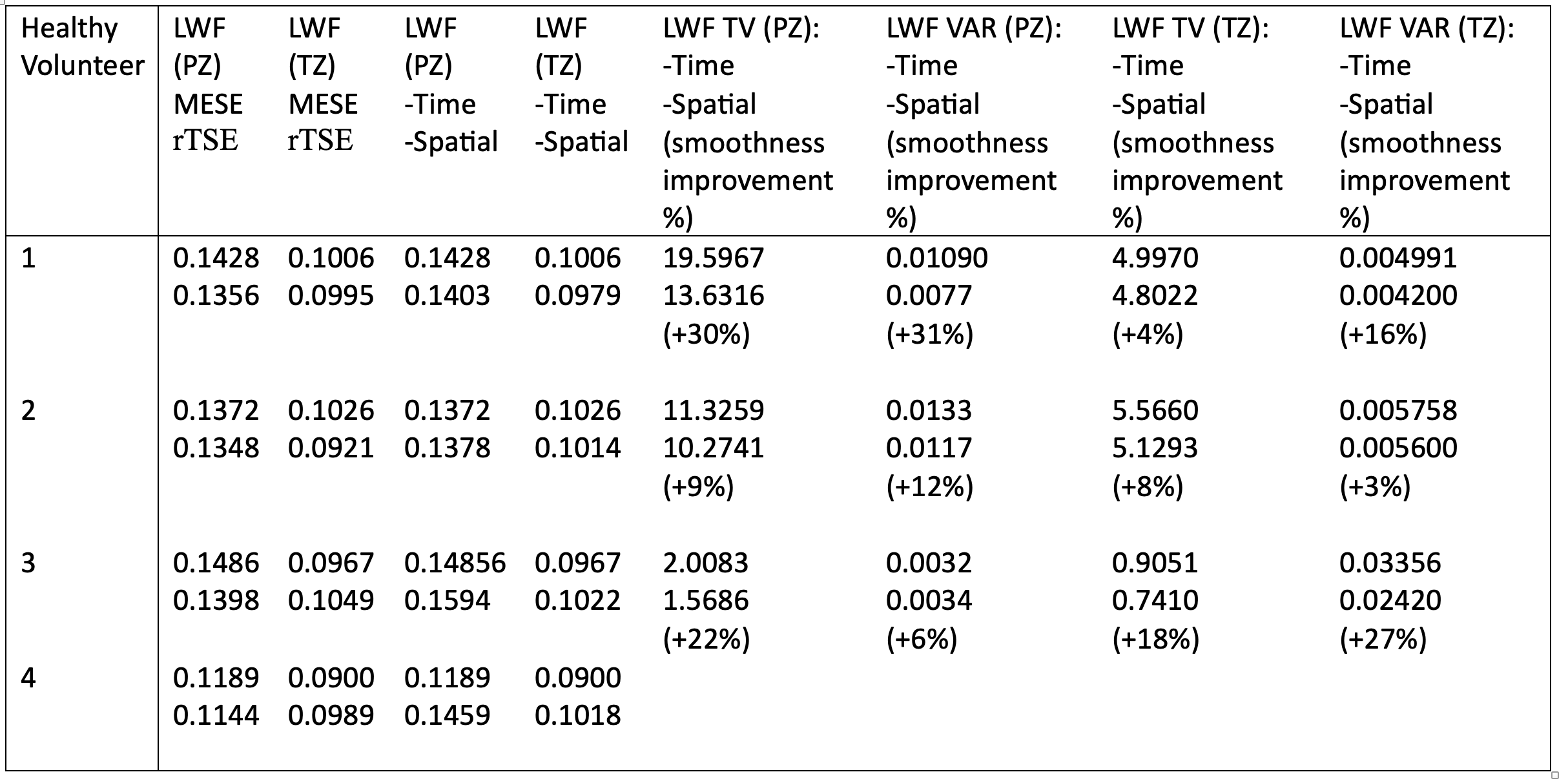
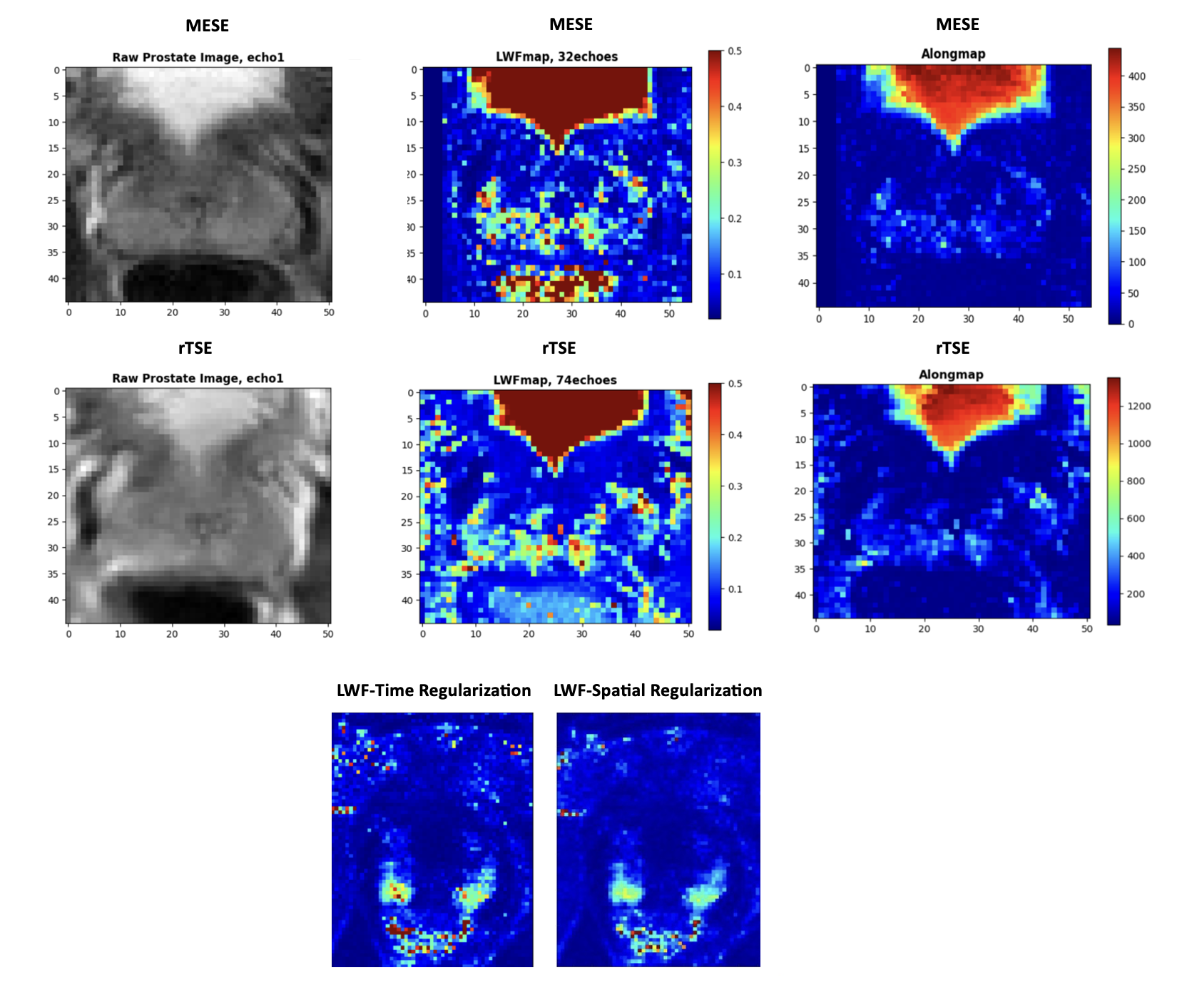
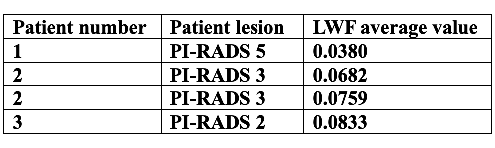
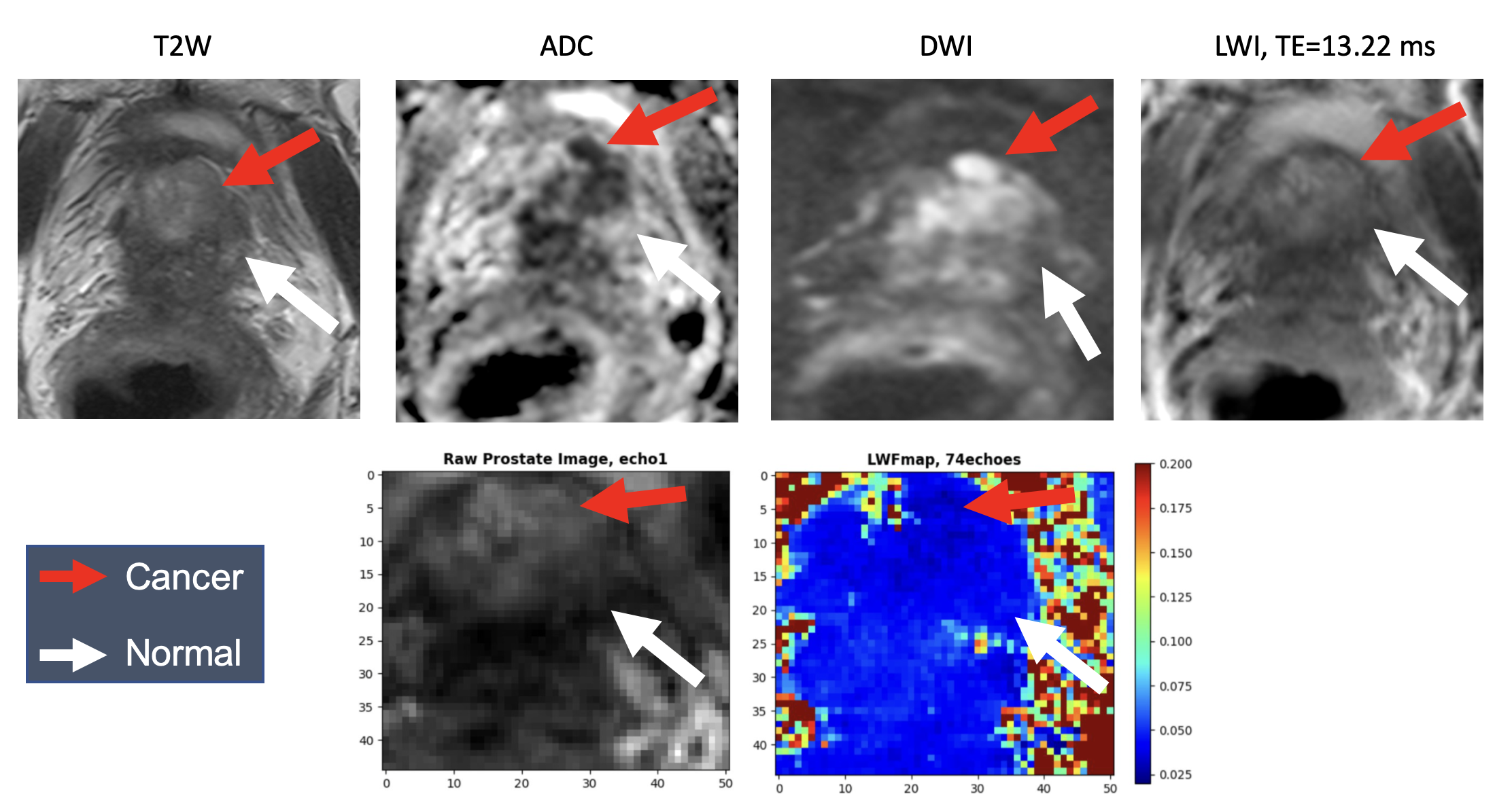
The shortened scan time effected by the rTSE protocol demonstrated no significant loss in image quality or diagnostic accuracy (Table 1, Table 2) when compared with traditional longer MESE sequence. rTSE demonstrated comparable capability in capturing luminal water imaging MR parameters (Fig. 2, Fig. 3).We achieved a smoother and more mathematically accurate representation of the T2 decay curves, substantiated by reduced total variation and variance values (common measures used in image processing to quantify the smoothness or roughness of an image) of LWF values across the prostate gland (Fig. 2). Given that local constraints are part of a global structure, disregarding the global effect leads to reduced efficiency. Therefore, considering all pixels simultaneously proves to be more efficient. However, the MATLAB-based AnalyzeNNLS package4 referenced in the LWI literature for producing a T2 distribution from multi-echo data, processes each pixel in isolation.
The spatial regularization technique, with detailed descriptions mentioned in a recent study5, enhanced the precision of the LWF maps, as evidenced by the smoother and more homogeneous appearance. The goal of adding a regularization term to T2-decay fitting is to enhance smoothing and impose smoothing constraints. Utilizing spatial regularization achieves a more homogeneously smooth LWF map.
Discussion
The rTSE sequence and spatial regularization method collectively address two major challenges in prostate MRI: the need for decreased scan times and improved image homogeneity. Our methodology not only fosters a more patient-friendly imaging experience by reducing time in the scanner but also enhances the radiologist's ability to discern subtle variations in tissue characteristics crucial for accurate diagnosis and treatment planning by providing more homogenous LWF maps.The reduction in scan time holds potential to increase patient throughput, a significant advantage in high-demand clinical settings. Moreover, the spatial regularization introduces a methodologically rigorous framework for adding a smoothing constraint to the LWI T2 decay fitting problem, which is a critical factor in the early detection and staging of prostate cancer.
Conclusion
This study marks a significant step forward in the domain of prostate cancer imaging. The rTSE sequence is a time-efficient alternative to traditional LWI protocols, halving the scan duration without a significant change in the quality of the LWF data. When coupled with spatial regularization, this method seems to not only preserve but also enhance the diagnostic value of LWI, providing smoother and more consistent LWF maps. Collectively, these advancements could lead to improved clinical workflows, enhanced patient experiences, and, ultimately, better patient outcomes in the management of prostate cancer.Acknowledgements
No acknowledgement found.References
1. Sabouri, Shirin, et al. "MR measurement of luminal water in prostate gland: Quantitative correlation between MRI and histology." Journal of Magnetic Resonance Imaging 46.3 (2017): 861-869.
2. Hectors, Stefanie J., et al. "Luminal water imaging: Comparison with diffusion‐weighted imaging (DWI) and PI‐RADS for characterization of prostate cancer aggressiveness." Journal of Magnetic Resonance Imaging 52.1 (2020): 271-279.
3. Altbach MI, Bilgin A, Li Z, Clarkson EW, Trouard TP, and Gmitro AF. Processing of radial fast spin-echo data for obtaining T2 estimates from a single k-space data set. Magnetic Resonance in Medicine, 54(3):549–559, 2005.
4. Bjarnason, Thorarin A., and J. Ross Mitchell. "AnalyzeNNLS: magnetic resonance multiexponential decay image analysis." Journal of Magnetic Resonance 206.2 (2010): 200-204.
5. Kim, Daeun, et al. "Diffusion‐relaxation correlation spectroscopic imaging: a multidimensional approach for probing microstructure." Magnetic resonance in medicine 78.6 (2017): 2236-2249.
Figures