0259
A deep learning-based approach to nuisance signal removal from MRSI data aqcuired without suppression1Radiology, Massachusetts General Hospital, Boston, MA, United States, 2Radiology, Harvard Medical School, Boston, MA, United States, 3School of Medicine, Yale University, Boston, MA, United States
Synopsis
Keywords: Spectroscopy, Data Processing, MRSI nuisance signal removal
Motivation: Unsuppressed water and lipid signals are several orders of magnitude stronger than the metabolite signals in MRSI, imposing significant challenges in MRSI data processing and image reconstruction.
Goal(s): To develop a novel deep learning-based method for nuisance signal removal from MRSI data acquired without suppression.
Approach: A neural network with a U-net structure was designed to remove nuisance signals in MRSI, where the input of the network was the Hankel matrix formed by the time-domain MRSI signal.
Results: The proposed method was validated using in vivo MRSI data, showing superior performance over the conventional method.
Impact: A deep learning-based method is proposed for nuisance signal removal in MRSI. It could enable MRSI without water or lipid suppression with robust performance in practical settings.
Introduction
A unique challenge in 1H-MRSI is the removal of nuisance water and lipid signals, which are several orders of magnitude stronger than the metabolite signals without suppression, imposing significant challenges in MRSI data processing and image reconstruction1. While the conventional wisdom is to suppress the water and lipid signals during MRSI data acquisition, it has been recently shown that it is feasible to remove the water and lipid signals from MRSI data acquired without any suppression using a union-of-subspaces (UOSS) model2-8. The unsuppressed water signals in turn not only provide B0 inhomogeneity and motion information that significantly improves the robustness of MRSI but also enable simultaneous multiparametric mapping (i.e., T1, T2, QSM, MWI) and metabolic imaging in a single imaging session3-8.The UOSS2 method uses a linear subspace model to represent the high-dimensional nuisance water and lipid signals and determine the model coefficients using the acquired data alone. The rapid development in deep neural networks (DNN)9-12 opens new opportunities to learn a nonlinear low-dimensional representation of the nuisance signals from population data to further improve the robustness of nuisance signal removal in practical settings. In this work, we propose a novel deep learning-based approach to nuisance signal removal in MRSI. The proposed network takes the Hankel matrix formed by the time-domain MRSI signal as an input and can be considered a nonlinear generalization of the UOSS model. We validated the performance of the proposed method using in vivo MRSI data acquired at 3T without suppression.
Methods
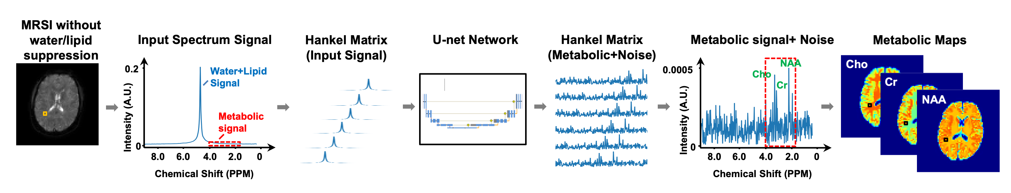
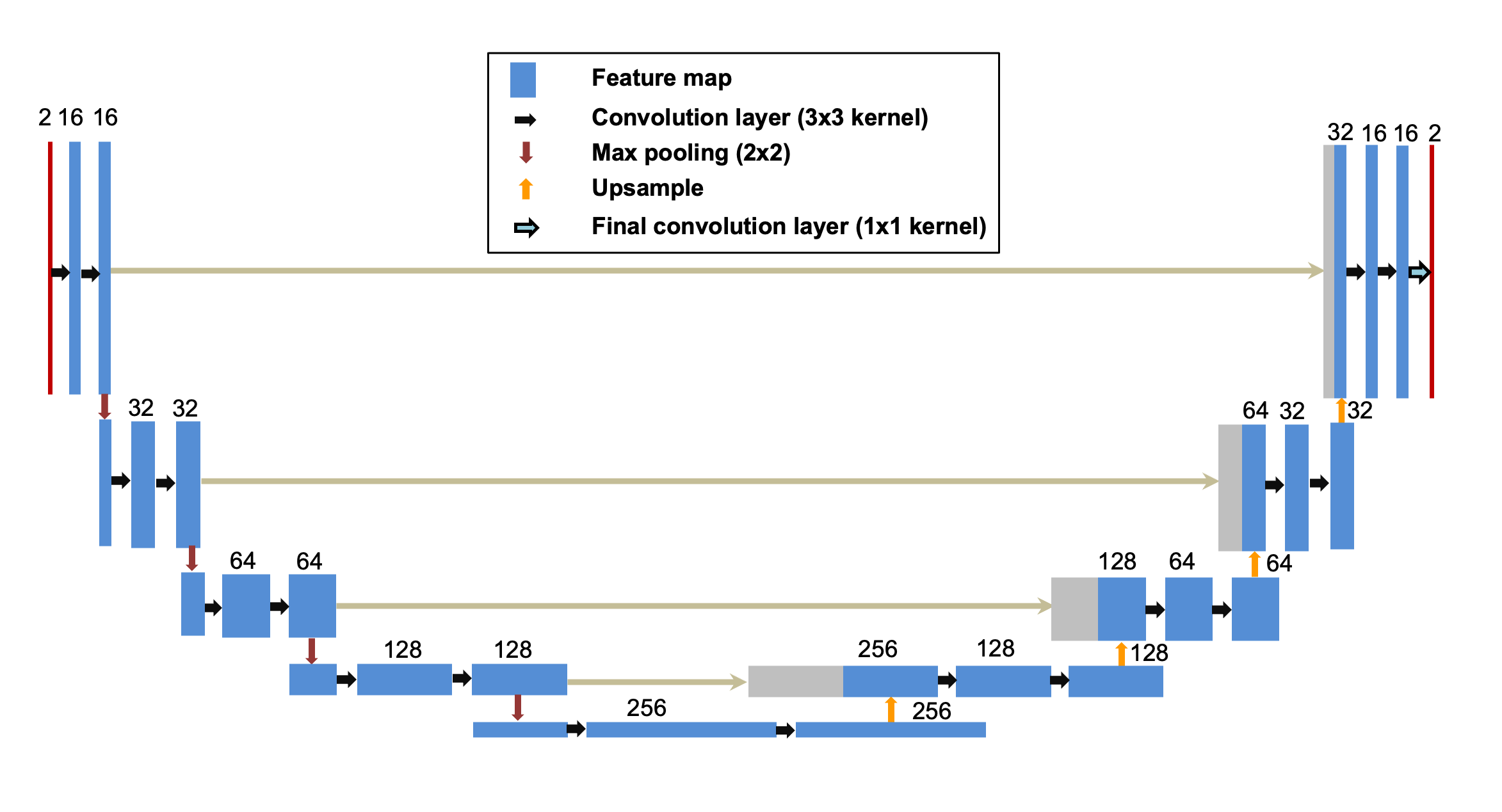
Network designFigure 1 shows the schematic diagram of the proposed method. First, the time-domain MRSI signal at each voxel was used to form a Hankel matrix, which served as the input of a 2D U-net. The structure of the proposed network is detailed in Figure 2. Second, a residual learning strategy was used to train the network to learn the nuisance signal removed MRSI signals using training labels created from in vivo data with a physics model (detailed below). Finally, the network-predicted nuisance signals were subtracted from the MRSI data for the consecutive image reconstruction and spectral quantification.
The novelty of the proposed method relies on the use Hankel matrix as the network input, which captures the low-rank structure of the nuisance signals as in the UOSS method. The neural network further learns the low-dimensional manifold structure of the nuisance signals from population data, which can thus be considered a nonlinear generalization of the UOSS method. In fact, the UOSS method is a special case of the proposed method if the 2D U-net is replaced by a single-layer auto-encoder.
Network training
Training labels were created using in vivo MRSI data acquired from 8 healthy subjects using a 3T Siemens Prisma scanner. All experiments were performed under a study protocol approved by our local institutional review board. The MRSI data were acquired using an EPSI sequence with the following imaging parameters were: spatial encodings = 72x72x32, spectral encodings = 380, resolution = 3.3x3.3x3.3 mm, TR/TE = 360/3.6 ms. No water or lipid suppression was used.
The ground truth signals were created from the in vivo data using an MR physics model. First, the ground truth lipid signals were extracted from the MRSI data using the UOSS model with spatial support constraints. Second, the ground truth water signals were extracted from the lipid-removed MRSI data by performing HSVD decomposition voxel by voxel. The rank of the HSVD was chosen based on the SNR. Note that no metabolite signals would be extracted in the above steps because their intensities were below the noise floor. Third, metabolite signals were created using quantum mechanic simulations with random concentrations, frequency shifts, lineshape distortions, and phase distortions. Finally, training data were created by adding the above signal components and i.i.d. Gaussian noise together. Each voxel of the 3D MRSI data was treated as independent labels.
Results
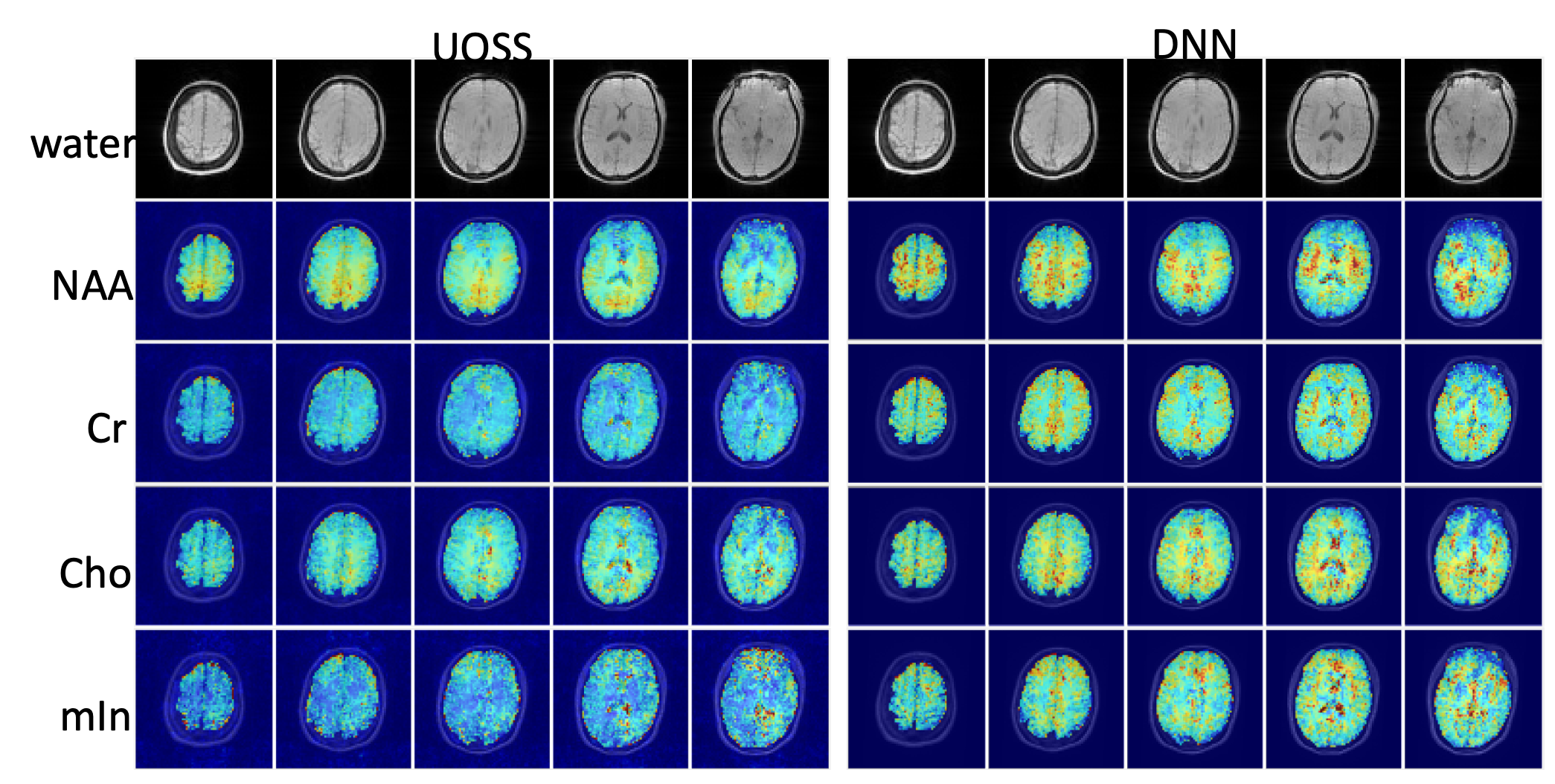
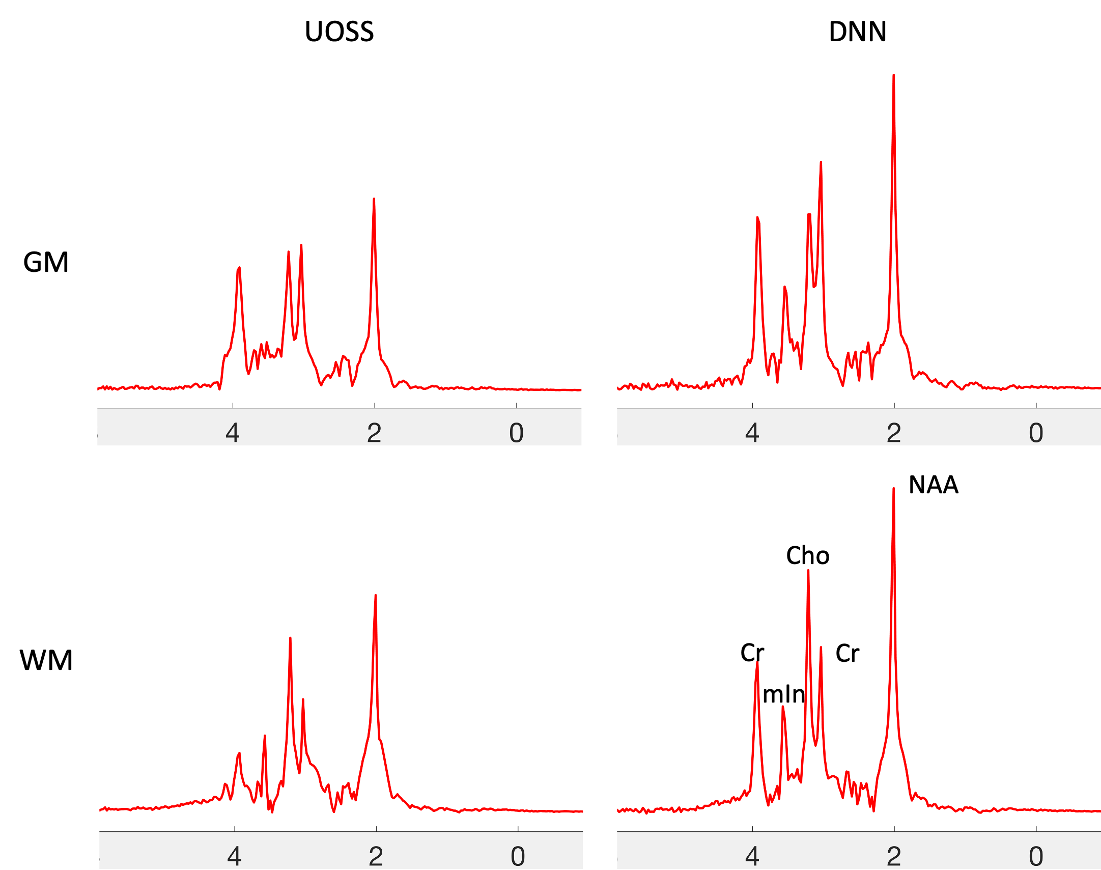
We validated the performance of the proposed method using in vivo MRSI data acquired from a different subject using the same imaging protocol. For comparison, the UOSS method was used to process the same dataset using a set of default parameters in2. After nuisance signal removal, the MRSI data were reconstructed using the SPICE method13-14. Figures 3 and 4 show representative metabolite maps and spectra. Compared to the UOSS method, the metabolite maps by the proposed method had less signal loss, especially in the Cr and mIn signals.Conclusions
A deep learning-based method is proposed for nuisance signal removal in MRSI. It could enable MRSI without water or lipid suppression with robust performance in practical settings.Acknowledgements
This work was supported in part by the National Institutes of Health (K01EB030045, P41EB022544, R01CA165221, and R01EB033582).References
1. Tkáč I, Deelchand D, Dreher W, Hetherington H, Kreis R, Kumaragamage C, de Graaf RA. Water and lipid suppression techniques for advanced 1H MRS and MRSI of the human brain: experts' consensus recommendations. NMR in Biomedicine. 2021;34(5):e4459.
2. Ma C, Lam F, Johnson CL, Liang ZP. Removal of nuisance signals from limited and sparse 1H MRSI data using a union-of-subspaces model. Magn Reson Med. 2016;75:488–497.
3. Peng X, Lam F, Li Y, Clifford B, Liang Z-P. Simultaneous QSM and metabolic imaging of the brain using SPICE. Magn Reson Med. 2018;79(1):13-21.
4. Guo R, Zhao Y, Li Y, Li Y, Liang Z-P. Simultaneous metabolic and functional imaging of the brain using SPICE. Magn Reson Med. 2019;82(6):1993-2002.
5. Guo R, Zhao Y, Li Y, et al. Simultaneous QSM and metabolic imaging of the brain using SPICE: Further improvements in data acquisition and processing. Magn Reson Med. 2021;85(2):970-977.
6. Li Y, Guo R, Zhao Y, et al. Rapid high-resolution simultaneous acquisition of metabolites, myelin water fractions, and tissue susceptibility of the whole brain using “SPICY” 1H-MRSI. In: Annual Meeting of International Society for Magnetic Resonance in Medicine. ; 2019:0946.
7. Li Y, Guo R, Zhao Y, et al. T1 and T2 mapping using highly sparse unsuppressed water signals from MRSI scans with generalized series-assisted low-rank tensor modelling. In: Annual Meeting of International Society for Magnetic Resonance in Medicine. ; 2023:1142.
8. Li Y, Xiong J, Guo R, Zhao Y, Li Y, Liang Z-P. Improved estimation of myelin water fractions with learned parameter distributions. Magn Reson Med. 2021;86(5):2795-2809.
9. Lee HH, Kim H. Intact metabolite Spectrum mining by deep learning in proton magnetic resonance spectroscopy of the brain. Magn Reson Med. 2019; 82: 33-48.
10. Lee, HH, Kim, H. Deep learning-based target metabolite isolation and big data-driven measurement uncertainty estimation in proton magnetic resonance spectroscopy of the brain. Magn Reson Med. 2020; 84: 1689–1706. https://doi.org/10.1002/mrm.28234
11. Louis MS, Coello E, Liao H, Joshi A, Lin A. Quantification of non-water-suppressed proton spectroscopy using deep neural networks. Proceedings of the Virtual 28th Annual Meeting of ISMRM, Toronto; 2021: 1298.
12. Lam F, Li Y, Peng X. Constrained magnetic resonance spectroscopic imaging by learning nonlinear low-dimensional models. IEEE Trans Med Imaging. 2020;39:545–555.
13. Lam F, Liang ZP. A subspace approach to high-resolution spectroscopic imaging. Magn Reson Med. 2014;71:1349–1357.
14. Lam F, Ma C, Clifford B, Johnson CL, Liang ZP. High-resolution 1H-MRSI of the brain using SPICE: Data acquisition and image reconstruction. Magn Reson Med. 2016;76:1059–1070.
Figures