0253
Robust Volumetric Diffusion-Weighted MRSI via Time-Resolved Phase Reconstruction and Correction1Department of Bioengineering, University of Illinois at Urbana-Champaign, Urbana, IL, United States, 2Beckman Institute for Advanced Science and Technology, University of Illinois at Urbana-Champaign, Urbana, IL, United States, 3Department of Electrical and Computer Engineering, University of Illinois at Urbana-Champaign, Urbana, IL, United States
Synopsis
Keywords: Spectroscopy, Spectroscopy, Diffusion, Quantitative Imaging
Motivation: To address the long-standing phase correction challenge and enhance the robustness for diffusion-weighted MRSI.
Goal(s): To correct the significant phase variations due to macroscopic and microscopic motions in in vivo diffusion-weighted MRSI acquisition.
Approach: We developed a novel fast diffusion-weighted MRSI sequence integrating time-resolved, sparsely sampled, volumetric phase navigators, a subspace-based phase image reconstruction, and a sensitivity-encoded phase-corrected reconstruction. The corrected diffusion-weighted MRSI data were processed by state-of-the-art subspace-based spatiospectral processing methods.
Results: Improved data quality, diffusion-weighted spatiospectral reconstruction and metabolite-specific diffusion parameter estimation achieved by the proposed method are demonstrated using in vivo data.
Impact: A novel integrative acquisition and reconstruction solution for robust, phase-corrected 3D in vivo diffusion-weighted MRSI was presented, an important step towards developing diffusion-weighted MRSI for its translation to quantitative, molecule-specific microstructural imaging.
Introduction
Diffusion-weighted (DW) MRSI is an emerging modality that allows for quantifying molecule-specific diffusion parameters, which promises to provide richer tissue microstructural information than diffusion MRI1-6. However, in vivo DW-MRSI is extremely challenging because of (1) poor SNR due to a combination of low metabolite abundance and diffusion encoding (DE) induced signal decay, (2) high dimensionality with the need to encode and decode both the spectroscopic and diffusion dimensions and (3) strong susceptibility to phase variations caused by confounding macroscopic and microscopic motions. Subspace imaging has demonstrated significant potential in addressing the first two issues for high-SNR DW-MRSI8,9. But the phase issue remains unsolved, particularly the spatially-varying inter-excitation phase variations. Limited correction methods have been described1,3,4,5. We present here a novel solution that integrated a fast DW-MRSI acquisition with interleaved sparsely sampled, time-resolved 3D phase navigators, a subspace-based phase image reconstruction, and a phase-corrected reconstruction strategy. The proposed method enabled significantly more robust, 3D DW-MRSI and metabolite-specific ADC mapping, demonstrated by in vivo brain data.Methods
We model DW-MRSI signals as:$$\rho_{b,s}(\boldsymbol{r},t)=e^{-i \phi_{b,s}(\boldsymbol{r})}\odot\rho_{b}(\boldsymbol{r},t),$$
where b-value-dependent $$$\rho_{b}(\boldsymbol{r},t)$$$ is the underlying DW spatiotemporal function of interest, from which metabolite diffusion can be quantified. $$$\rho_{b,s}(\boldsymbol{r},t)$$$represents the function at each shot/TR with spatially varying phases $$$\phi_{b,s}(\boldsymbol{r})$$$ ($$$b$$$ and $$$s$$$ being DE and shot/TR indices) that need to be corrected. For DW-MRSI, only very limited k-space can be acquired for each $$$\rho_{b,s}(\boldsymbol{r},t)$$$, a more extreme multi-shot scenario (e.g., one phase encoding each TR) than DW-MRI, making self-navigation infeasible and additional navigator acquisition more difficult. One-dimensional navigator has been used, ignoring spatial dependence of $$$\phi_{b,s}(\boldsymbol{r})$$$4,5. Here, we propose a new integrative acquisition and reconstruction strategy to better capture and correct for $$$\phi_{b,s}(\boldsymbol{r})$$$.
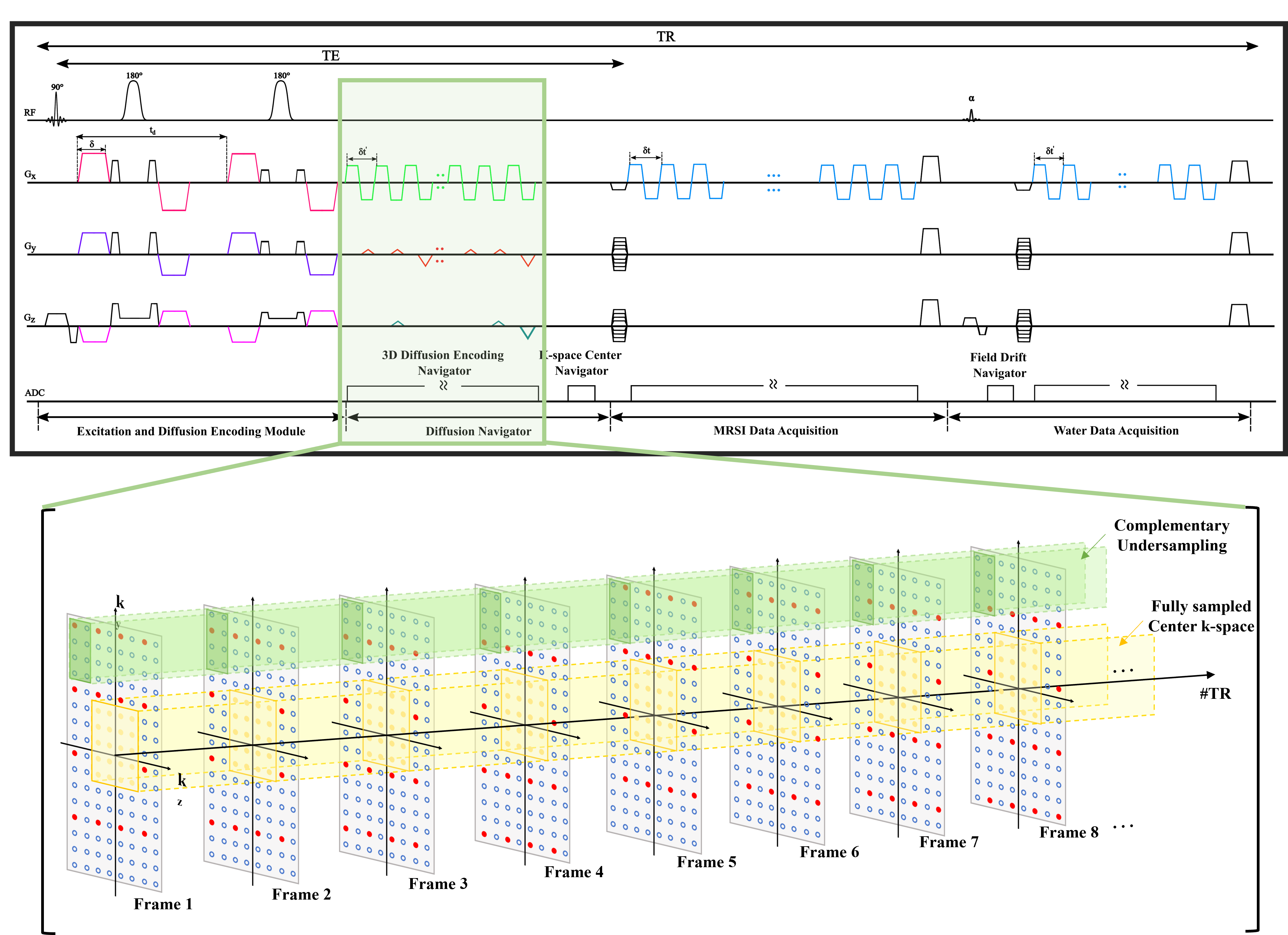
Phase navigators with sparse sampling: We interleaved an echo-volume-imaging (EVI) navigator7 into a double-spin-echo DW-MRSI sequence8 to fully capture $$$\phi_{b,s}(\boldsymbol{r})$$$ at each excitation (Fig. 1). With the limited window to collect this data prior to phase encoding and TE, sparse sampling is needed. Specifically, we designed a segmented sampling strategy with the same portion of center (ky,kz)-region fully sampled and the outer region sampled with a complementary 8× sparse pattern each TR (see Fig.1,bottom). Every 8 TRs formed a fully-sampled k-space. A short 1D phase navigator was also included for zeroth-order phase correction. No water suppression was used.
Time/TR-resolved EVI navigator reconstruction: Reconstructing the TR-resolved EVI navigator images from sparse data was enabled by a subspace model. Specifically, a DE-dependent, coil-independent subspace $$$V_b\in\mathcal{C}^{L \times N_{TR}}$$$($$$N_{TR}$$$ the total TR number) can be estimated from the center portion. Then, (coil,DE)-dependent spatial coefficients $$$U_{c,b}$$$ were obtained by solving
$$\hat{U}_{c,b}=\arg\min_{U_{c,b}}\left\|d_{c,b}-F_{\Omega_{b}}U_{c,b}V_b\right\|_F^2+\lambda\left\|D U_{c,b}V_b\right\|_F^2+\frac{\lambda}{100}\left\|U_{c,b}\right\|_F^2$$
where $$$d_{c,b}$$$ is the undersampled EVI data ($$$c$$$ the coil index), $$$F_{\Omega_{b}}$$$ a Fourier operator with (k,s)-sampling pattern $$$\Omega_{b}$$$, $$$D$$$ a finite-difference operator and $$$\lambda$$$ the spatial smoothness regularization parameter. 3D phase maps $$$\{\varphi_{b,s}\}$$$ were extracted from the reconstructed images $$$\{\hat{U}_{c,b}V_b\}$$$ for the correction described below.
Phase-corrected reconstruction for 3D DW-MRSI: We first applied a coil-dependent, zeroth-order phase correction to the DW-MRSI and EVI data using the 1D navigator4,5,8,9. A second spatially-varying phase correction was performed by solving
$$\hat{\rho}_{b}=\arg\underset{\rho_{b}}{\min}\sum_{s}\sum_c\left\|d_{c,b,s}-F_{\Omega_{b,s}}S_c\varphi_{b,s}\rho_{b}\right\|_F^2+\lambda\left\|\rho_{b}\right\|_F^2$$
where $$$\{d_{c,b,s}\}$$$ are zeroth-order phase-corrected, multicoil DW-MRSI data with sampling pattern $$$\Omega_{b,s}$$$ (one phase encoding per shot/TR). $$$S_c$$$ are coil sensitivity maps and $$$\{\varphi_{b,s}\}$$$ are coil-independent phases after zeroth-order correction. Note that the DW-MRSI data can have a higher resolution than the navigators assuming smoothness of the phase. After obtaining $$$\hat{\rho}_{b}$$$, subsequent nuisance water/lipid removal, spatiospectral reconstruction, and parameter fitting were applied. These processing steps for high-resolution DW-MRSI are similar to those presented in [8,9] and omitted due to the word limit.
Results
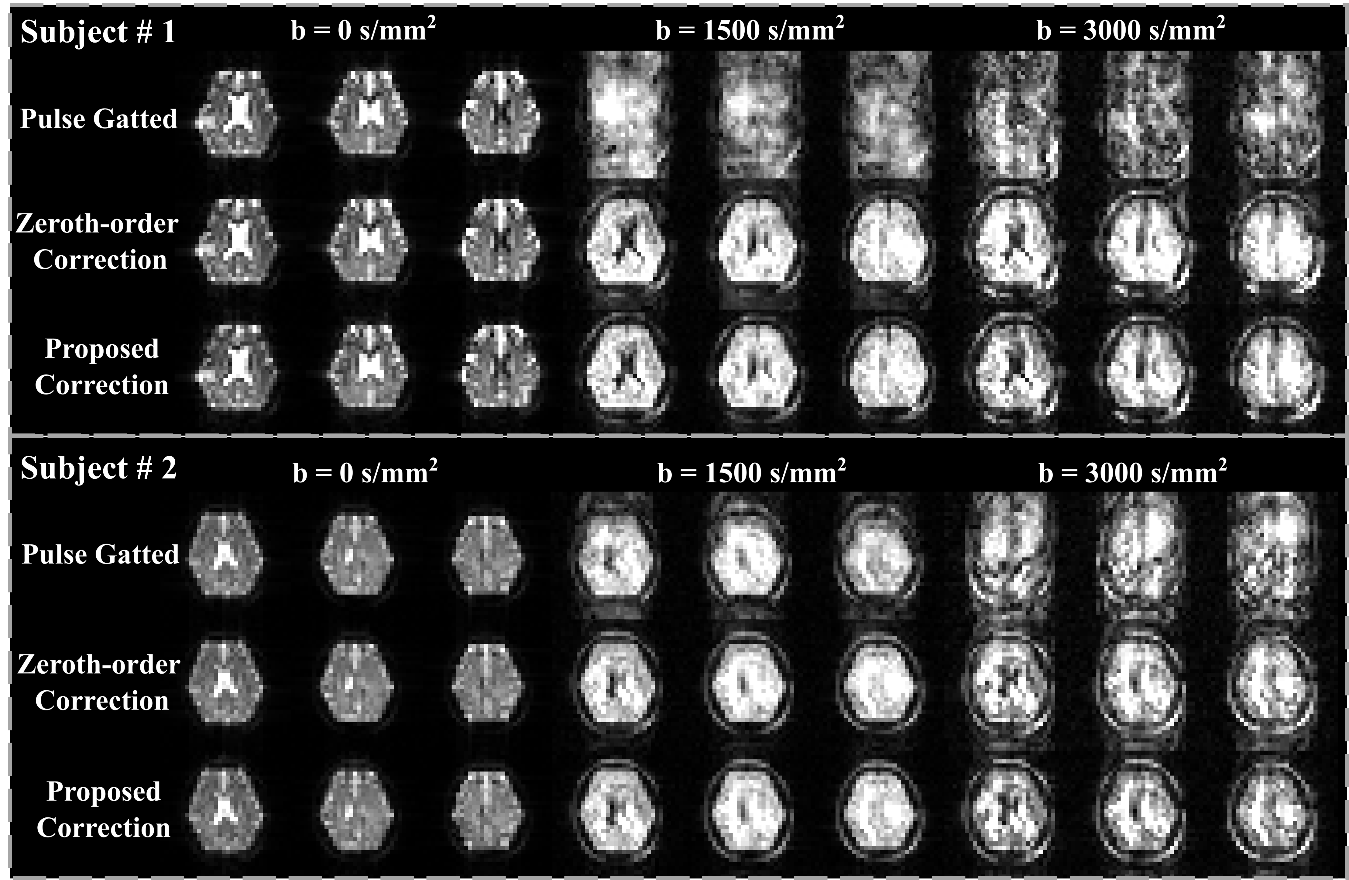
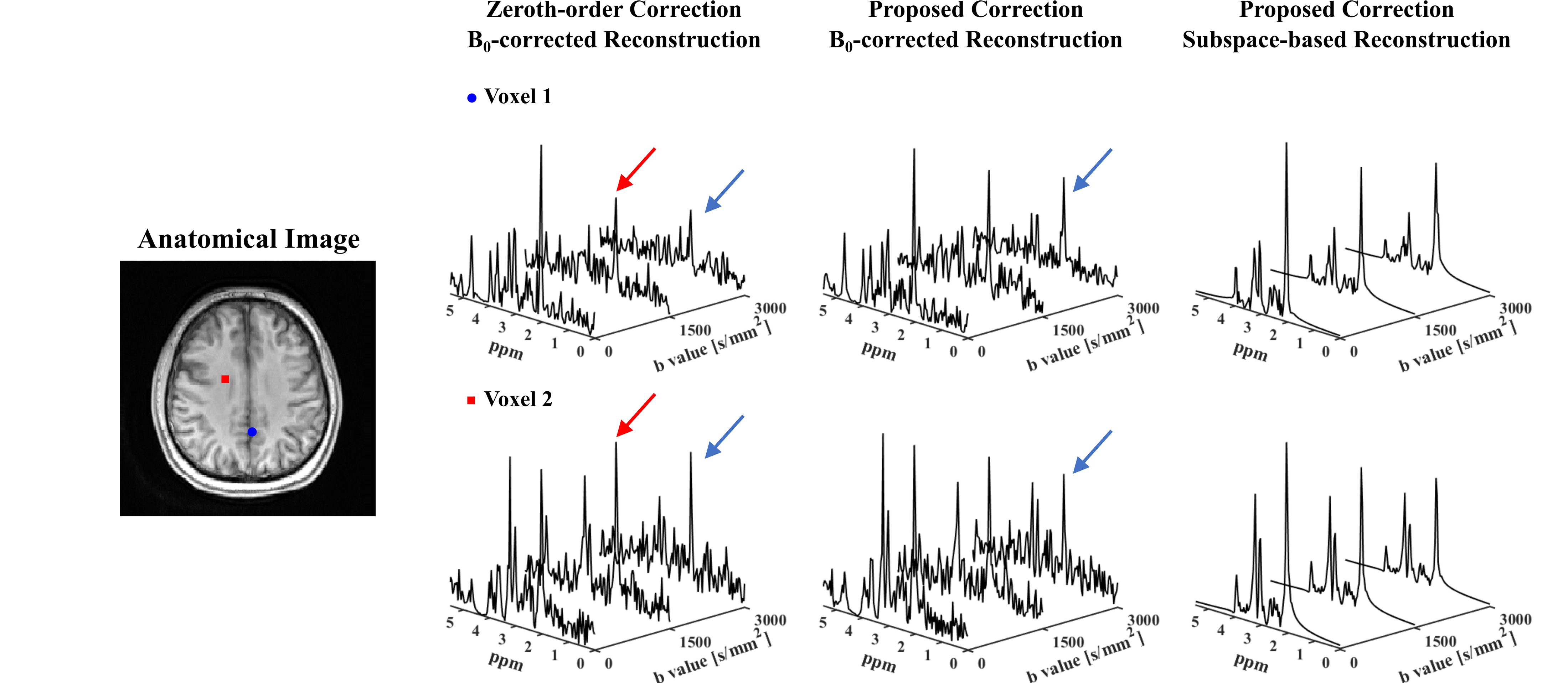
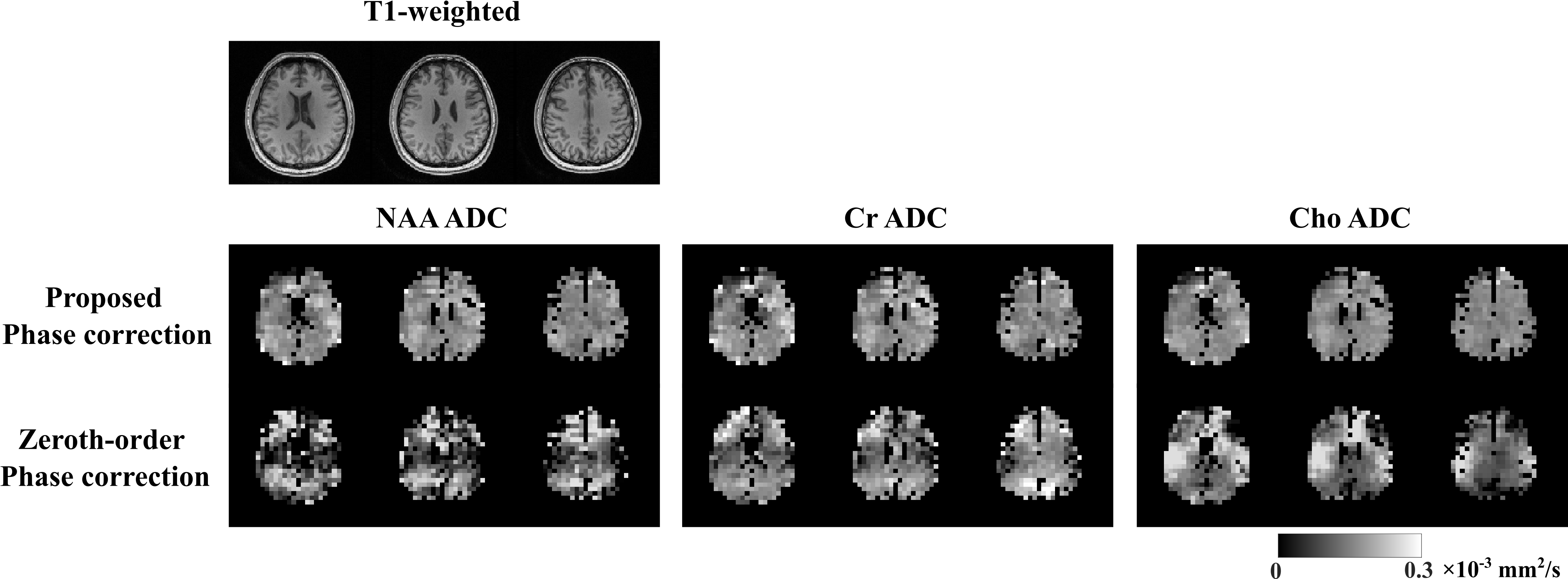
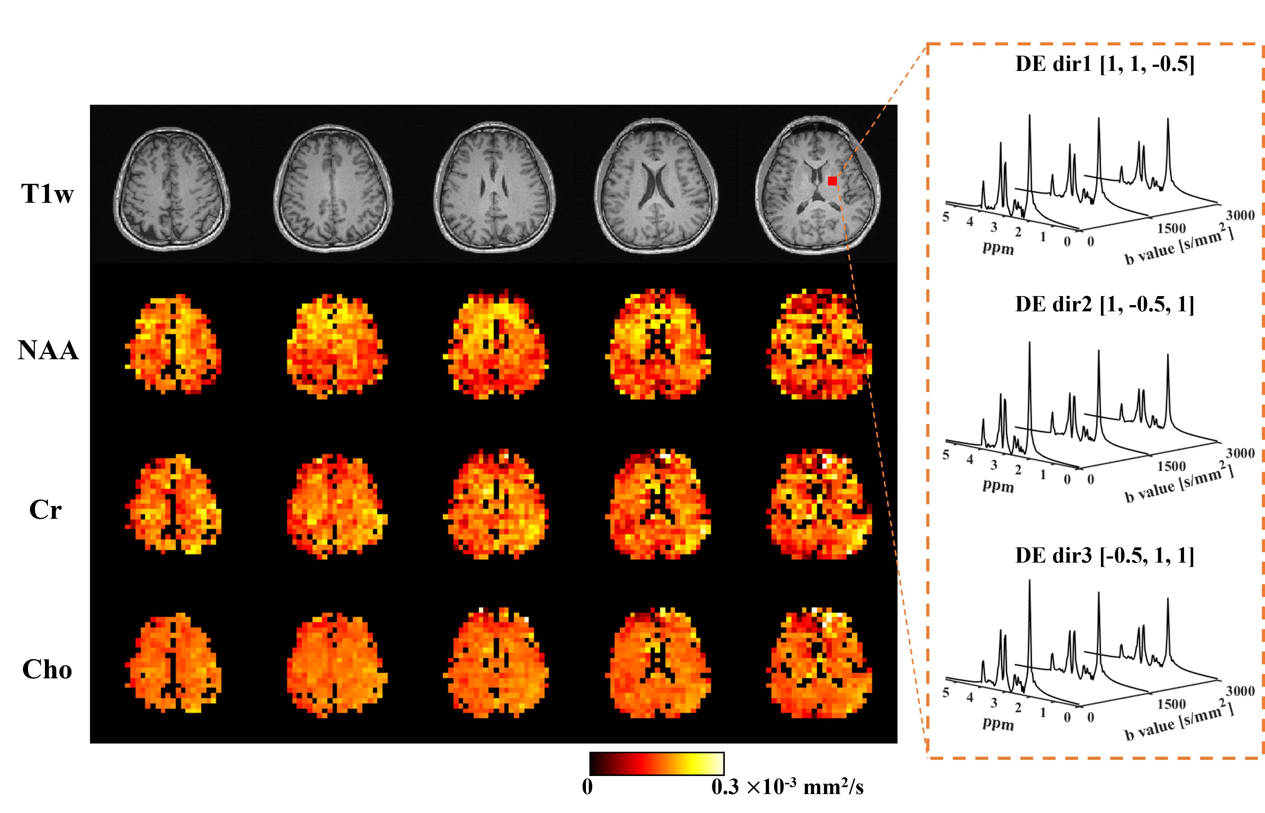
In vivo experiments were conducted on a Prisma 3T system with IRB approval. Pulse triggering was used with a 20ms trigger delay. Other key parameters include: 220×220×56mm3 FOV, 650/160ms TR/TE, 32×32×8 matrix size for the DW-MRSI data and EVI navigators (6.9×6.9×7mm3 voxels), and b-values = [0,1500,3000] s/mm2 with a 78.8ms diffusion time. The overall acquisition was ~10 mins for 3 b-values at one DE direction. Data with both isotropic and 3 orthogonal DE directions were acquired9.Significant and robust artifact reduction were observed for the proposed method (Fig. 2, reconstruction of unsuppressed water spectroscopic images), which led to improved spatiospectral reconstruction (Fig. 3). Higher-quality metabolite ADC maps were produced by the proposed method (Fig. 4), with robust performance on mean diffusivity (MD) estimation and interesting tissue microstructural information revealed. (Fig. 5)
Conclusion
We presented a novel integrative volumetric phase navigator acquisition and reconstruction strategy for robust phase-corrected, subspace-based 3D DW-MRSI. Significantly improved data quality, spatiospectral reconstruction, and metabolite diffusion parameter mapping were demonstrated using in vivo data.Acknowledgements
This work was supported in part by NSF-CBET 1944249 and NIH-NIBIB 1R21EB029076.References
[1] Ronen I et al. Diffusion‐weighted magnetic resonance spectroscopy. EMagRes. 2007;15:733-50.
[2] Posse S et al. Human brain: proton diffusion MR spectroscopy. Radiology. 1993; 188:719-25.
[3] Palombo M et al. Insights into brain microstructure from in vivo DW-MRS. NeuroImage. 2018; 182:97-116.
[4] Ercan AE et al. Diffusion‐weighted chemical shift imaging of human brain metabolites at 7T. Magn. Reson. Med. 2015; 73:2053-61.
[5] Fotso K et al. Diffusion tensor spectroscopic imaging of the human brain in children and adults. Magn. Reson. Med. 2017; 78:1246-56.
[6] Cao P et al. Diffusion magnetic resonance monitors intramyocellular lipid droplet size in vivo. Magn. Reson. Med. 2015; 73:59-69.
[7] Liu C, et al. Simultaneous phase correction and SENSE reconstruction for navigated multi‐shot DWI with non‐cartesian k‐space sampling. Magn. Reson. Med. 2005; 54:1412-22.
[8] Wang et al. High-resolution volumetric diffusion-weighted MRSI using a subspace approach. In Proc. of ISMRM, 2021; pp. 37.
[9] Wang et al. Fast volumetric diffusion-weighted MRSI: Improved acquisition and data processing. In Proc. of ISMRM, 2022; pp. 4642.
[10] Ellegood et al. Considerations for measuring the fractional anisotropy of metabolites with diffusion tensor spectroscopy. NMR in Biomedicine, 2011; 24:270-280.
Figures