0244
Evaluating MRE-Tract Integrity in HIV-CSVD Cohort: A Comprehensive Analysis with Functionally Defined Atlases and Neurocognitive Assessment1University of Rochester, Rochester, NY, United States, 2Charité - Universitätsmedizin Berlin, Berlin, Germany
Synopsis
Keywords: Data Processing, Elastography
Motivation: MR-Elastography and diffusion-MRI represents complementary modalities with mechanical-and-structural information of the brain. Structural-connectomes alone lack tract-integrity information. Tissue viscoelastic-measures can provide valuable insights for tract-integrity when combined with connectomes.
Goal(s): To aid the studies with neurodegeneration(HIV/CSVD), an analytical approach to combine viscoelastic-measures with diffusion-tractography is proposed which shows promise in targeted-analysis of functionally-defined-networks.
Approach: For 14 functionally-defined brain-networks, viscoelastic measures from MRE are mean-sampled with the dMRI derived tracts. Then, the significantly-affected viscoelastic alterations are studied in-accord with cognitive-changes.
Results: In a cohort of HIV-CSVD, by MRE-Tract-Integrity analysis, we reported significantly affected network connectivity that follows the cognitive decline(p<0.05) in processing-speed and motor-skills.
Impact: The MRE-Tract-Integrity analysis enables us to study the missing mechanical properties of the structural connections from diffusion-tractography. This will help researchers performing targeted cognitive performance analysis with brain connectivity aided with mechanical basis which is a prominent marker for neural-change.
Introduction
MR-Elastography(MRE) extracts the physical and mechanical state of tissue indeterminable by any other MR modalities, thus it holds the potential to reflect a unique biomarker of brain-injury(1). Diffusion MRI(dMRI), another well-established imaging modality, enables tractography and connectome analysis(2). Therefore, MRE and dMRI could provide complementary information about the brain health. To the best of our knowledge, this is the first MRE-Tract-Integrity (MTI) study, where we combine these two complementary modalities to probe into the connectivity of 14 functionally defined networks of the human brain(3). Further, we examine the relationships between MRE measures with neurocognitive-scores throughout these networks. The study was implemented in a cohort of people with HIV(PWH) at risk for Cerebrovascular Small Vessel Disease(CSVD) and Controls.Materials & Methods
Informed consent was obtained for scanning 30 HIV infected individuals(HIV+) (age=55.1±10.1years, range=31-76years, female=8) and 36-Controls(age=54.4±15.7years, range=19-76years, female=8) according to the institutional protocol. Siemens MAGNETOM Prisma 3T whole-body scanner, equipped with a 64-channel head coil (receive-only) and body coil transmission was used for image acquisition. The protocol included T1-weighted images using a 3D MPRAGE-sequence[inversion time(TI)=962ms, repetition time/echo time(TR/TE) =1840ms/2.34ms, 1 mm isotropic resolution] for anatomical reference. Multifrequency MRE was performed using 20, 40 and 50 Hz frequencies with 8 wave dynamics and 3 motion-encoding gradients (MEG, amplitude=34mT/m, frequency=31.81Hz) directions (TE/TR=76ms/5100ms, 2 mm isotropic resolution). Multifrequency dual elasto-visco(MDEV)(4,5) inversion was used to calculate magnitude (|G*|, stiffness) and phase (Ø, tissue viscosity) of complex shear modulus. Additional dMRI scan was performed using 2D single-shot spin echo echo-planar imaging (SE-EPI) sequence (TR/TE= 4300 ms/69.0 ms; 1.5 mm isotropic resolution, 64 gradients directions for each b values (b=1,000 and 2,000 s/mm2) with 7 b=0 s/mm2 reference images). Preprocessing included top-up and eddy correction with FSL (v6.0.0/b1)(6). Diffusion and advanced diffusion metrics were calculated with MRtrix3(7). The UManitoba-JHU functional atlases and MRE maps were transformed to diffusion native space using ANTs(8). Then we generated the tractography and connectomes using MRtrix3 for 14 different functionally defined networks(3, 9).MRE-Tract-Integrity is assessed via mean-sampling of viscoelastic maps from the tracts generated with diffusion tractography. MRtrix3-“tck2connectome” and “tcksample” commands were used to generate and sample the stiffness-|G*| and viscosity-Ø data. Hebbian law states that the neurons that fire together wire together, this leads to our hypothesis to observe integrity loss in MTI in neurodegenerative cases such as HIV-infection with predefined functional atlases. Relying on the probabilistic functional maps of 14 networks e.g., dorsal and ventral Default Mode Network (DMN_D, DMN_V), left and right Executive Control Network (ECN_L, ECN_R), anterior and posterior Salience-Network (SN_A, SN_P), Auditory Netwrok (AUD), Basal Ganglia Network (BGN), Higher Visual Network (HVN), Language-Network (LN), Precuneus-Network (PN), Primary Visual Network (PVN), Sensorimotor-Network(SMN), Visuospatial Network (VSN).
For each functional atlas, we performed connectivity-based group comparison and p-value<0.05 was reported as significant. Pearson correlations were conducted to evaluate the associations between MRE metrics within functionally defined networks and neurocognitive total z-scores, along with individual domain z-scores. Cognitive domains included Speed of Information Processing, Executive function, Verbal/Language Skill, Learning, Memory, Attention/Working Memory and Motor Skill.
Results & Discussion
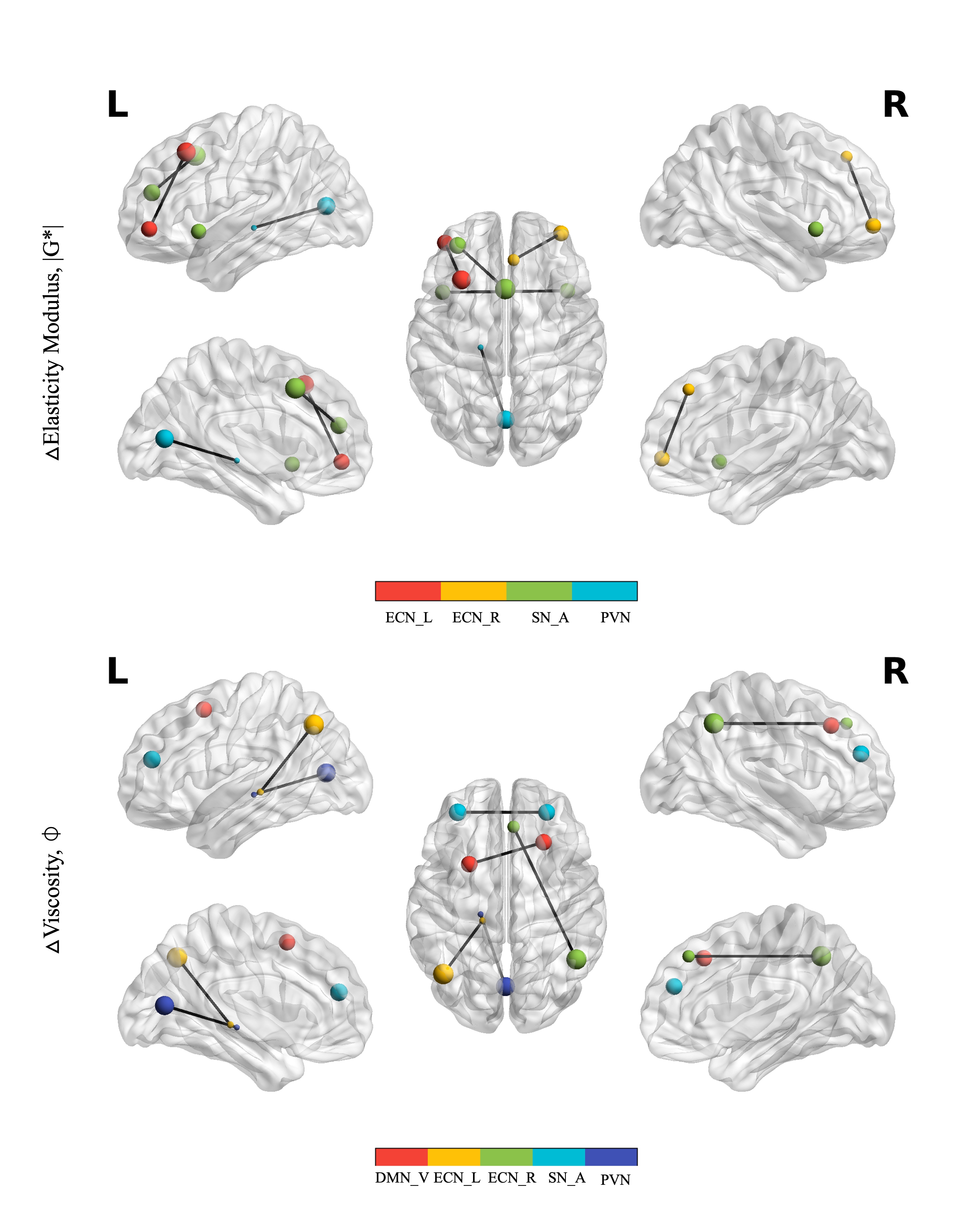
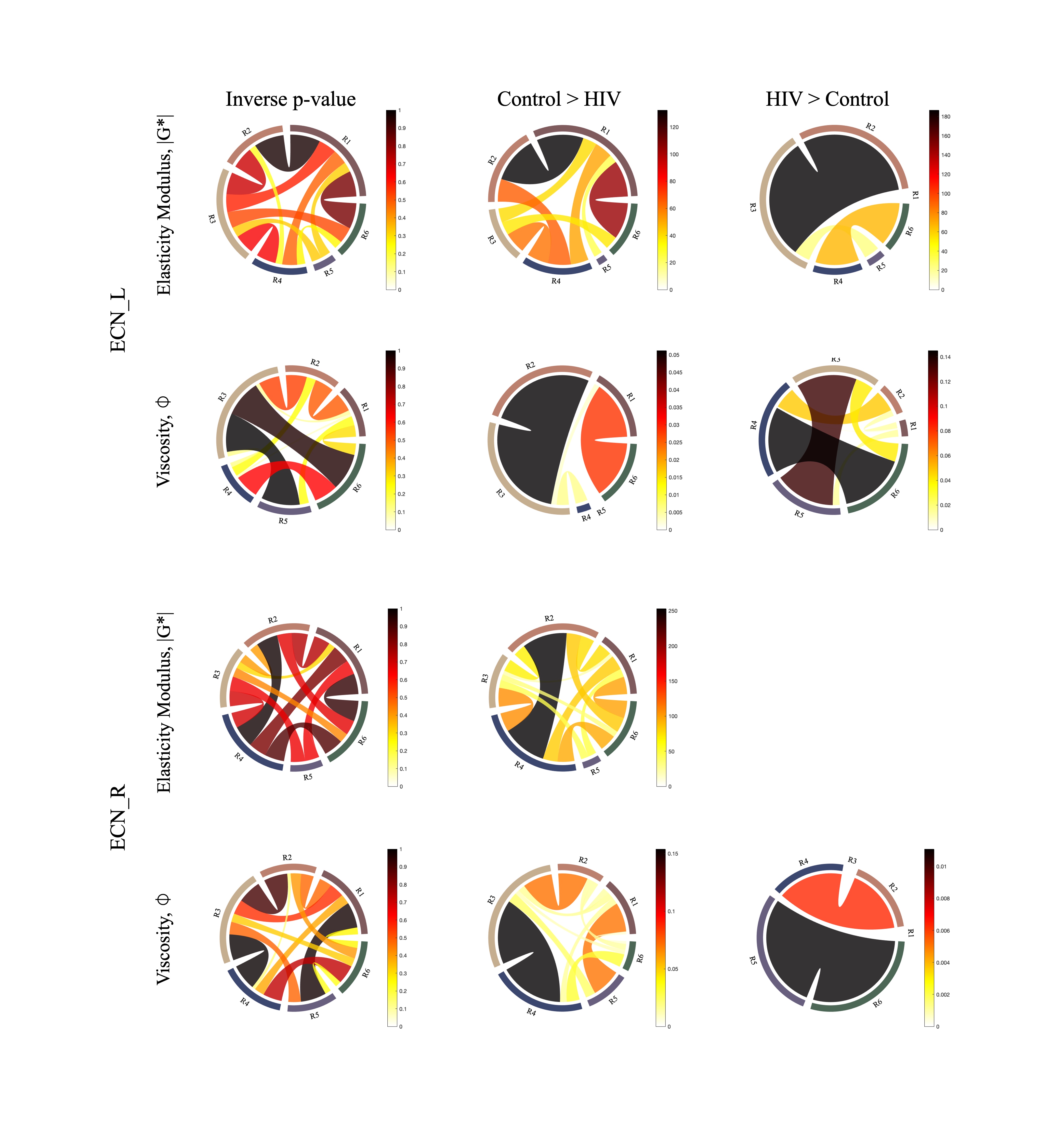
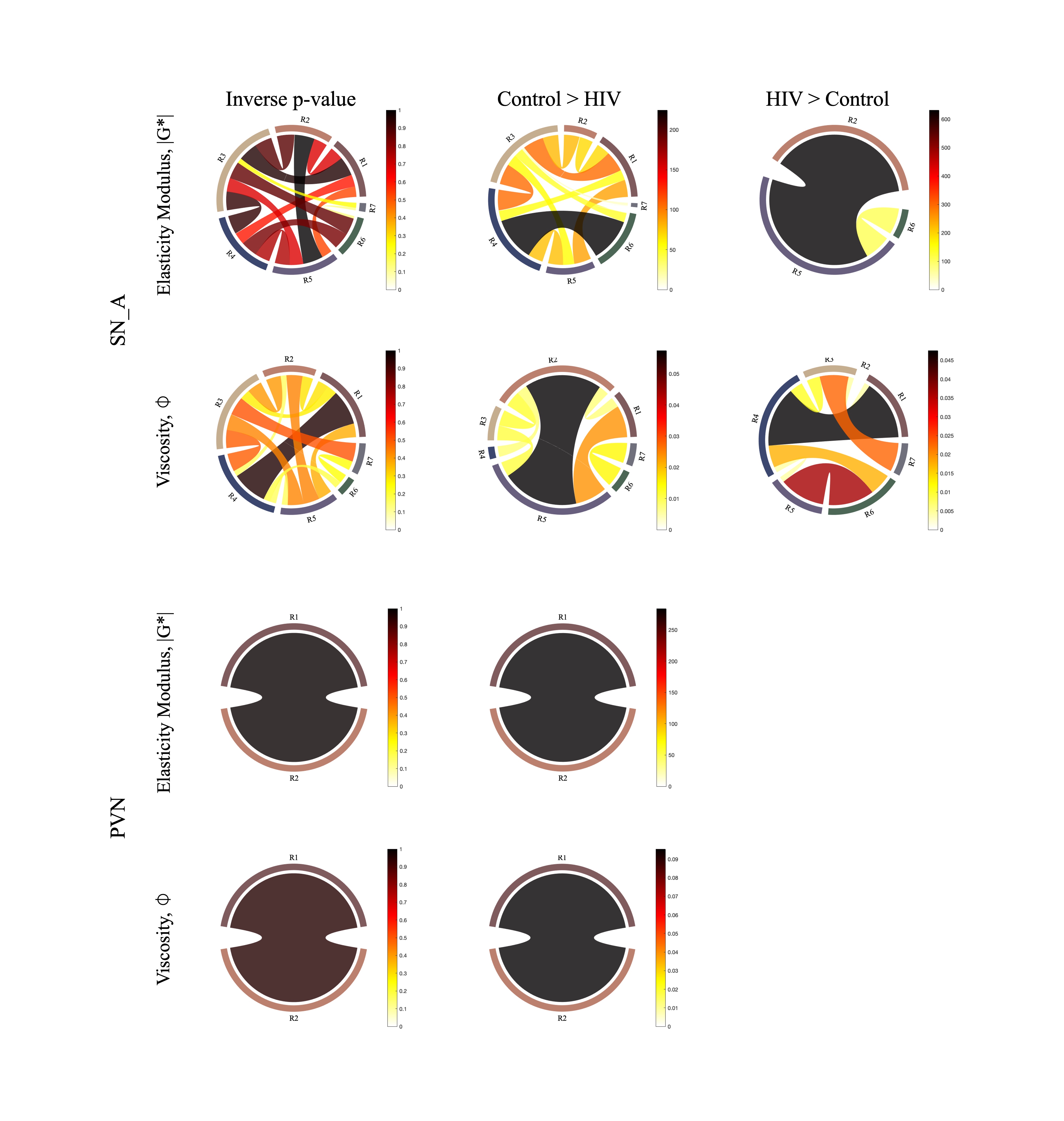
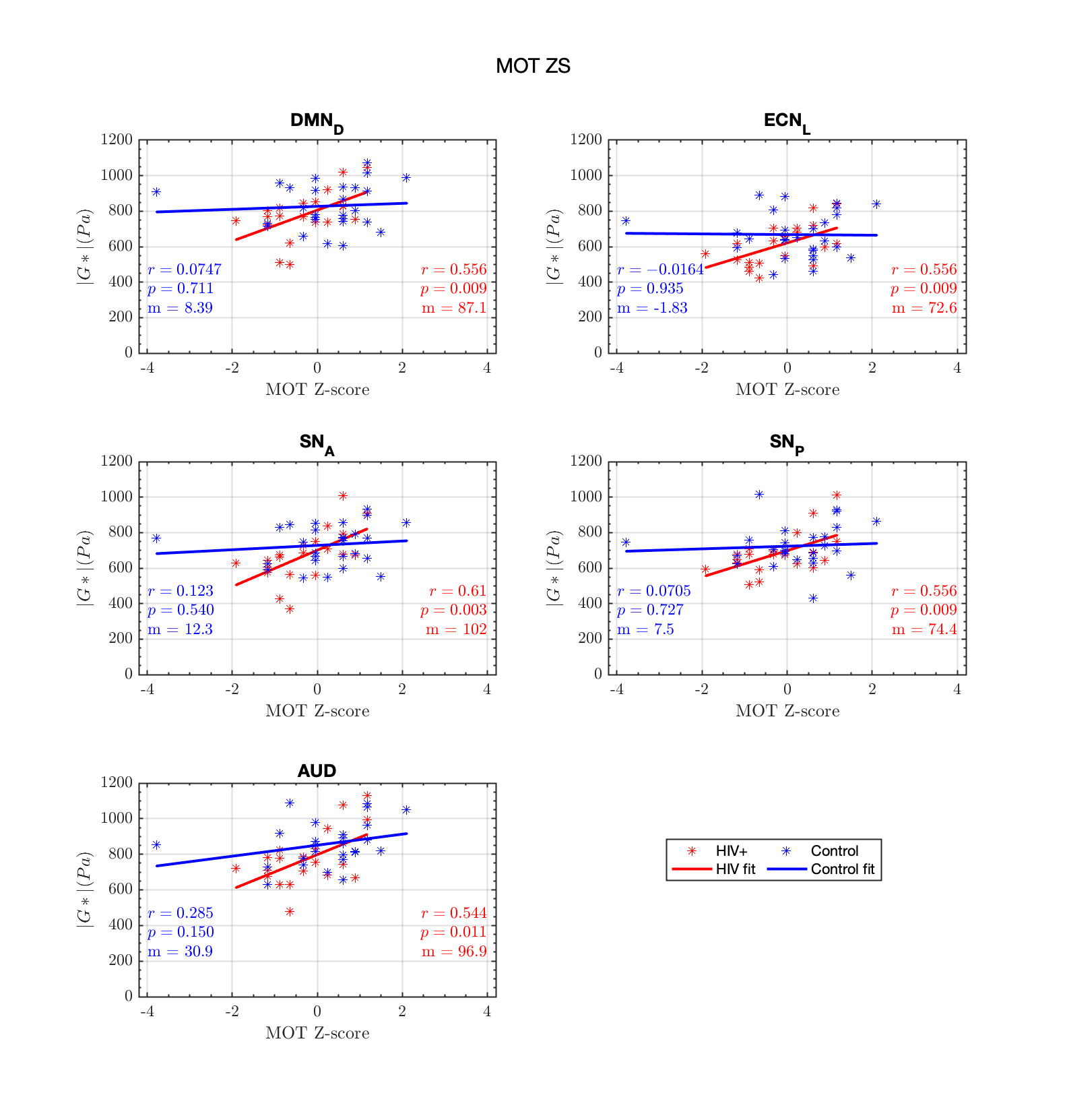
The following functionally defined networks are observed to have significantly changed viscoelasticity properties i.e. ECN_L, ECN_R, SN_A, PVN and DMN_D in Figure-1 in HIV+. Further , in the HIV-infected-cohort, we observe motor related cognitive-scores to be significantly correlated(p<0.05) with the connectivity of following networks: ECN_L, SN_A, DMN_D, SN_P and AUD. Among these networks ECN_L, SN_A and DMN_D are observed to hold significant viscoelastic changes. Figure-2 and Figure-3 show the summary of significantly ((1 – p-value) > 0.95) altered-|G*| and Ø for the HIV+ in their ROI connectivity of ECN_L, ECN_R, SN_A and PVN. It has been reported that, increasing or decreasing stiffness can be an effect of pathological conditions(4) and thus be responsible for reduced cognitive performance. This is finally reflected in Figure-4, which observes correlation of the significantly affected MTI with |G*| vs MOT_Z-score in PWH (HIV+). It should be noted that the Grooved-Pegboard test assess fine motor-skill, measures also speed of information processing involving multiple brain-networks. Correlation coefficients and p-values are insignificant for the Control group, reflecting role of HIV-infection for cognitive decline; congruent with previous report of known brain injury associated with HIV-infection(10).Conclusion
The study demonstrates the usefulness of MRE-dMRI tract integrity analysis in studying brain injury as in this cohort of PWH and CSVD. The proposed pipeline focuses on distinguished viscoelastic connectivity of the functionally defined networks and helps in analysis of cognitive performance. Our results suggest that early viscoelastic changes in the HIV-CSVD population are associated with fine motor skills and speed of information processing.Acknowledgements
This work was supported by the National Institutes of Health (Grant Numbers: R01 AG054328, and R21 NS113674)References
1. Coelho A, Sousa N. Magnetic resonance elastography of the ageing brain in normal and demented populations: A systematic review. Hum Brain Mapp. 2022;43(13):4207–18.
2. Hagmann P, Kurant M, Gigandet X, Thiran P, Wedeen VJ, Meuli R, et al. Mapping Human Whole-Brain Structural Networks with Diffusion MRI. PLOS ONE. 2007 Jul 4;2(7):e597.
3. Figley TD, Mortazavi Moghadam B, Bhullar N, Kornelsen J, Courtney SM, Figley CR. Probabilistic White Matter Atlases of Human Auditory, Basal Ganglia, Language, Precuneus, Sensorimotor, Visual and Visuospatial Networks. Front Hum Neurosci. 2017;11:306.
4. Streitberger KJ, Reiss-Zimmermann M, Freimann FB, Bayerl S, Guo J, Arlt F, et al. High-Resolution Mechanical Imaging of Glioblastoma by Multifrequency Magnetic Resonance Elastography. PLOS ONE. 2014 Oct 22;9(10):e110588.
5. Hirsch S, Guo J, Reiter R, Papazoglou S, Kroencke T, Braun J, et al. MR Elastography of the Liver and the Spleen Using a Piezoelectric Driver, Single-Shot Wave-Field Acquisition, and Multifrequency Dual Parameter Reconstruction. Magn Reson Med. 2014;71(1):267–77.
6. Woolrich MW, Jbabdi S, Patenaude B, Chappell M, Makni S, Behrens T, et al. Bayesian analysis of neuroimaging data in FSL. NeuroImage. 2009 Mar;45(1 Suppl):S173-186.
7. Tournier JD, Smith R, Raffelt D, Tabbara R, Dhollander T, Pietsch M, et al. MRtrix3: A fast, flexible and open software framework for medical image processing and visualisation. NeuroImage. 2019 Nov 15;202:116137.
8. Avants BB, Tustison NJ, Song G, Cook PA, Klein A, Gee JC. A reproducible evaluation of ANTs similarity metric performance in brain image registration. NeuroImage. 2011 Feb 1;54(3):2033–44.
9. Figley TD, Bhullar N, Courtney SM, Figley CR. Probabilistic atlases of default mode, executive control and salience network white matter tracts: an fMRI-guided diffusion tensor imaging and tractography study. Front Hum Neurosci. 2015;9:585.
10. Elicer IM, Byrd D, Clark US, Morgello S, Robinson-Papp J. Motor function declines over time in human immunodeficiency virus and is associated with cerebrovascular disease, while HIV-associated neurocognitive disorder remains stable. J Neurovirol. 2018 Aug;24(4):514–22.
Figures