0241
The Welsh Advanced Neuroimaging Database: an open-source state-of-the-art resource for brain research1Cardiff University Brain Research Imaging Centre, Cardiff University, Cardiff, United Kingdom, 2Department of Psychology, Royal Holloway, University of London, Surrey, United Kingdom, 3School of Psychology, Aston University, Birmingham, United Kingdom, 4Department of Psychology, Durham University, Durham, United Kingdom, 5Washington Singer Laboratories, University of Exeter, Exeter, United Kingdom, 6Leeds Institute of Cardiovascular and Metabolic Medicine, University of Leeds, Leeds, United Kingdom, 7The Russell H. Morgan Department of Radiology and Radiological Science, Johns Hopkins University, Balitmore, MD, United States, 8Computer Science, University College London, London, United Kingdom, 9University of Chieta-Pescara, Chieti, Italy, 10School of Medicine, Centre for Neuropsychiatric Genetics and Genomics, Cardiff University, Cardiff, United Kingdom
Synopsis
Keywords: Data Processing, Brain, data release
Motivation: Advances in MRI have increased our understanding of the human brain but are frequently limited by single modality study designs. Combining data from multiple modalities/MR contrasts can enhance our understanding of the complex multi-scale neural relationships that underpin human behaviour.
Goal(s): Our goal was to create an open-access multi-scale, multi-modal imaging database of the healthy human brain.
Approach: The Welsh Advanced Neuroimaging Database (WAND) includes micro and macro-structural, functional and spectroscopic MRI, MEG and cognitive data from over 150 healthy volunteers.
Results: WAND is free, open-source, organised using the Brain Imaging Data Structure (BIDS), and now available for download.
Impact: The Welsh Advanced Neuroimaging Database takes steps toward democratising magnetic resonance research by making multi-modal, multi-scale neuroimaging data freely and easily available, enhancing opportunities for collaboration and development of novel analysis techniques, further progressing the field of neuroimaging.
Introduction
The development of neuroimaging techniques, such as MRI and magnetoencephalography (MEG), that non-invasively manipulate or exploit the magnetic properties of tissue, has revolutionised neuroscience and our understanding of the living human brain. Furthermore, combining these modalities allows researchers to capitalise on their different spatial and temporal scales, allowing for multi-scale investigations of brain structure and function. Despite the value of high-tech multi-modal imaging, however, few researchers have access to the necessary resources, limiting scientific advancements in brain and imaging science.The Welsh Advanced Neuroimaging Database (WAND) is a multi-scale, multi-modal imaging database of the healthy human brain. It includes non-invasive in vivo brain data from 3T MRI with ultra-strong (300mT/m) magnetic field gradients (especially designed for evaluating tissue microstructure), 7T and 3T MRI and nuclear magnetic resonance spectroscopy (MRS), and MEG. The dataset also includes cognitive and behavioural data that allow for investigation of brain-behaviour interactions at multiple spatial and temporal scales. By employing cutting-edge technologies that target specific features of brain structure and function, WAND’s objective is to generate an integrated description of brain coupling over multiple domains that can be shared with researchers around the world.
Methods
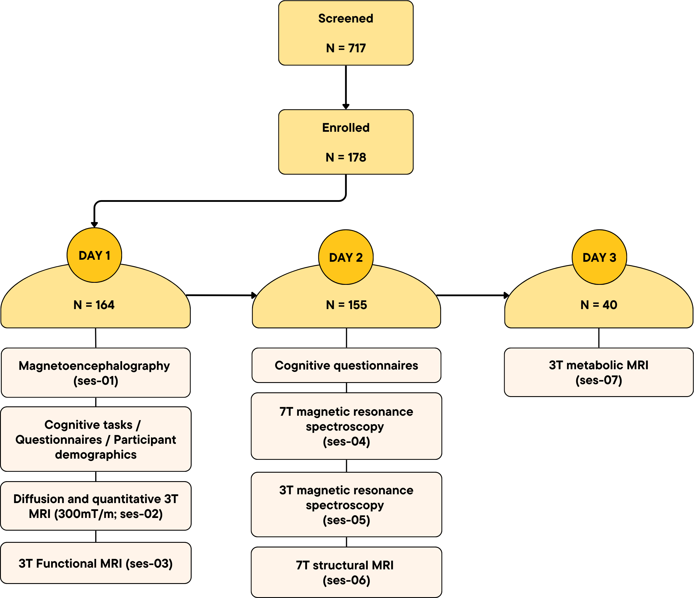
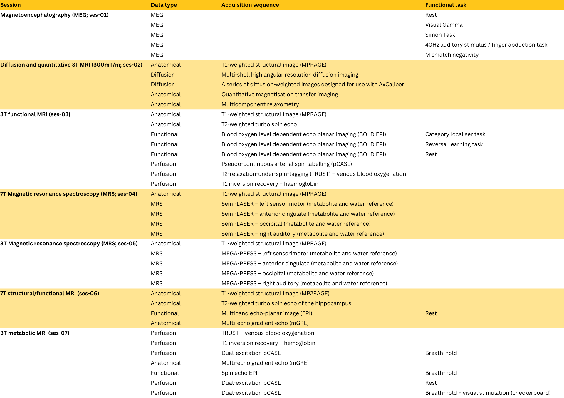
Healthy volunteers (N=178; 61% female) between 18 and 63 (median 25) years of age were recruited from Cardiff and surrounding areas between February 2019 and May 2023. Participants were invited to Cardiff University Brain Research Imaging Centre (CUBRIC), to take part in up to 7 neuroimaging sessions plus additional cognitive testing (see Figure 1). The study was approved by the Cardiff University School of Psychology Ethics Committee; all participants gave permission for their anonymised data to be shared with researchers in other organisations and deposited in publicly accessible databases.Details of acquisition sequences and tasks are provided in Figure 2, and were collected using the following state-of-the-art equipment:
Ultra-strong gradient 3T MRI
Diffusion, relaxometry and quantitative magnetisation transfer imaging data were acquired using an ultra-strong gradient (300mT/m) 3T Siemens Connectom MRI scanner, using a 32-channel head coil, and sequences previously described by Koller et al1. Ultra-strong gradients allowed for stronger diffusion weighting per unit time, shortening the minimum echo time, improving signal to noise ratio, and increasing sensitivity to small volume displacement2.
3T MRI
Functional, metabolic and edited spectroscopy data were acquired using a Siemens 3T MAGNETOM Prisma system, fit with a 32-channel head coil.
7T MRI
High-field structural, functional and spectroscopy data were acquired using 7T MAGNETOM "Classic" (Siemens Healthineers), fit with a 32-channel receive, volume transmit head coil (Nova Medical, Wilmington, MA, United States).
Magnetoencephalography
Whole-head MEG recordings were acquired at a 1200 Hz sampling rate on a 275-channel CTF radial gradiometer system. An additional 29 reference channels were recorded for noise cancellation purposes and the primary sensors were analysed as synthetic third-order gradiometers3. Head digitization was performed prior to the participant entering the magnetically shielded room, using the Polhemus device.
Demographic, Questionnaire and Cognitive data
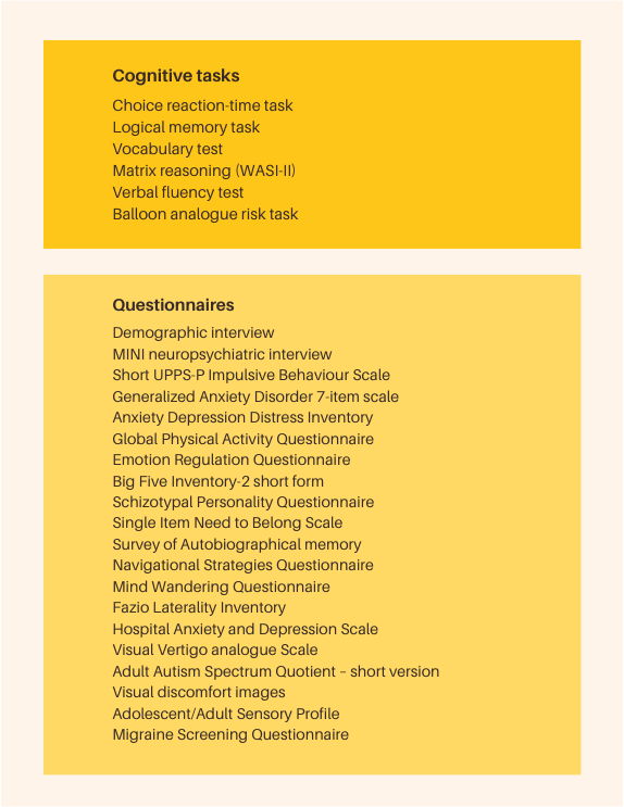
In addition to neuroimaging procedures, each participant completed a collection of cognitive tasks and questionnaires, outlined in Figure 3.
Results, data sharing and availability
All data are organised using the Brain Imaging Data Structure (BIDS)4. Within each participant’s directory, data are first arranged by neuroimaging session, then data type (see Figure 2). All MPRAGE data have been defaced using FSL’s5 deface tool, to protect the identity of participants. In addition to raw (NIFTI and JSON) data, we provide brain-extracted T1w images for all MPRAGE data, as well as quality reports for multishell high angular resolution diffusion imaging (using FSL’s eddy-qc), T1w and T2w structural data, and blood-oxygen level dependent functional tasks (using MRIqc6 and MRIQCEPTION; https://github.com/elizabethbeard/mriqception).The data are hosted on Cardiff University’s OwnCloud server. Users can access and download the whole dataset or can interact with, retrieve and link data (and metadata) using DataLad7, allowing for improved storage management and working practices.
Discussion
WAND is a multi-modal dataset containing data from state-of-the-art neuroimaging tools, not widely available to the neuroimaging community. The dataset consists of macro- and micro-structural, functional, metabolic, chemical and behavioural information, uniquely designed to improve our understanding of multi-scale coupling in the human brain.Conclusion
By making these data publicly available, we intend to bolster research into neuroimaging and related fields, contribute to the democratisation of MRI, and facilitate collaboration in MRI and MEG research.WAND can be accessed using the following link: https://git.cardiff.ac.uk/cubric/wand
Acknowledgements
WAND data were acquired at the UK National Facility for In Vivo MR Imaging of Human Tissue Microstructure funded by the EPSRC (grant EP/M029778/1), and The Wolfson Foundation, and supported by a Wellcome Trust Investigator Award (096646/Z/11/Z) and a Wellcome Trust Strategic Award (104943/Z/14/Z). Olivier Mougin (University of Nottingham) developed the phase-sensitive inversion recovery and T1 code. William Clarke (Oxford University) provided the image reconstruction code for the MP2RAGE.References
- Koller K, et al. MICRA: Microstructural image compilation with repeated acquisitions. NeuroImage 225, 117406 (2021).
- Jones DK, et al. Microstructural imaging of the human brain with a ‘super-scanner’: 10 key advantages of ultra-strong gradients for diffusion MRI. NeuroImage 182, 8–38 (2018).
- Vrba J & Robinson SE. Signal processing in magnetoencephalography. Methods San Diego Calif 25, 249–271 (2001).
- Gorgolewski KJ, et al. The brain imaging data structure, a format for organizing and describing outputs of neuroimaging experiments. Sci. Data 3, 160044 (2016).
- Jenkinson M, Beckmann CF, Behrens TE, Woolrich MW, Smith SM. FSL. Neuroimage. 62(2):782-90 (2012).
- Esteban O, Birman D, Schaer M., Koyejo OO, Poldrack RA, Gorgolewski KJ. MRIQC: Advancing the Automatic Prediction of Image Quality in MRI from Unseen Sites. PLOS ONE. 12(9):e0184661; doi:10.1371/journal.pone.0184661.
- Halchenko, YO, et al., (2021). DataLad: distributed system for joint management of code, data, and their relationship. Journal of Open Source Software, 6(63), 3262, https://doi.org/10.21105/joss.03262.
Figures