0240
NeoAudi Tract: An Automated Tool for Identifying Auditory Fiber Bundles in Infants1School of Information and Technology, Northwest University, Xi'an, China, 2School of Biomedical Engineering & State Key Laboratory of Advanced Medical Materials and Devices, ShanghaiTech University, Shanghai, China, 3Department of Otolaryngology-Head and Neck Surgery, Shanghai Ninth People's Hospital, Shanghai Jiao Tong University School of Medicine, Shanghai, China, 4Ear Institute, Shanghai Jiao Tong University School of Medicine, Shanghai, China, 5Shanghai Key Laboratory of Translational Medicine on Ear and Nose Diseases, Shanghai, China, 6Shanghai United Imaging Intelligence Co., Ltd., Shanghai, China, 7Department of Radiology, Shanghai Ninth People's Hospital, Shanghai Jiao Tong University School of Medicine, Shanghai, China, 8Shanghai Clinical Research and Trial Center, Shanghai, China
Synopsis
Keywords: Data Processing, Pediatric, Neuroimage computing, Auditory pathway, Normal development
Motivation: Charting the development of infant auditory system is vital for understanding language acquisition and hearing disorders.
Goal(s): Extracting auditory fiber bundles from diffusion MRI data and overcoming the processing difficulties due to tiny and complex structures, as well as very low tissue contrast in the structural MRI data.
Approach: We propose an NAT framework with three core processes: 1) constructing a high-resolution atlas, 2) segmenting tissues and regions of interest (ROIs), and 3) applying a hierarchical registration framework.
Results: A high-resolution auditory fiber bundle template is constructed, and 12 auditory fiber bundles are successfully extracted.
Impact: Our approach is the first toolbox to identify individual auditory fiber bundles in infants, thus effectively mitigating the processing challenges caused by spatial-temporal asynchrony during the development of the first two postnatal years.
Purpose
Charting the development of infant auditory system is vital for understanding language acquisition and hearing disorders. Extracting fiber bundles in auditory pathway from diffusion MRI (dMRI) data presents significant difficulty due to tiny and complex structures, as well as very low tissue contrast in the structural MRI (sMRI) data. Recent methodologies have been proposed to enhance data resolution by utilizing adult post-mortem specimens examined ex vivo [1] or 7T MRI data captured in vivo [2]. Furthermore, [3] leverages both tract density imaging (TDI) and sMRI data to construct an auditory pathway atlas specifically for children. However, extracting individual auditory pathway in infants remains challenging due to substantial spatial-temporal variability of infant brain MRI data in the first two postnatal years. In this study, we propose NeoAudi Tract (NAT), an automated toolbox designed to identify auditory fiber bundles in infants, by incorporating segmentation [4] and registration [5] techniques specifically tailored to address the pronounced variability in infant brain MRI data.Methods
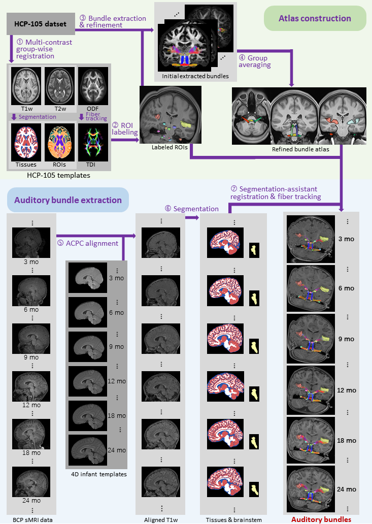
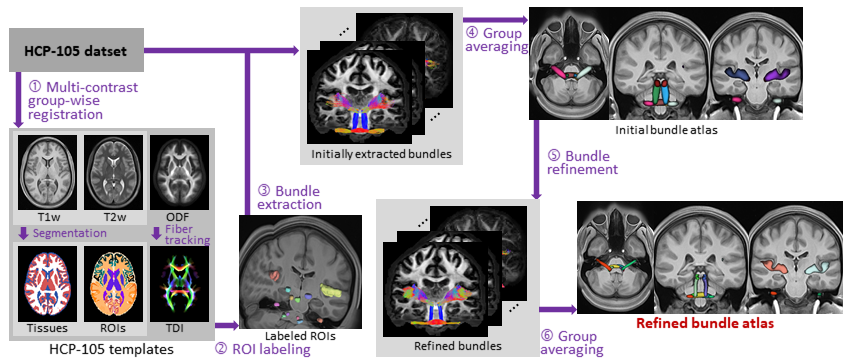
We propose the NAT framework with three core processes: i) constructing a high-resolution atlas, ii) segmenting tissues and regions of interest (ROIs), and iii) applying a hierarchical registration framework. Fig. 1 illustrates NAT and showcases the fiber bundles extracted from infants at 3, 6, 9, 12, 18, and 24 months of age, highlighting effectiveness of NAT.We utilize the HCP-105 dataset to construct high-resolution templates aligned to the dMRI data space, encompassing T1w, T2w, and constrained spherical deconvolution fiber orientation distribution function (ODF) [6]. Tissue segmentation is performed using a deep learning model [7], and the TDI map is derived through the whole brain tracts generated by the probabilistic fiber tracking algorithm [3,8]. Working in parallel with six templates, two experienced clinicians manually delineate and validate the processing and relay stations of acoustic stimulus, encompassing a total of twelve ROIs, as shown in Fig. 2.
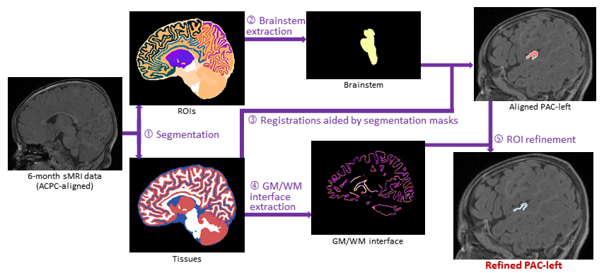
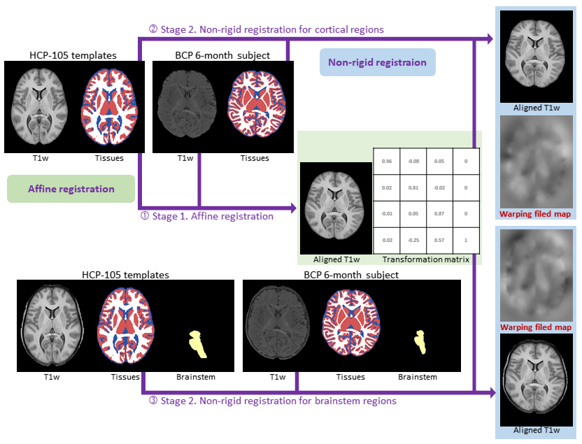
To accurately align adult templates with infant data, our initial step involves using 4D infant templates for ACPC alignment [5] to facilitate the subsequent non-linear image registration. In addition, the precise tissue maps and ROIs, segmented by deep learning technique, are utilized to assist registration procedures. As shown in Fig. 3, brain tissues, as well as subcortical and cortical ROIs defined in Desikan-Killiany-Tourville (DKT) atlas, are segmented and employed to refine the aligned ROIs, including the primary auditory cortex (PAC). Next, both the brainstem and interface between gray matter (GM) and white matter (WM) are extracted. Following this, as depicted in Fig. 4, the segmentation results are incorporated into the registration process, thereby managing the variable intensity ranges of GM and WM and enhancing the sMRI data. NAT conducts a hierarchical alignment between templates and infant data, i.e., employing initially an affine registration and subsequently a two-branch non-rigid approach to cortical and subcortical regions, respectively.
Results
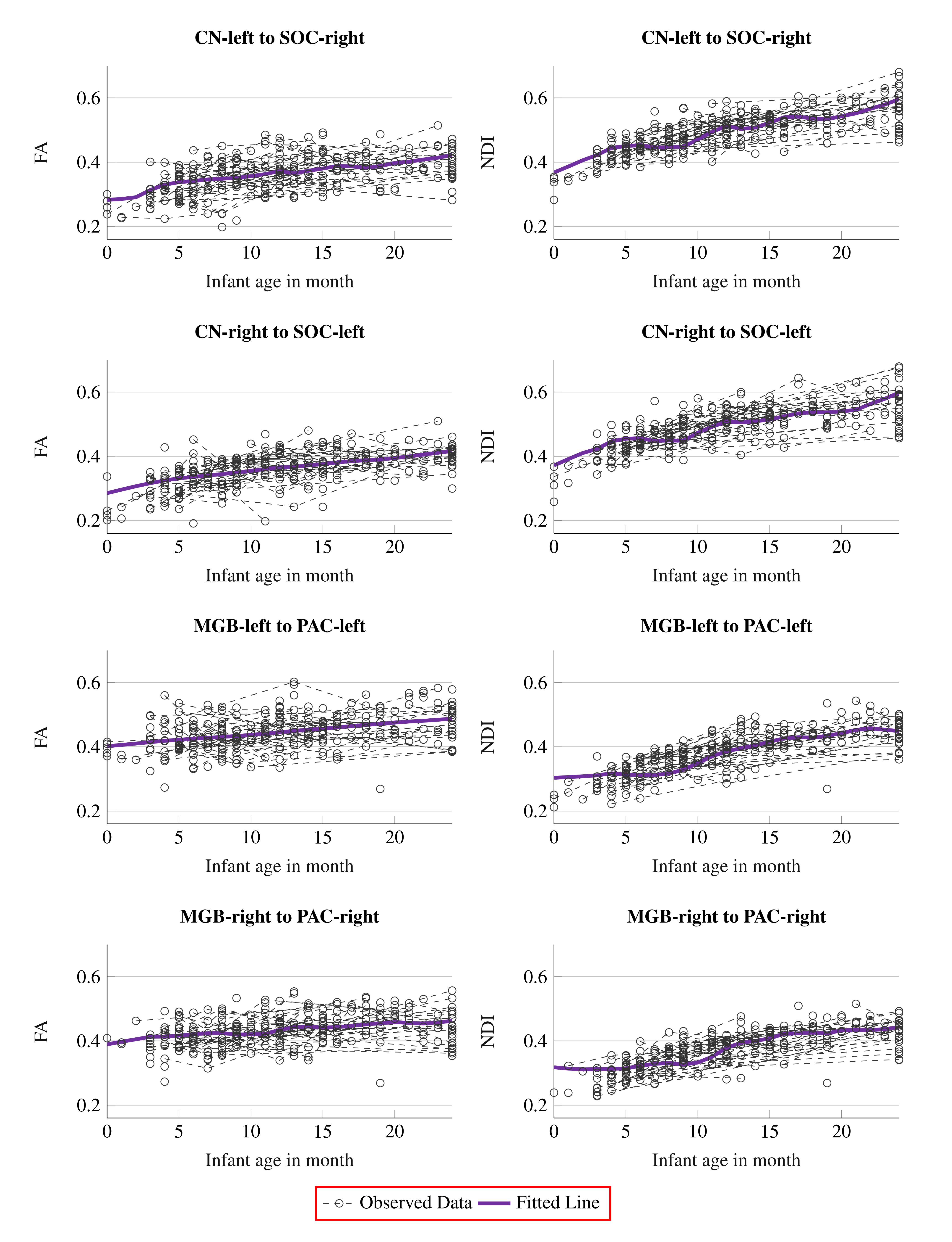
We employ the Baby Connectome Project (BCP) dataset to identify the auditory fiber bundles and monitor the development of the auditory system during the first two postnatal years. Fig. 5 demonstrates the trajectories of two fiber bundles, contralateral CN-to-SOC (located in the brainstem), and ipsilateral MGB-to-PAC (situated in the cortical regions). For each fiber bundle, we calculate average fractional anisotropy (FA) and neurite density index (NDI), with individual subjects represented as distinct points in Fig. 5. A linear mixed-effects model (LMM) is used to model developmental trajectory for these bundles. We can observe that the NDI of MGB-to-PAC is lower than that of CN-to-SOC, which is consistent with previous neuroscientific findings [9]. On the other hand, the CN-to-SOC shows a lower FA value, which may reflect the inadequacy of FA to accurately characterize dense crossing fibers in the brainstem. Additionally, the NDI of CN-to-SOC exhibits a more pronounced increase compared to MGB-to-PAC, implying developmental emphasis on the maturation of fundamental acoustic processing in the early stages.Conclusion
We present NeoAudi Tract (NAT), an automated and accurate toolbox, for extracting auditory fiber bundles in infant brains. By integrating specialized segmentation and registration techniques tailored for the infant brain, NAT successfully delineates six pairs of fiber bundles across developmental spectrum from newborns to 24-month-old infants. Our findings highlight earlier maturation of the CN-to-SOC, which is instrumental in processing fundamental acoustic stimuli. This underlines the importance of CN-to-SOC in the initial stages of auditory system development, supporting its potential impact on early auditory and language acquisition.Acknowledgements
This work was supported in part by the National Natural Science Foundation of China (No.62131015 and 62203355), Science and Technology Commission of Shanghai Municipality (STCSM) (No.21010502600), the Key R & D Program of Guangdong Province (No.2021B0101420006), and the National Key Technology R & D Program (No.2022ZD0209000).References
[1] Zanin, J., Dhollander, T., Farquharson, S., Rance, G., Connelly, A., Nayagam, B.A., 2019. Using diffusion-weighted magnetic resonance imaging techniques to explore the microstructure and connectivity of subcortical white matter tracts in the human auditory system. Hearing Research 377, 1–11.
[2] Sitek, K.R., Gulban, O.F., Calabrese, E., Johnson, G.A., Lage-Castellanos, A., Moerel, M., Ghosh, S.S., De Martino, F., 2019. Mapping the human subcortical auditory system using histology, postmortem MRI and in vivo MRI at 7T. eLife 8, e48932.
[3] Wang, Y., Jiang, M., Zhu, Y., Xue, L., Shu, W., Li, X., Chen, H., Li, Y., Chen, Y., Chai, Y., et al., 2023. Impact of inner ear malformation and cochlear nerve deficiency on the development of auditory-language network in children with profound sensorineural hearing loss. eLife 12, e85983.
[4] Liu, J., Liu, F., Sun, K., Liu, M., Sun, Y., Ge, Y., Shen, D., 2023. Adult-like phase and multi-scale assistance for isointense infant brain tissue segmentation, in: International Conference on Medical Image Computing and Computer-Assisted Intervention, Springer. pp. 56–66.
[5] Chen, L., Wu, Z., Hu, D., Wang, Y., Zhao, F., Zhong, T., Lin, W., Wang, L., Li, G., 2022. A 4D infant brain volumetric atlas based on the UNC/UMN baby connectome project (BCP) cohort. NeuroImage 253, 119097.
[6] Wasserthal, J., Neher, P., Maier-Hein, K.H., 2018. TractSeg-Fast and accurate white matter tract segmentation. NeuroImage 183, 239–253.
[7] Xiao, B., Cheng, X., Li, Q., Wang, Q., Zhang, L., Wei, D., Zhan, Y., Zhou, X.S., Xue, Z., Lu, G., 2019. Weakly supervised confidence learning for brain mr image dense parcellation, in: Proceedings of International Workshop on Machine Learning in Medical Imaging, Springer. pp. 409–416.
[8] Tournier, J.D., Calamante, F., Connelly, A., et al., 2010. Improved probabilistic streamlines tractography by 2nd order integration over fibre orientation distributions, in: Proceedings of the ISMRM, John Wiley & Sons, Inc, New Jersey.
[9] Silver, E., Korja, R., Mainela-Arnold, E., Pulli, E.P., Saukko, E., Nolvi, S., Kataja, E.L., Karlsson, L., Karlsson, H., Tuulari, J.J., 2021. A systematic review of MRI studies of language development from birth to 2 years of age. Developmental Neurobiology 81, 63–75.
Figures