0238
Harmonizing Multi-Modality Biases in Infant Development Analysis with an Integrated MRI Data Processing Pipeline1School of Biomedical Engineering & State Key Laboratory of Advanced Medical Materials and Devices, ShanghaiTech University, Shanghai, China, 2School of Information and Technology, Northwest University, Xi'an, China, 3Shanghai United Imaging Intelligence Co., Ltd., Shanghai, China, 4Department of Computer Science and Technology,Nanjing University of Aeronautics and Astronautics, Nanjing, China, 5School of Computer Science and Engineering, Northwestern Polytechnical Universit, Xi'an, China, 6The Key Laboratory of Biomedical Information Engineering of Ministry of Education, School of Life Science and Technology, Xi’an Jiaotong University, Xi'an, China, 7School of Mathematics and Statistics, Xi’an Jiaotong University, Xi'an, China, 8Shanghai Clinical Research and Trial Center, Shanghai, China
Synopsis
Keywords: Data Processing, Brain, Neuroimage computing,pipeline
Motivation: Understanding infant neurodevelopment is pivotal for unraveling the anatomical underpinnings of psychomotor and cognitive functions, as well as pinpointing the origins of various disorders.
Goal(s): Introduce an integrated multi-modality MRI data processing pipeline tailored for infant development studies, with the goal of reliablly discerning relationship across brain anatomy and cognitive functions.
Approach: Incorporating precise deep learning tools specifically designed for infant brain, structural, functional, diffusion MRI data can be accurately analyzed, w.r.t. surface attributes for group-level study and network attributes for individual-level study.
Results: We introduce an integrated multi-modal infant MRI data processing pipeline toolkit with dedicated processing results.
Impact: We introduce the first infant multi-modal atlas and parcellation map
Purpose
Understanding infant neurodevelopment is pivotal for unraveling the anatomical underpinnings of psychomotor and cognitive functions, as well as pinpointing the origins of various disorders. Previous studies typically encountered two primary challenges: i) the scarcity of data due to unpredictable infant head movements during acquisition and ii) the difficulty of data processing stemming from non-synchronous spatial-temporal maturation during brain development. With the advancements in the acceleration of data acquisition, there has been a significant increase of high-quality infant MRI data. However, precise processing of infant MRI data remains challenging. In practice, discrepancies in atlas definitions and metric computations across different pipelines introduce notable biases in the analysis, which hinder the cross-validation of findings and the full exploitation of their complementary information. In this study, we introduce an integrated multi-modality MRI data processing pipeline for infant development studies, which incorporates high-quality deep learning-based tools specifically designed for infant MRI data processing, can be employed to construct multi-modality consistent atlases, and eventually facilitates to the delineation of more precise developmental trajectories.Methods
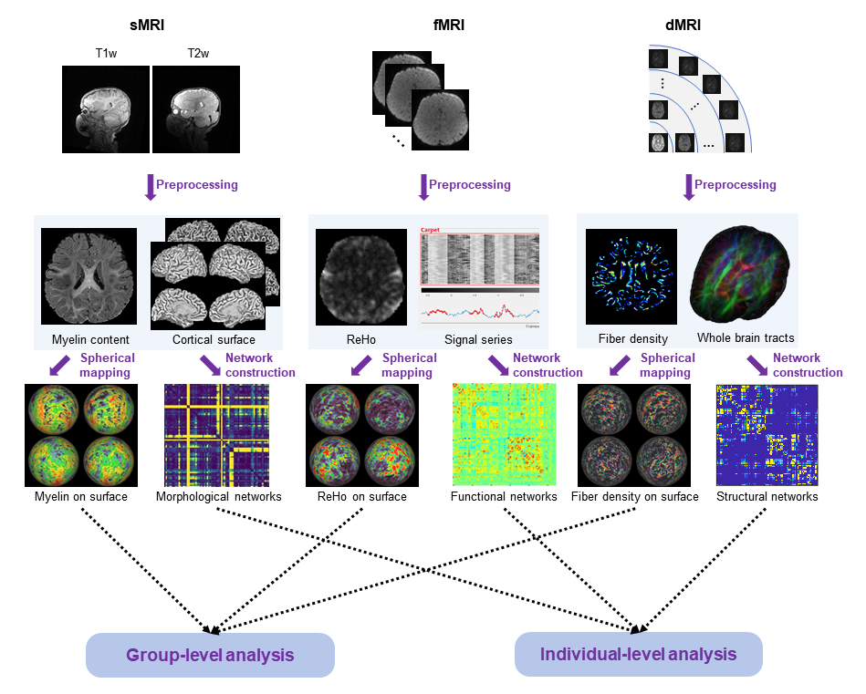
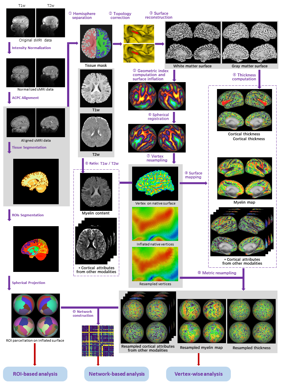
This processing pipelines are settled down largely according to HCP and dHCP pipelines [1,2], which are further optimized in two crucial aspects, i) the incorporation of more recently published tools [3], ii) catering to accommodate the unique characteristics of the infant brain [4]. The inputs of this pipeline are the raw structural MRI (sMRI), functional MRI (fMRI), and diffusion MRI (dMRI) data, and the outputs include two aspects, i.e., surface attributes for group-level analysis and network attributes for individual-level analysis, as shown in Fig.1.Specifically, sMRI primarily comprises T1w and T2w, which delineate the profiles of white matter (WM), gray matter (GM), and cerebrospinal fluid (CSF). We employ advanced deep learning models [5] for accurate segmentation of infant brain tissues, cortical and subcortical region of interest (ROI), as shown in Fig.2. Then, standard inner (white) and outer (pial) cortical surfaces are reconstructed, with vertex of corresponding morphological attributes mapped (e.g. thickness and curvature). The cortical surface is further inflated to a sphere to facilitate the precise alignment of infant brains for longitudinally consistent analysis. A group-level surface template is utilized for vertex-wise analysis, while the attributes in individual surface could be leveraged to construct morphological networks.
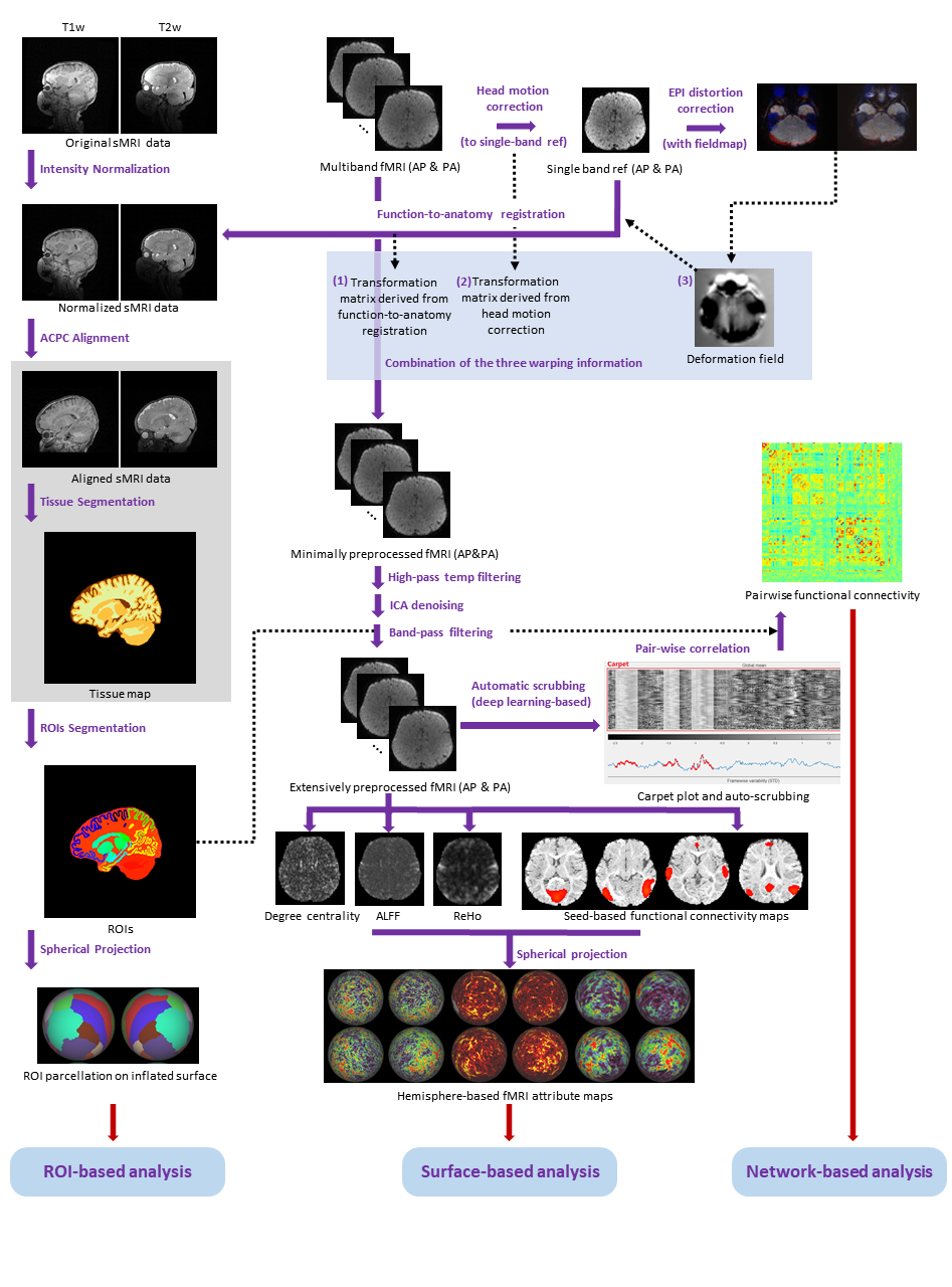
As illustrated in Fig.3, the fMRI processing pipeline includes several critical steps: head motion correction, distortion correction, functional-anatomical alignment, high-pass temporal filtering, independent component analysis (ICA) denoising, and automated scrubbing procedures. The registration, ICA denoising, and scrubbing steps integrate sophisticated deep learning-based methods tailored to the specific characteristics of infant brain tissues.
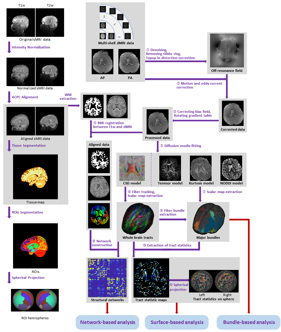
The dMRI pipeline mainly includes denoising, bias and distortion correction, co-registration, and rotation of B-matrix, as shown in Fig.5. For whole-brain tractography, multi-shell, and multi-tissue constrained spherical deconvolution techniques are employed to derive fiber orientation distribution functions. Probabilistic tracking is then conducted, with seeds located on the interface of gray matter and white matter. After screening false positive streamlines, a brain connectome is established from obtained fibers. Both the registration and fiber bundle segmentation stages are enhanced with deep learning-based methods catering to infant tissue characteristics.
Results
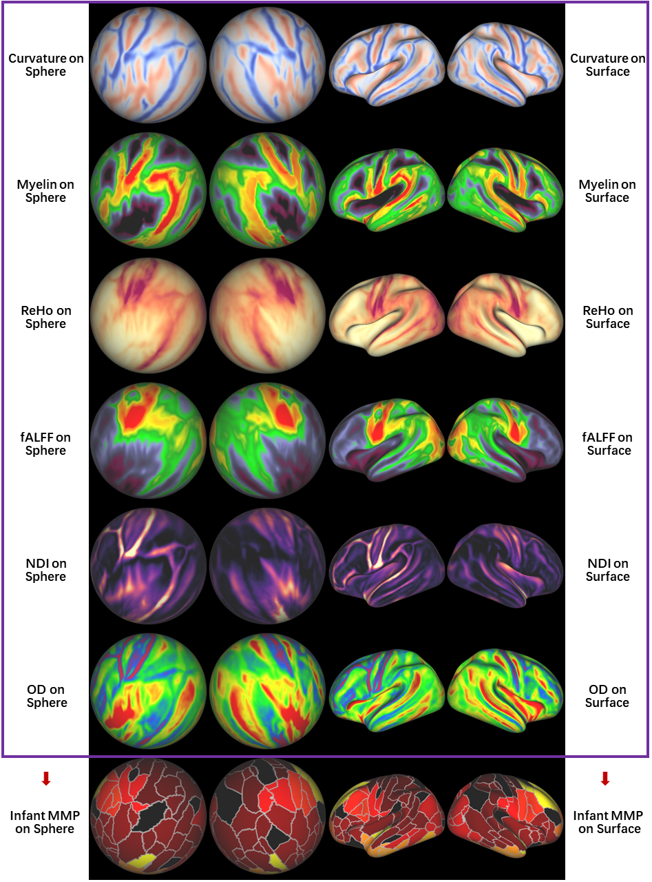
Fig.5 demonstrates an application of our proposed pipeline. By employing a specialized group-wise registration strategy [5], we initially construct a longitudinally consistent infant cortical surface template through the alignment of geometric patterns of cortical folding. Individual surfaces are then mapped onto this infant surface atlas to establish group-level vertex correspondence. Subsequently, multi-modality cortical attributes are aggregated across both hemispheres to profile multi-modal cortical attributes. Finally, we derive whole cortex connectivity patterns based on vertex-wise similarity across multi-modal attributes. Utilizing a local-gradient approach, we delineate the first infant multi-modal parcellation (MMP) map [6].Conclusion
In this study, we introduce an integrated multi-modal MRI data processing pipeline toolkit, which incorporates advanced deep learning-based methods for facilitating and guaranteeing the precision of infant MRI data processing. Our proposed pipeline has been meticulously adjusted and incorporated into a unified package aimed at reducing discrepancies stemming from diverse processing tools and, therefore, could essentially harmonize infant development investigations.Acknowledgements
This work was supported in part by the National Key Technology R&D Program (No.2022ZD0209000), the National Natural Science Foundation of China (No.62131015 and 62203355), Science and Technology Commission of Shanghai Municipality (STCSM) (No.21010502600), and the Key R&D Program of Guangdong Province (No.2021B0101420006).References
[1] Glasser, M.F., Sotiropoulos, S.N., Wilson, J.A., Coalson, T.S., Fischl, B., Andersson, J.L., Xu, J., Jbabdi, S., Webster, M., Polimeni, J.R., et al., 2013. The minimal preprocessing pipelines for the Human Connectome Project. NeuroImage 80, 105–124.
[2] Fitzgibbon, S.P., Harrison, S.J., Jenkinson, M., Baxter, L., Robinson, E.C., Bastiani, M., Bozek, J., Karolis, V., Grande, L.C., Price, A.N., et al., 2020. The developing Human Connectome Project (dHCP) automated resting-state functional processing framework for newborn infants. NeuroImage 223, 117303.
[3] Chen, L., Wu, Z., Hu, D., Wang, Y., Zhao, F., Zhong, T., Lin, W., Wang, L., Li, G., 2022. A 4D infant brain volumetric atlas based on the UNC/UMN baby connectome project (BCP) cohort. NeuroImage 253, 119097.
[4] Liu, J., Liu, F., Sun, K., Liu, M., Sun, Y., Ge, Y., Shen, D., 2023. Adult-like phase and multi-scale assistance for isointense infant brain tissue segmentation, in: International Conference on Medical Image Computing and Computer-Assisted Intervention, Springer. pp. 56–66.
[5] Ahmad, S., Wu, Z., Li, G., Wang, L., Lin, W., Yap, P.T., Shen, D., 2019. Surface-constrained volumetric registration for the early developing brain. Medical image analysis 58, 101540.
[6] Gordon, E.M., Laumann, T.O., Adeyemo, B., Huckins, J.F., Kelley, W.M., Petersen, S.E., 2016. Generation and evaluation of a cortical area parcellation from resting-state correlations. Cerebral cortex 26, 288–303.
Figures