0230
Evaluating the Immunotherapy Efficacy of Lung Cancer by 129Xe MRI1Innovation Academy for Precision Measurement Science and Technology, Chinese Academy of Sciences, Wuhan, China
Synopsis
Keywords: Visualization, Hyperpolarized MR (Gas), 129Xe MRI, Lung cancer, immunotherapy, ferroptosis
Motivation: The evaluation of lung cancer immunotherapy progress using non-invasive methods is challenging. Also, multiple scans of CT during the treatment period expose patients to more radiation.
Goal(s): To confirm 129Xe MRI is a potentially robust technology for monitoring immunotherapy effects.
Approach:
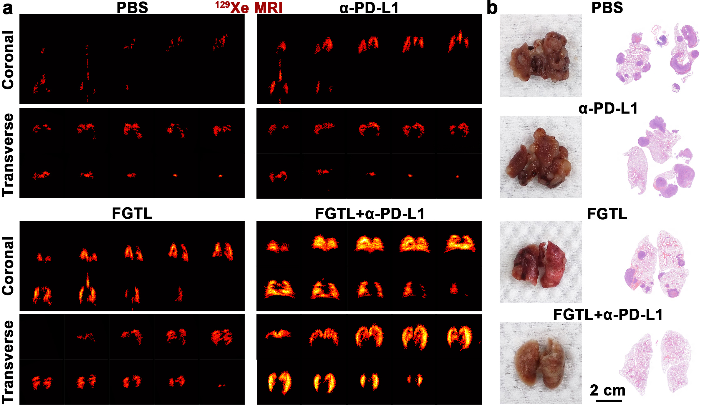
Results: The 129Xe MRI displayed a complete ventilatory image of the lung in the probe plus α-PD-L1 group while severe ventilation deficiency was observed in the control group.
Impact: The 129Xe MRI results of the lung showed significant differences in ventilation defects among different treatment groups, revealing the excellent tumor immunotherapy efficiency of nanoprobe-mediated immunotherapy, which represents a potential protocol for the evaluation of immunotherapy against tumors.
Cancer immunotherapy based on the immune checkpoint blockade (ICB) strategy has great potential in the therapy of lung cancer, but only 10-20% of patients respond due to the immunosuppressive tumor microenvironment (TME) [1]. Although several imaging techniques including MRI, PET, and CT have been widely used in tumor diagnosis, novel protocols to effectively monitor the efficiency of immunotherapy in real-time are precious. Hyperpolarized 129Xe MRI with ultrahigh sensitivity has received intense attention for monitoring lung diseases such as evaluating pulmonary function in COVID-19 patients [2-3]. Therefore, monitoring immunotherapy of lung cancer by using 129Xe MRI has great potential.
Methods
To reverse the immunosuppressive TME for the improvement of the ICB immunotherapy efficacy, a Fe3O4 nanoprobe that responds to reactive oxygen species (ROS) was created. Glucose oxidase was loaded into nanoprobe (FG) to convert glucose into hydrogen peroxide. The loading of IR820 endows the nanoprobe with T2 MR/fluorescence dual-modal imaging ability (FGI). The immune-activating peptide Tuftsin was loaded into the nanoprobe (FGIT) to reverse the immunosuppressive TME. To improve biocompatibility, the Fe3O4 nanoprobe was further coated with lipids (FGITL). The successful preparation of FGTL was characterization with TEM, XPS, and XRD analysis. The T2 MRI/fluorescence imaging was carried out on a 9.4 T imaging system and an IVIS Spectrum imaging system, respectively. The tumor immune activation effect of FGTL was analyzed by flow cytometry. Finally, the 129Xe ventilation MRI of the lung was performed on a 7 T imaging system.
Results and discussion
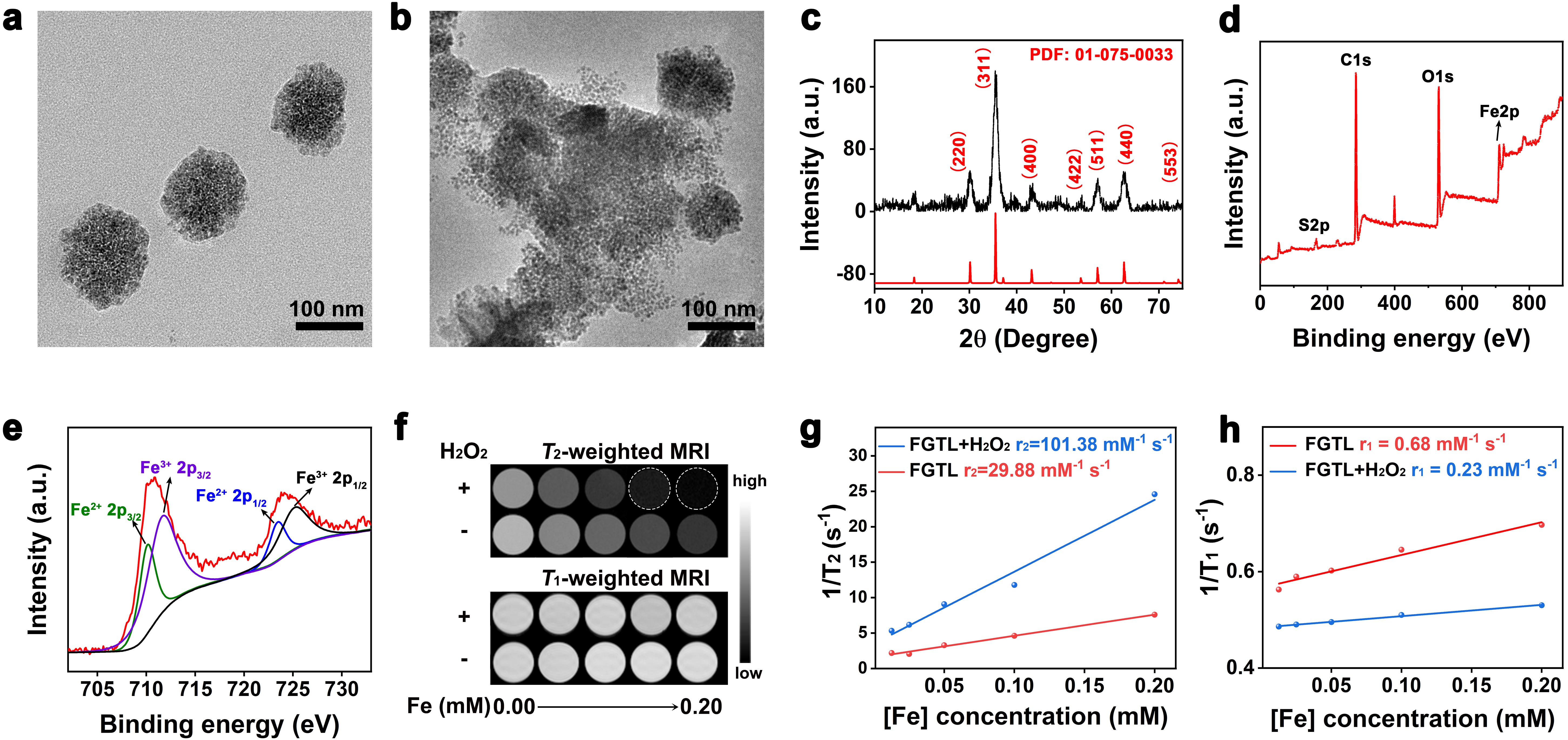
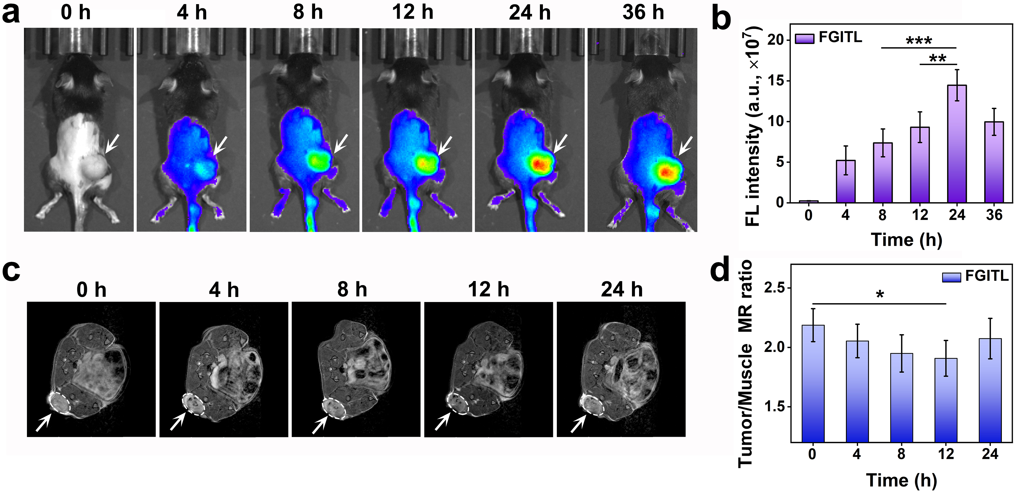
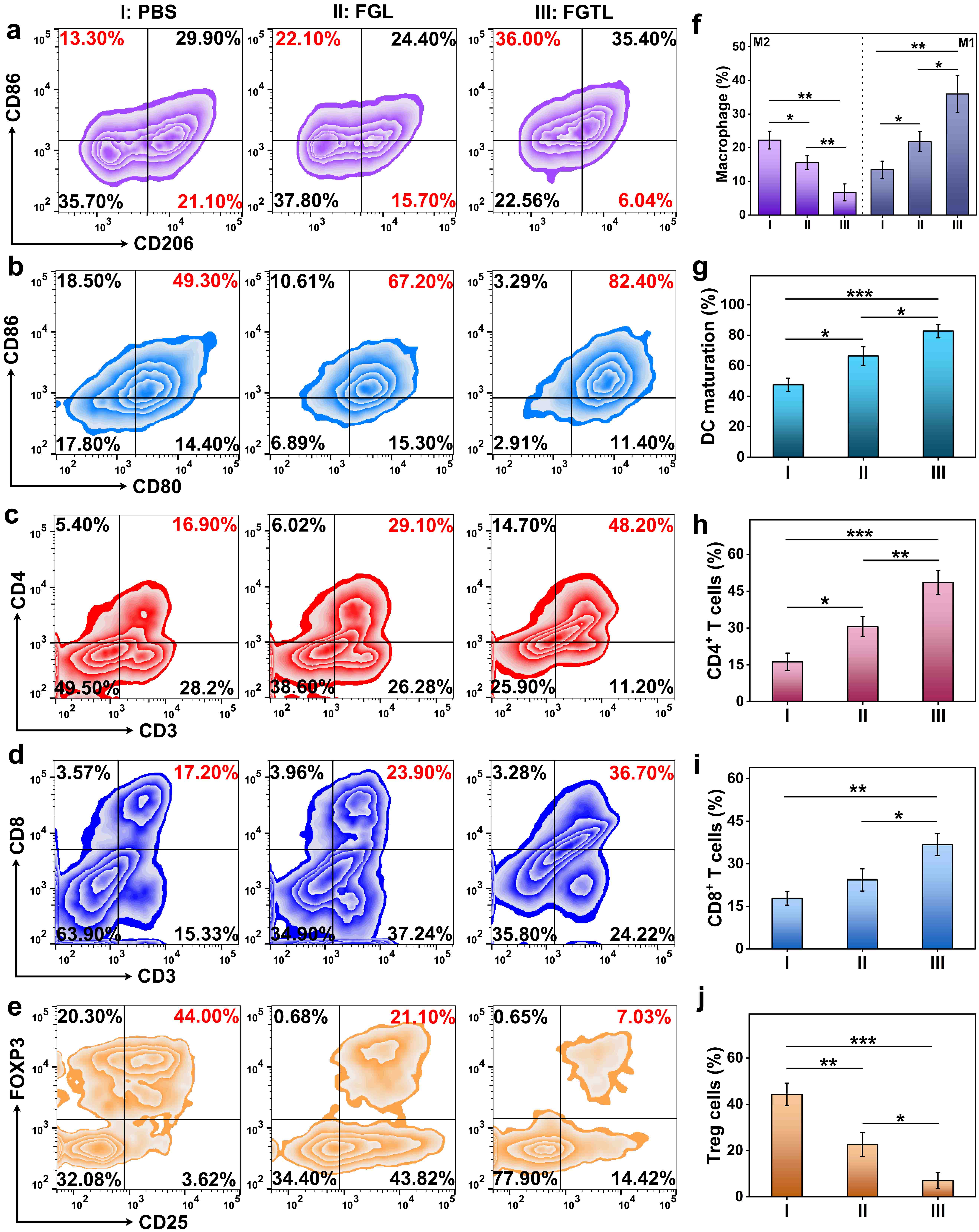
The diameter of the ROS-responsive FGTL nanoprobe with the spherical structure was about 100 nm (Fig 1a). After treatment with H2O2, the FGTL was disintegrated into small particles (Fig 1b), promoting the uptake of FGTL by tumor cells. X-ray diffraction (XRD) results demonstrated that the prepared nanoprobe was Fe3O4 (Fig 1c). The presence of Fe2+ and Fe3+ was verified by the XPS analysis of Fe3O4 nanoprobe (Fig 1d,e). The results of 1H MRI revealed that FGTL was equipped with a superior capacity of T2 MRI and the enhanced T2-weighted MRI contrast was obtained after treatment with H2O2. (Fig 1f-h). After i.v. injection of FGITL into mice, the nanoprobe could accumulate in the tumor region via the enhanced permeability and retention effect, confirmed by the T2-weighted MR/fluorescence dual-modal imaging (Fig 2). Immunosuppressive TME including low immunogenicity T cell infiltration and insufficient antigen presentation efficacy would hinder the ICB immunotherapy of lung cancer. Thanks to the ferroptosis of lung cancer cells induced by the FGTL nanoprobe under mildly acidic and overexpressed H2O2 in TME and the loading of immune-activating peptide Tuftsin, the immunosuppressive TME could be reversed by promoting immunogenic death of tumor cells (Fig 3), finally achieving the enhancement of response rate after i.v. injection of α-PD-L1. After treatment with I) PBS, II) α-PD-L1, III) FGTL, and IV) FGTL plus α-PD-L1, respectively, the immunotherapy efficacy of lung metastatic cancer model mice was evaluated through hyperpolarized 129Xe ventilation MRI. Significantly, the 129Xe MRI signal intensity of the lung in the FGTL plus α-PD-L1 group was higher than that of groups I-III. The 129Xe MRI displayed the complete shape of the lung. In contrast, there were some ventilation defects in groups I-III (Fig 4a). Furthermore, the immunotherapy efficacy of lung metastatic cancer was confirmed by the photos and H&E staining of the lung in all the treatment groups (Fig 4b), indicating the precise evaluation of immunotherapy efficiency by 129Xe ventilation MRI.
Conclusion
Compared to CT and PET imaging, 129Xe MRI not only possesses the advantage of non-ionizing radiation but also fulfills the high spatial resolution of CT and high sensitivity of PET imaging. Herein, we successfully apply 129Xe ventilation MRI to evaluate the immunotherapy efficacy of lung metastatic cancer. Due to the overgrowth of lung metastatic cancer, leading to obvious ventilation defects in the lung were observed by 129Xe ventilation MRI. Upon being treated with FGTL plus α-PD-L1, the lung 129Xe ventilation MRI revealed the amelioration of ventilation defects, which could be used as a non-destructive imaging method for medication guidance in immunotherapy.
Acknowledgements
This work is supported by National Key R&D Program of China (2018YFA0704000), and National Natural Science Foundation of China (U21A20392, 82127802, 21921004, and 81901737).References
[1] Riley R, June C, Langer R, et al. Delivery technologies for cancer immunotherapy. Nat Rev Drug Discov. 2019;18(3):175-196.
[2] Kadlecek S, Friedlander Y, Virgincar R. Preclinical MRI using hyperpolarized 129Xe. Molecules. 2022;27(23):8338.
[3] Kooner H, McIntosh M, Matheson A, et al. More data about 129Xe MRI ventilation defects in long COVID-19. Radiology. 2023;307(4):e230479.
Figures