0218
6 MeV electron irradiated 13C-alanine as a sterile, transportable probe with long-lived radicals for dissolution Dynamic Nuclear Polarization1Department of Physiology, Anatomy and Genetics, University of Oxford, Oxford, United Kingdom, 2GE Healthcare, Schenectady, NY, United States, 3Department of Cardiovascular Medicine, University of Oxford, Oxford, United Kingdom, 4The National Research Facility for Electron Paramagnetic Resonance, The University of Manchester, Manchester, United Kingdom, 5Department of Chemistry, University of Oxford, Oxford, United Kingdom, 6Oxford Institute for Radiation Oncology, University of Oxford, Oxford, United Kingdom, 7Department of Material Science, University of Oxford, Oxford, United Kingdom, 8The MR Research Centre, Dept Clinical Medicine, Aarhus University, Aarhus, Denmark, 9The PET Research Centre, Dept Clinical Medicine, Aarhus University, Aarhus, Denmark
Synopsis
Keywords: Hyperpolarized MR (Non-Gas), Hyperpolarized MR (Non-Gas)
Motivation: Human hyperpolarized metabolic imaging relies upon unstable exogenous radicals like the trityl radical EPA, necessitating clean rooms, pharmacy staff, and filters.
Goal(s): We wished to avoid EPA by using an ultrahigh-dose-rate 6 MeV electron accelerator, generating endogenous [1-13C]alanine radicals for DNP.
Approach: We studied irradiated samples up to 100 kGy at two polariser field-strengths (3.35/6.7T), characterised radical species formed by EPR, X-ray diffraction, and numerical quantum-mechanical simulations.
Results: Radicals from biologically sterilising doses were stable for months when stored anhydrously, quenching rapidly with dissolution. Comparable nuclear polarisation to pyruvate at 6.7T was observed in a partially-ordered glycerol/alanine mix, potentially via a cross-effect mechanism.
Impact: This has several novel impacts – it: (1) makes centralised manufacturing & storage possible with dual-purpose irradiation sterilising a sealed fluid-path; (2) demonstrates electron irradiation feasible for DNP; and (3) highlights how molecular environments could be partially controlled for polarisation optimisation.
Introduction
Dissolution Dynamic Nuclear Polarization (d-DNP) is a revolutionary metabolic imaging technique undergoing worldwide clinical trials. Conventionally it requires exogenous chemical radicals to provide electrons to pump nuclear spins at low temperature, of which the trityl radical EPA/AH111501 is used most. EPA is expensive owing to a seven-step organic synthesis, quenches in visible light, cannot be sterilised without filtration, and is chemically unstable when mixed with most 13C-labelled molecules. Consequently, sites conducting human trials require local access to pharmacy staff, clean rooms, and the manufacturing of sterile fluid paths that are transported frozen. The toxicity of EPA necessitates its removal by filtration, complicating fluid path design. This has led to approaches to replace exogenous radicals with endogenous radicals formed via UV irradiation.[1–4] Here, we explore ultra-high-dose-rate irradiation with 6$$$\,$$$MeV electrons as a novel method for the generation of stable-until-dissolved endogenous radicals. As proof-of-concept, we used 13C-alanine, a hyperpolarised metabolic probe[5,6] used as a radiation dosimeter with a linear relationship between dose and radical concentration.[7]Methods
Polycrystalline natural-abundance (NA) and [1-13C]L-alanine (Sigma/Isotec) was irradiated using an in-house 6$$$\,$$$MeV ultra-high-dose rate electron linear accelerator[8] with doses between 10-100$$$\,$$$kGy. Anhydrous irradiated 13C-alanine samples were investigated at 3.35$$$\,$$$T/94$$$\,$$$GHz (Hypersense), and a 2:1-glycerol/13C-alanine mixture. This was additionally studied at 6.7$$$\,$$$T/188$$$\,$$$GHz (SpinAligner). Comparable samples containing trityl radicals were made as described previously.[6] Electron Paramagnetic Resonance (EPR) spectra were collected on a Bruker EMXmicro X-band CW spectrometer at room temperature (powders) or at 5K (glycerol mix); TEMPO was used as a spin-counting concentration reference. X-ray diffraction experiments were undertaken (Malvern Panalytical) with NA-alanine powder or NA-alanine/glycerol mix. NMR spectra were quantified by peak integration; EPR spectra were compared to models built with the ‘pepper’ function in EasySpin, a validated QM spectral simulation library.[9] XRD data was compared to a previously-reported crystal structure of alanine, obtained via neutron scattering,[10] and a Rietveld refinement undertaken in Maud using a spherical-harmonic texture basis.[11]Results
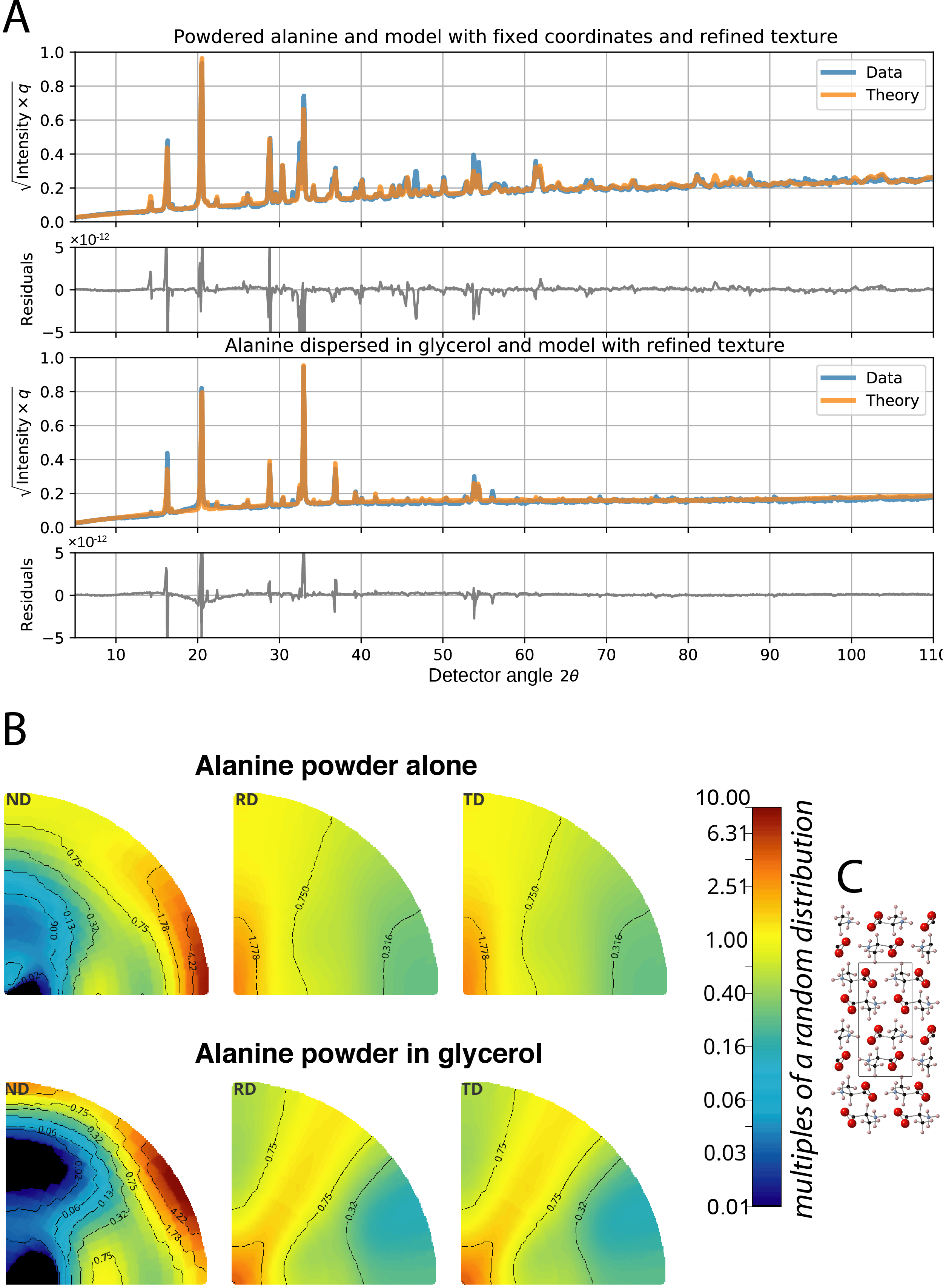
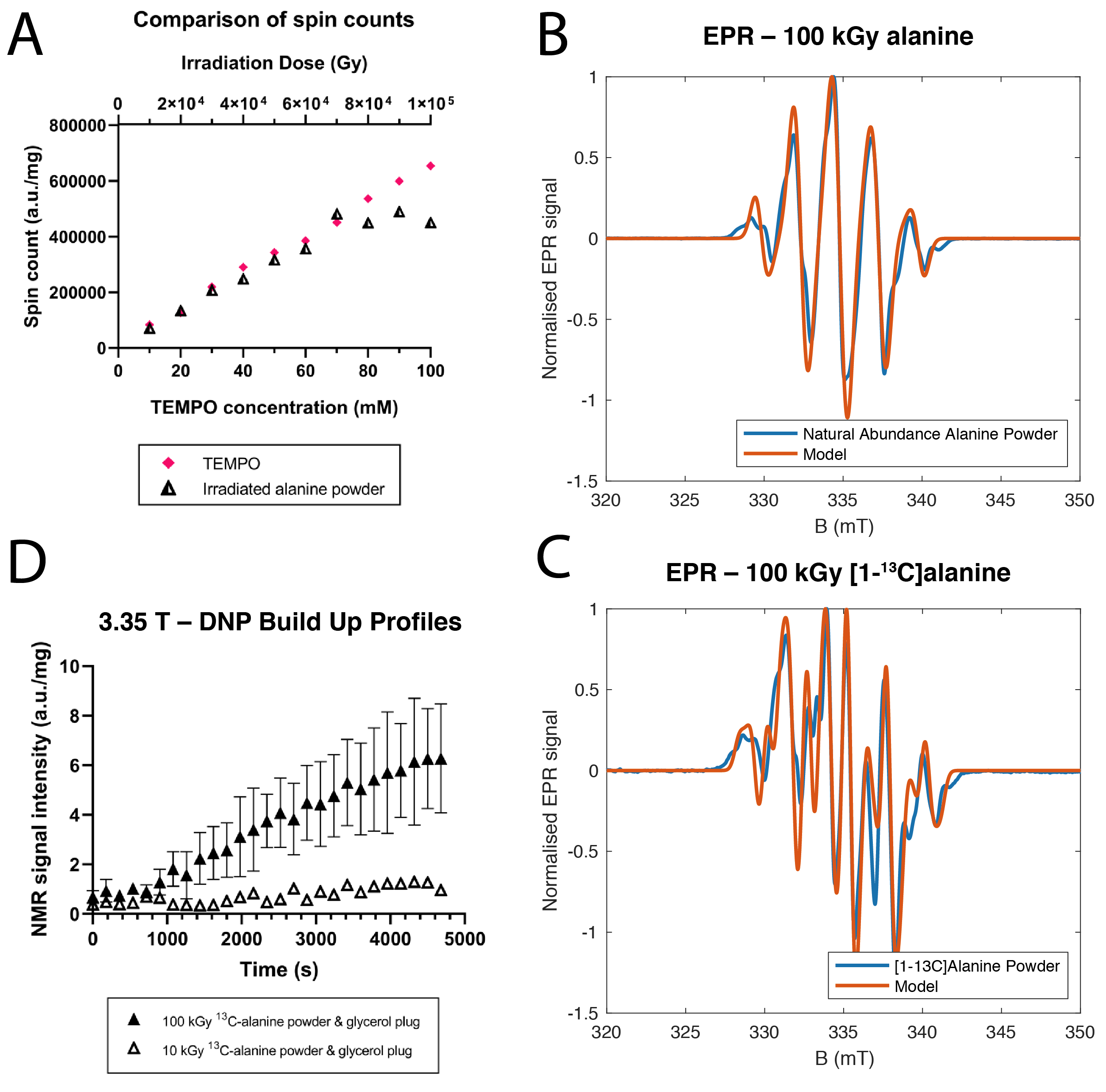
EPR signal from NA-alanine showed each 10$$$\,$$$kGy of irradiation corresponded to an increase in spin concentration of $$$\sim6.022\times10^{21}\text{spins}$$$; approximately $$$\equiv10\,$$$mM TEMPO, saturating at 70$$$\,$$$kGy (Fig.1A). Spectra were comparatively well described by the previously- identified g-tensor and hyperfine interaction constants for the stable alanine radical (R1).[12] The 13C-alanine spectrum was different, occurring from an expected hyperfine interaction, which we fitted via a constrained Bayesian optimisation routine,[13] obtaining the principal values of $$$\mathbf{A}$$$ as $$$[−21.1679,\,2.4736,\,85.7337]\,$$$MHz with Euler angles in the previously-reported molecular frame[14] as $$$[4.9476,\,1.4290,\,2.7288]\,$$$rad (Fig.1B-C). No signal was observed from hydrated samples; it was not possible to measure a radical recombination time. EPR spectra were measured 8 weeks later and comparable for all samples. Despite being approximately an order of magnitude wider than trityl, these lineshapes were sufficient to hyperpolarize 13C-alanine powder at 3.35$$$\,$$$T (Fig. 1D).The glycerol/alanine mix yielded substantial improvements in polarisation at 3.35$$$\,$$$T, within a factor of 10 of exogenous radicals (Fig. 2A). The EPR spectrum of the glycerol-alanine mix (Fig. 2B) was not readily explainable in terms of previously-published alanine radicals, indicating either conformational changes, the formation of other radical species, crystal fields or preferential ordering. X-ray diffraction indicated that polycrystalline alanine does not dissolve in glycerol and is well explained by the known neutron structure but has distinct preferential orientation (Fig. 3).
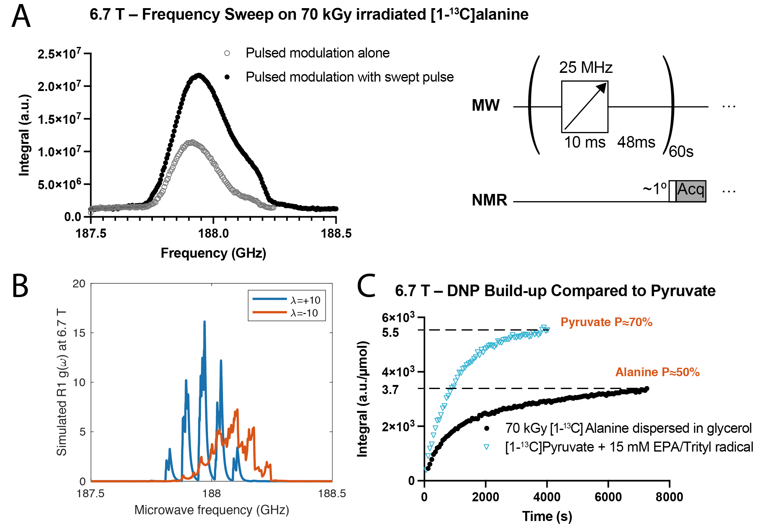
At 6.7$$$\,$$$T, we observed a non-bimodal enhancement curve, significantly increased by frequency modulation (Fig.4A). We note that the simulated alanine R1 radical has distinct spectral density functions g(ω) depending on preferred orientation, quantified by the order parameter $$$\lambda$$$ where the angle $$$\alpha$$$ between the molecular z-axis and $$$B_0$$$ is distributed by $$$p(\alpha)=\exp\left(-\lambda\left(3\cos^2\alpha{}-1\right)/2\right)$$$, with the preferred orientation found via XRD corresponding approximately to $$$\lambda=+\text{ve}$$$ (Fig.4B).Under these conditions, approximately 50% nuclear polarisation was achieved in comparison to pyruvate's 70% measured on identical hardware under identical conditions.[15]
Discussion and Conclusion
This is the first reported study of using electron-beam irradiation to simultaneously sterilise a hyperpolarized contrast agent for DNP and generate radicals for it. We have demonstrated that nuclear polarisation obtained is compatible with in vivo imaging, and by XRD that glycerol alters ordering of the system. We posit that ordering, together with high electron concentrations, may permit a cross-effect type mechanism as the rate of spectral diffusion within each line is faster than that between them.Further work will quantitatively explore both the mechanism of DNP occurring in this system at high field and the exact identity of the solvated radical species formed. Finally, we note the myriad uses of alanine as a hyperpolarized molecular probe: pyruvate and lactate produced from it are visible downstream and their ratio reports on the redox potential of the cell.[5]
Acknowledgements
We would like to acknowledge support from the Novo Foundation (ref: NNF21OC0068683). This work was supported by funding from the Engineering and Physical Sciences Research Council (EPSRC) and Medical Research Council (MRC) [grant number EP/L016052/1], as well as by the British Heart Foundation (FS/19/18/34252).References
[1] A. Capozzi, S. Patel, C. P. Gunnarsson, I. Marco-Rius, A. Comment, M. Karlsson, M. H. Lerche, O. Ouari, J. H. Ardenkjær-Larsen, Angewandte Chemie International Edition 2019, 58, 1334–1339.
[2] S. Patel, A. C. Pinon, M. H. Lerche, M. Karlsson, A. Capozzi, J. H. Ardenkjær-Larsen, The Journal of Physical Chemistry C 2020, 124, 23859–23866.
[3] C. C. Zanella, A. Capozzi, H. A. Yoshihara, A. Radaelli, L. P. Arn, R. Gruetter, J. A. Bastiaansen.
[4] C. C. Zanella, A. Capozzi, H. A. I. Yoshihara, A. Radaelli, A. L. C. Mackowiak, L. P. Arn, R. Gruetter,J. A. M. Bastiaansen, NMR in Biomedicine 2021, 34, e4584.
[5] P. M. Nielsen, C. Ø. Mariager, M. Mølmer, N. Sparding, F. Genovese, M. A. Karsdal, R. Nørregaard,L. B. Bertelsen, C. Laustsen, Magnetic Resonance in Medicine 2020, 84, 2063–2073.
[6] S. Hu, M. Zhu, H. A. Yoshihara, D. M. Wilson, K. R. Keshari, P. Shin, G. Reed, C. von Morze, R. Bok,P. E. Larson, Magnetic resonance imaging 2011, 29, 1035–1040.
[7] W. W. Bradshaw, D. G. Cadena, G. W. Crawford, H. A. W. Spetzler, Radiation research 1962, 17,11–21.
[8] A. Berne, K. Petersson, I. D. Tullis, R. G. Newman, B. Vojnovic, Physics in Medicine & Biology 2021,66, 045015.
[9] S. Stoll, A. Schweiger, Journal of magnetic resonance 2006, 178, 42–55.
[10] C. C. Wilson, D. Myles, M. Ghosh, L. N. Johnson, W. Wang, New Journal of Chemistry 2005, 29, 1318.
[11] L. Lutterotti, H. Wenk, S. Matthies in Proceeding of the Twelfth International Conference on Textures of Materials (ICOTOM-12), Vol. 2, NRC Research Press, 1999, pp. 1599–1604.
[12] E. Sagstuen, E. O. Hole, S. R. Haugedal, W. H. Nelson, The Journal of Physical Chemistry A 1997, 101, 9763–9772.
[13] R. Martinez-Cantin, J. Mach. Learn. Res. 2014, 15, 3735–3739.
[14] E. Pauwels, H. D. Cooman, M. Waroquier, E. O. Hole, E. Sagstuen, Physical Chemistry ChemicalPhysics 2014, 16, 2475–2482.
[15] J. H. Ardenkjær-Larsen, S. Bowen, J. R. Petersen, O. Rybalko, M. S. Vinding, M. Ullisch, N. C. Nielsen,Magnetic Resonance in Medicine 2019, 81, 2184–2194.
Figures
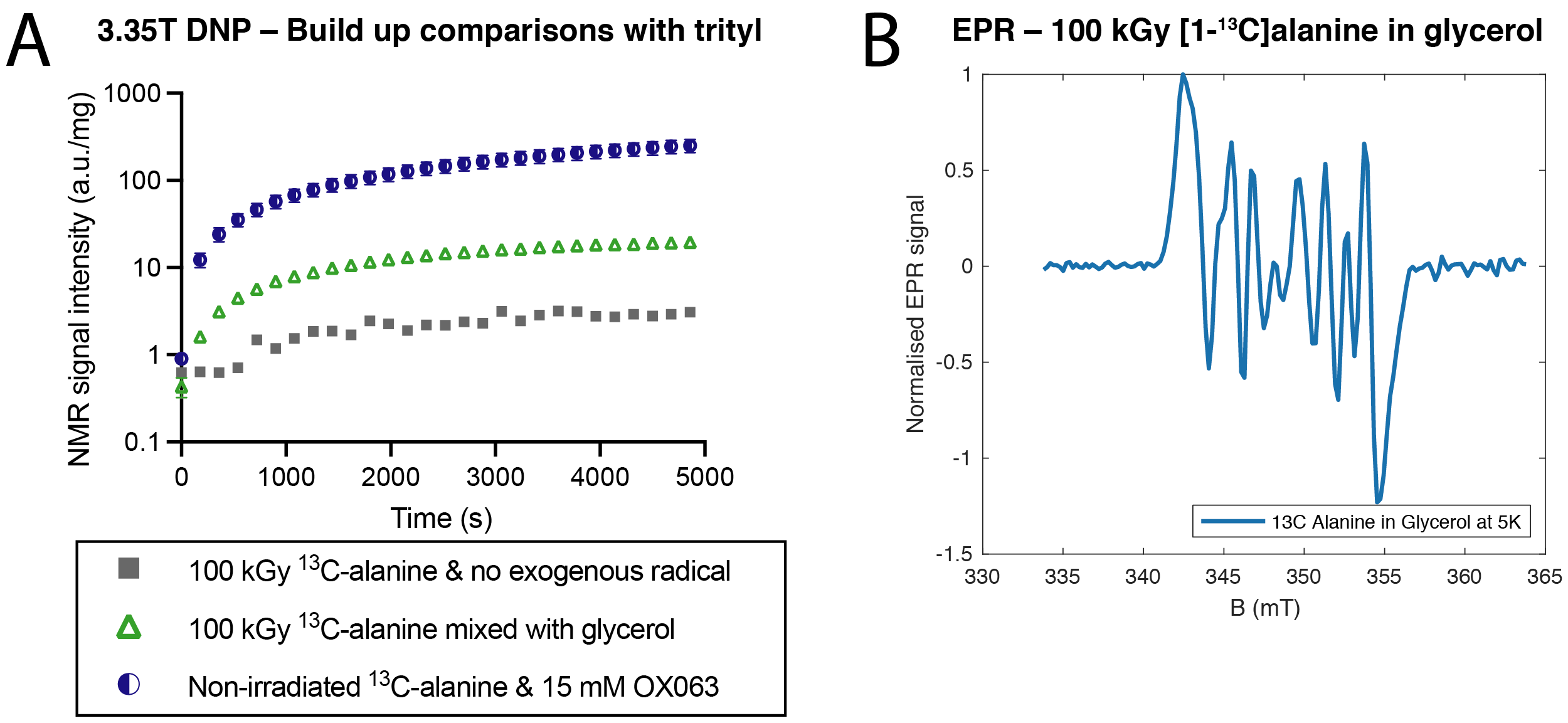
The effect of dispersing polycrystalline alanine in glycerol. A: At 3.35 T, a significant enhancement was seen in obtainable polarization, within an order of magnitude of that obtainable with trityl. B: A profound shift was seen in the resulting EPR spectrum, which was not able to be adequately fitted by modulating the hyperfine interaction tensor on all protons and carbon spins present. Glycerol radicals are predicted (via DFT) to have a substantially different g-tensor and do not appear at the predicted location in the spectrum.