0217
Hyperpolarized [2-13C]pyruvate-d3 detects hepatic gluconeogenesis in vivo1Advanced Imaging Research Center, UTSW Medical Center, Dallas, TX, United States, 22. Department of Biomedical Engineering, UTSW Medical Center, Dallas, TX, United States, 3Department of Radiology, UTSW Medical Center, Dallas, TX, United States
Synopsis
Keywords: Probes & Targets, Hyperpolarized MR (Non-Gas), pyruvate, hyperpolarization, hepatic gluconeogenesis, liver
Motivation: Assessing gluconeogenesis using hyperpolarized [1-13C]pyruvate is technically challenging because [13C]bicarbonate can be produced from both oxidative and gluconeogenic pathways and spectrally resolving the gluconeogenic products from large, neighboring peaks is non-trivial at 3T.
Goal(s): This study examines the utility of deuterated hyperpolarized [2-13C]pyruvate in assessing gluconeogenesis.
Approach: Sodium [2-13C]pyruvate-d3 was synthesized to prolong the T1. Hepatic metabolism was investigated using hyperpolarized [2-13C]pyruvate-d3with D2O dissolution under normal fed and fasted conditions.
Results: The T1 of [2-13C]pyruvate-d3 was ~80 s when dissolved with D2O. Gluconeogenic products such as [2-13C]oxaloacetate and [2-13C]phosphoenolpyruvate were observed from fasted rats only, highlighting clear advantages over [1-13C]pyruvate in investigating gluconeogenesis.
Impact: Hyperpolarization technology is rapidly being translated to humans. With the proven safety and feasibility, hyperpolarized [2-13C]pyruvate-d3 will facilitate its utilization in underexplored liver and kidney metabolism, illuminating mechanistic understanding for several disorders that are believed to depend on altered gluconeogenesis.
Introduction
While its application to glycolytic diseases (e.g., cancer) or oxidative organs (e.g., myocardium, brain) has been clearly demonstrated via appearing [1-13C]lactate and [13C]bicarbonate production, respectively, the interpretation of HP [1-13C]pyruvate studies in the liver and the kidneys has been obscured by gluconeogenesis since [13C]bicarbonate can be generated from either pyruvate oxidation via pyruvate dehydrogenase (PDH) or gluconeogenesis via pyruvate carboxylase (PC) and subsequent phosphoenolpyruvate carboxykinase (PEPCK)1–3. Resolving PC-specific products such as [1-13C]malate, [4-13C]malate, [1-13C]aspartate, and [4-13C]aspartate from large [1-13C]pyruvate-hydrate and [1-13C]alanine peaks in the adjacent resonance was demonstrated in high fields (> 7T)4 but is challenging in conventional 3T or lower magnetic field due to limited chemical shift dispersion2,5.Methods
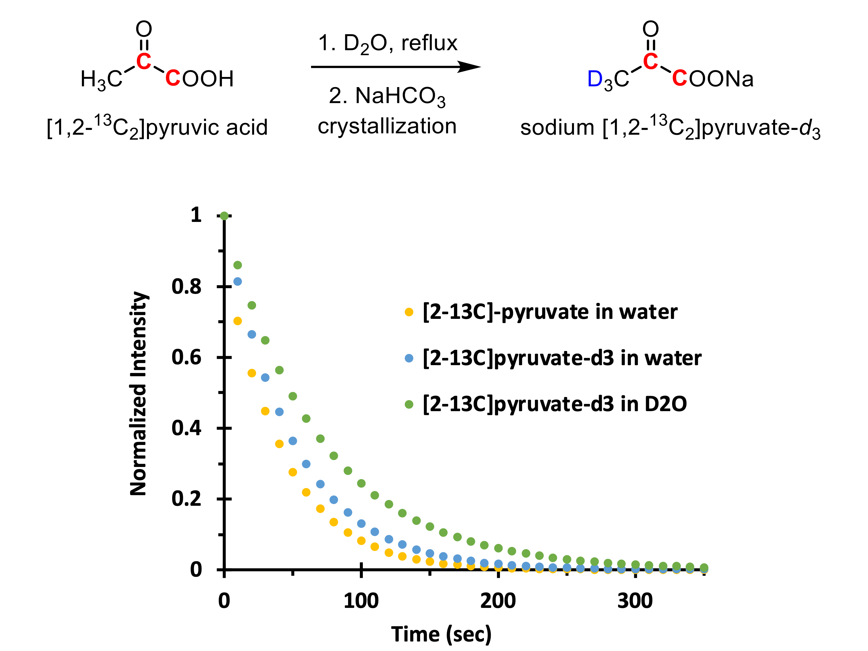
Commercially available [2-13C]pyruvic acid was deuterated by dissolving the labeled pyruvic acid in D2O and refluxing the solution for 5 h, followed by neutralization with NaCO3 and crystallization of the sodium salt (Figure 1) as reported in the literature.6 The synthesized probe, sodium [2-13C]pyruvate-d3, was polarized using the 5T SPINlabTM DNP polarizer (GE Healthcare) for ~4 h. Two groups of male Wistar rats were prepared for the study (332 ± 30 g) and each rat was injected with 80-mM hyperpolarized [2-13C]pyruvate-d3 (1 mmol/kg body weight) under either 24 h fasting condition (n = 4) and normal fed condition (n = 4). Hyperpolarized [2-13C]pyruvate was dissolved in D2O and injected intravenously. The transfer time from dissolution to injection was 20.4 ± 1.7 s. A 3T 750w clinical MRI scanner (GE Healthcare) and a custom-made 13C transmit/receive surface RF coil was used for in vivo study. FID signals were acquired with 10o nominal flip angle every 3 s. In parallel, 0.3 mL of hyperpolarized [2-13C]pyruvate solution was scanned using a benchtop 1T 13C Spinsolve NMR spectrometer (Magritek) for estimating in vitro T1 relaxation and polarization level. Peaks were quantified from time-averaged (0 – 90 s) absorption-mode 13C spectra after 0th and 1st phase corrections.Results and Discussion
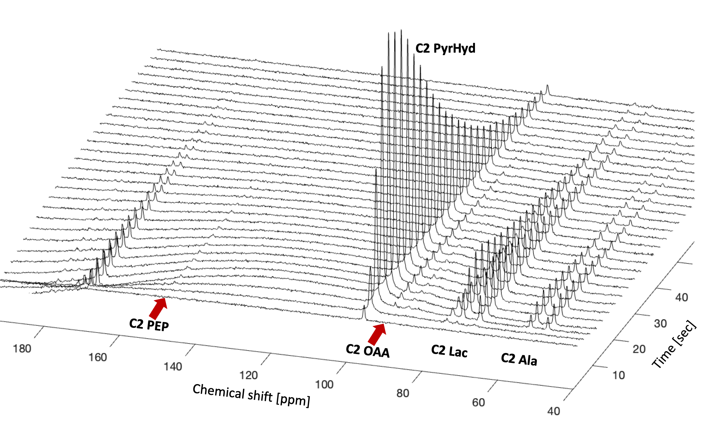
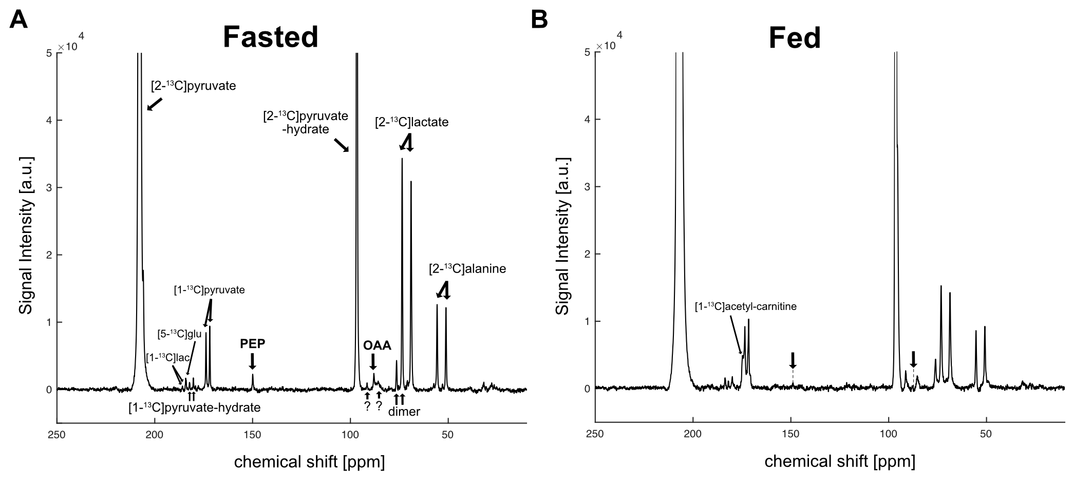
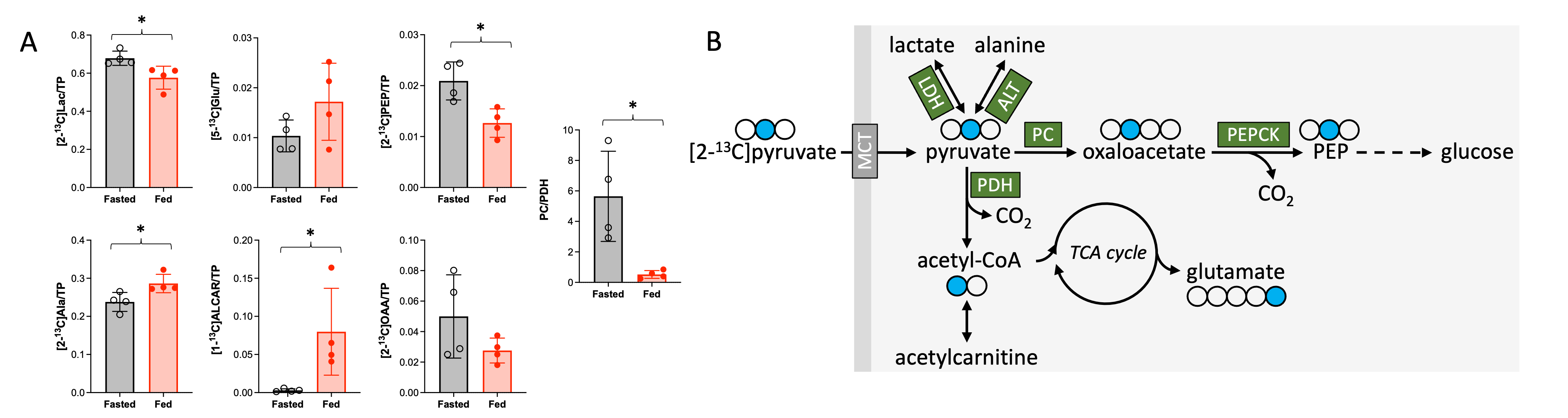
When dissolved in D2O, the liquid-state polarization level of [2-13C]pyruvate-d3 at the time of dissolution was estimated as 41.8 ± 3.3 % (n = 10). In vitro T1 relaxation time was 79.6 ± 2.6 s at 1T (n = 10) and 74.5 s at 3T (n = 1), which is longer than H2O-dissolved [2-13C]pyruvate-d3 (56 s) and non-deuterated [2-13C]pyruvate (45 s) at 1 T, Figure 1. This observation is comparable with what was previously reported by Chen, et al.7 From in vivo studies, additional peaks were detected at 184.1 ppm ([5-13C]glutamate), 174.8 ppm ([1-13C]acetylcarnitine), 149.9 ppm ([2-13C]phosphoenolpyruvate), and 88.0 ppm ([2-13C]oxaloacetate) in addition to [2-13C]pyruvate (207.7 ppm), [2-13C]pyruvate-hydrate (96.6 ppm), [2-13C]lactate (71.2 ppm) and [2-13C]alanine peaks (53.3 ppm), Figure 2 and Figure 3. Natural abundance of [1-13C]pyruvate (172.8 ppm), [1-13C]pyruvate-hydrate (181.2 ppm), [1-13C]lactate (184.8 ppm), and [3-13C]pyruvate (29.7 ppm) were detected as doublets due to the 13C-13C coupling. Notably, the products along oxidative pathway such as [5-13C]glutamate (P = 0.15) and [1-13C]acetylcarnitine (P = 0.036) decreased after 24 h fasting. Conversely, the products along gluconeogenesis such as [2-13C]phosphoenolpyruvate (P = 0.013) and [2-13C]oxaloacetate (P = 0.17) were larger under fasted condition, Figure 4A. The metabolite ratio of PC-products (OAA and PEP) and PDH-products (acetylcarnitine and glutamate) was higher under fasted condition (5.65 ± 2.95) than fed condition (0.52 ± 0.26, P = 0.014). The appearance of [2-13C]oxaloacetate and [2-13C]phosphoenolpyruvate is likely via PC and PEPCK, respectively, key regulatory enzymes in glucogenesis, Figure 4B.Conclusions
In conclusion, we showed that hyperpolarized [2-13C]pyruvate-d3, dissolved in D2O, extends T1 (79.6 s) by 76.9 % (at 1T) and could detect [2-13C]oxaloacetate and [2-13C]phosphoenolpyruvate from the rat liver in vivo after 24 h fasting. This study demonstrates unique and exciting opportunities of hyperpolarized [2-13C]pyruvate-d3 in investigating hepatic gluconeogenesis.Acknowledgements
This study was supported by the National Institutes of Health of the United States (R01 NS107409, P41 EB015908, S10 OD018468, P30 DK127984, R21 EB034413, R21 EB030765, R21 EB031367); U.S. Army Medical Research Acquisition Activity (W81XWH2210485); Cancer Prevention and Research Institute of Texas (RP210099).References
1. Merritt ME, Harrison C, Sherry AD, Malloy CR, Burgess SC. Flux through hepatic pyruvate carboxylase and phosphoenolpyruvate carboxykinase detected by hyperpolarized 13C magnetic resonance. Proc Natl Acad Sci U S A. 2011;108(47):19084-19089. doi:10.1073/pnas.11112471082.
2. Jin ES, Moreno KX, Wang JX, et al. Metabolism of hyperpolarized [1-13C]pyruvate through alternate pathways in rat liver. NMR Biomed. 2016;29(4):466-474. doi:10.1002/nbm.34793.
3. Can E, Bastiaansen JAM, Couturier D laurent, Gruetter R, Yoshihara HAI, Comment A. [13C]bicarbonate labelled from hyperpolarized [1-13C]pyruvate is an in vivo marker of hepatic gluconeogenesis in fasted state. Commun Biol. 2022;5(1):1-9. doi:10.1038/s42003-021-02978-24.
4. Lee P, Leong W, Tan T, Lim M, Han W, Radda GK. In Vivo hyperpolarized carbon-13 magnetic resonance spectroscopy reveals increased pyruvate carboxylase flux in an insulin-resistant mouse model. Hepatology. 2013;57(2):515-524. doi:10.1002/hep.260285.
5. Chen J, Hackett EP, Kovacs Z, Malloy CR, Park JM. Assessment of hepatic pyruvate carboxylase activity using hyperpolarized [1-13C]-l-lactate. Magn Reson Med. 2021;85(3):1175-1182. doi:10.1002/mrm.284896.
6. Shchepin RV, Coffey AM, Waddell KW, Chekmenev EY. Parahydrogen induced polarization of 1-(13)C-phospholactate-d(2) for biomedical imaging with >30,000,000-fold NMR signal enhancement in water. Anal Chem. 2014;86(12):5601-5605. doi:10.1021/ac500952z7.
7. Chen AP, Keshari KR, Cunningham CH. Slowing T1 relaxation of hyperpolarized [2-13C]pyruvate with deuterium enrichment. Proc Intl Soc Magn Reson Med. 2021. #3803
Figures