0214
First in-human MR Metabolic Imaging of the Brain Using Hyperpolarized [1-13C]alpha-ketoglutarate1Department of Radiology and Biomedical Imaging, University of California, San Francisco, CA, United States, 2Department of Neurological Surgery, University of California, San Francisco, CA, United States, 3ISOTEC Stable Isotope Division, MilliporeSigma, Merck KGaA, Miamisburg, OH, United States
Synopsis
Keywords: Hyperpolarized MR (Non-Gas), Contrast Agent
Motivation: Isocitrate dehydrogenase (IDH) mutational status is crucial for accurate diagnosis and prognosis of malignant gliomas. However, the current clinical assessment of IDH mutation requires an invasive brain biopsy for pathological testing.
Goal(s): We aimed to perform first in-human MR studies using hyperpolarized [1-13C]alpha-ketoglutarate as a new probe of IDH mutational status via cancer metabolic reprogramming, along with cerebral bioenergetics.
Approach: We acquired 13C MRS data from healthy brain volunteers (N=6) and glioma patients (N=6) who received hyperpolarized aKG.
Results: Feasibility and safety were demonstrated in these 12 studies, with signals observed from [1-13C]alpha-ketoglutarate and its metabolite glutamate in the obtained 13C MRS data.
Impact: MR molecular imaging with the new probe hyperpolarized [1-13C]alpha-ketoglutarate provided novel measurements of aKG metabolism and can investigate glioma IDH mutational status by detecting glutamate or the oncometabolite, 2-hydroxyglutarate.
Introduction
Low-grade gliomas account for 20-25% of gliomas in adults and 40% of all pediatric brain tumors1. Currently, accurate diagnosis of these tumors requires an invasive brain biopsy to assess isocitrate dehydrogenase (IDH) mutation. Hyperpolarized carbon-13 (HP 13C) MRI using [1-13C]alpha-ketoglutarate (aKG) has been shown in animal models to detect aKG and its conversion to glutamate and the oncometabolite 2-hydroxyglutarate (2HG) catalyzed by mutant IDH2,3. A recent study has also shown that this technique can be used to monitor therapeutic response to IDH-inhibitor treatment in preclinical IDH-mutant models4. To explore the potential of HP [1-13C]aKG to non-invasively assess IDH status in patients, our group developed an FDA IND approved HP [1-13C]aKG approach for patient studies. In this project, we investigated for the first time HP aKG imaging in healthy volunteers and glioma patients.Methods
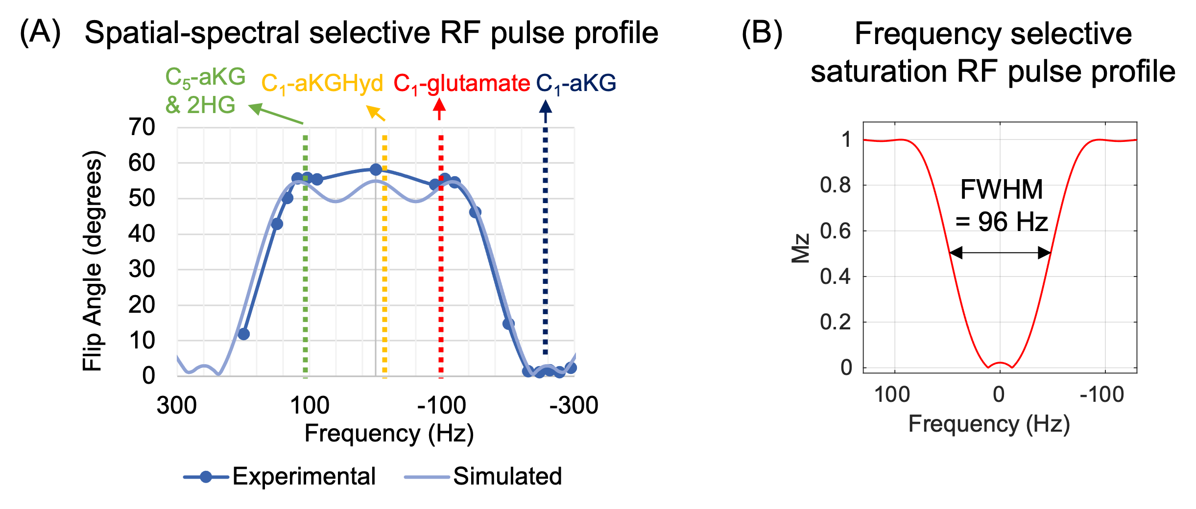
We conducted 12 clinical research studies (6 volunteer and 6 glioma patient scans) to assess the feasibility of using HP [1-13C]aKG to study aKG metabolism in human brains. To generate HP [1-13C]aKG, samples containing 5.6 M [1-13C]aKG and 15 mM electron paramagnetic agent in ethanol and water mixture (60:40 v/v) were polarized for 3 hours in a 5T GE SPINlab at 0.8 K. Samples were then rapidly dissolved with superheated water and neutralization buffer. HP [1-13C]aKG MR data were acquired from a clinical GE 3T scanner using a Tx/Rx 8-ch 1H/ 24-ch 13C birdcage head coil. A dynamic slab MRS was used with a slab thickness of 6 cm in volunteer scans or the size of the lesion in patient scans. A 0.67ml/kg sterile dose was injected at a rate of 5 mL/s, followed by a 20 mL sterile saline flush. Data acquisition began 3 s after the saline flush, and spectra were acquired every 3 s for 60 s. A spectral-spatial RF pulse was used to apply a small flip angle (2°) to 13C1-aKG and a higher flip angle (55°) to metabolites while accounting for chemical shift slice displacement (Figure 1A). A frequency-selective RF saturation pulse (Figure 1B) was applied before the first 4 excitation pulses to suppress the 13C5-aKG signal at natural abundance which overlaps with 2HG. Higher-order shimming was performed before 13C MRS to improve B0 field homogeneity. In addition to HP MRI, single-voxel 1H MRS was also acquired to detect 2HG. The multi-channel HP 13C MR data were apodized, pre-whitened, denoised5, and coil-combined6. Data were phased individually for each metabolite and substrate signal, and baseline corrected for signal quantification.Results
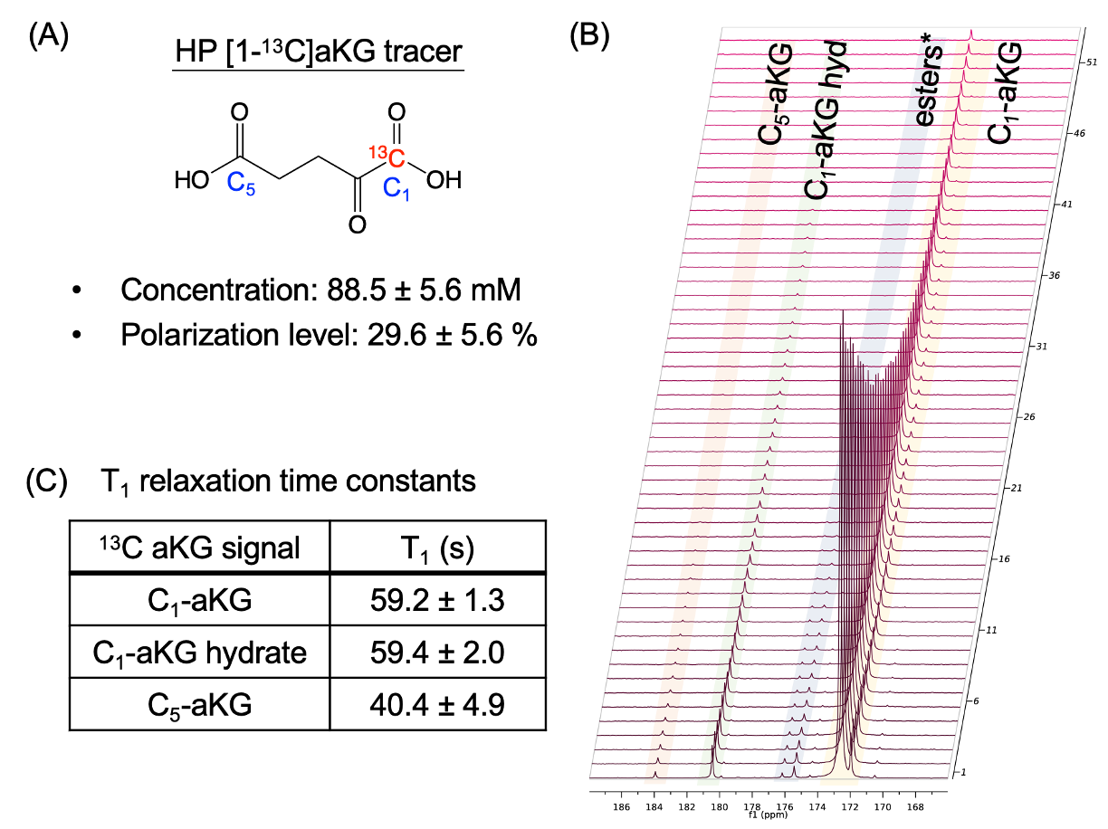
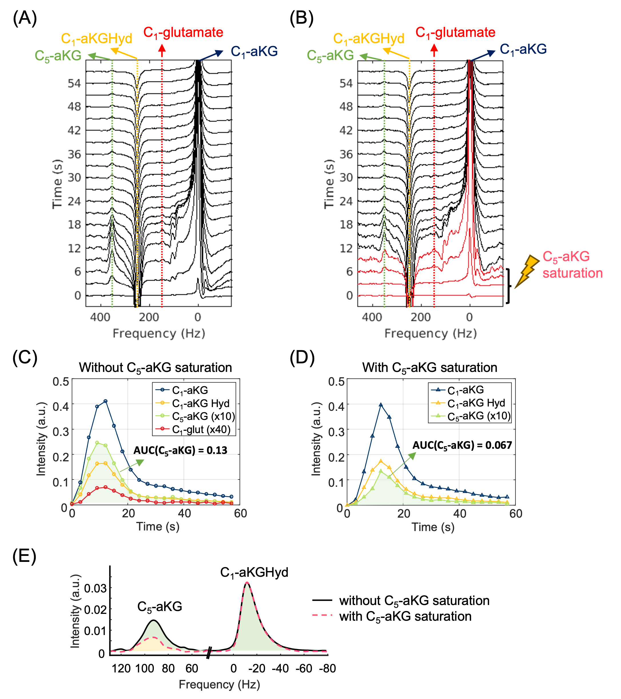
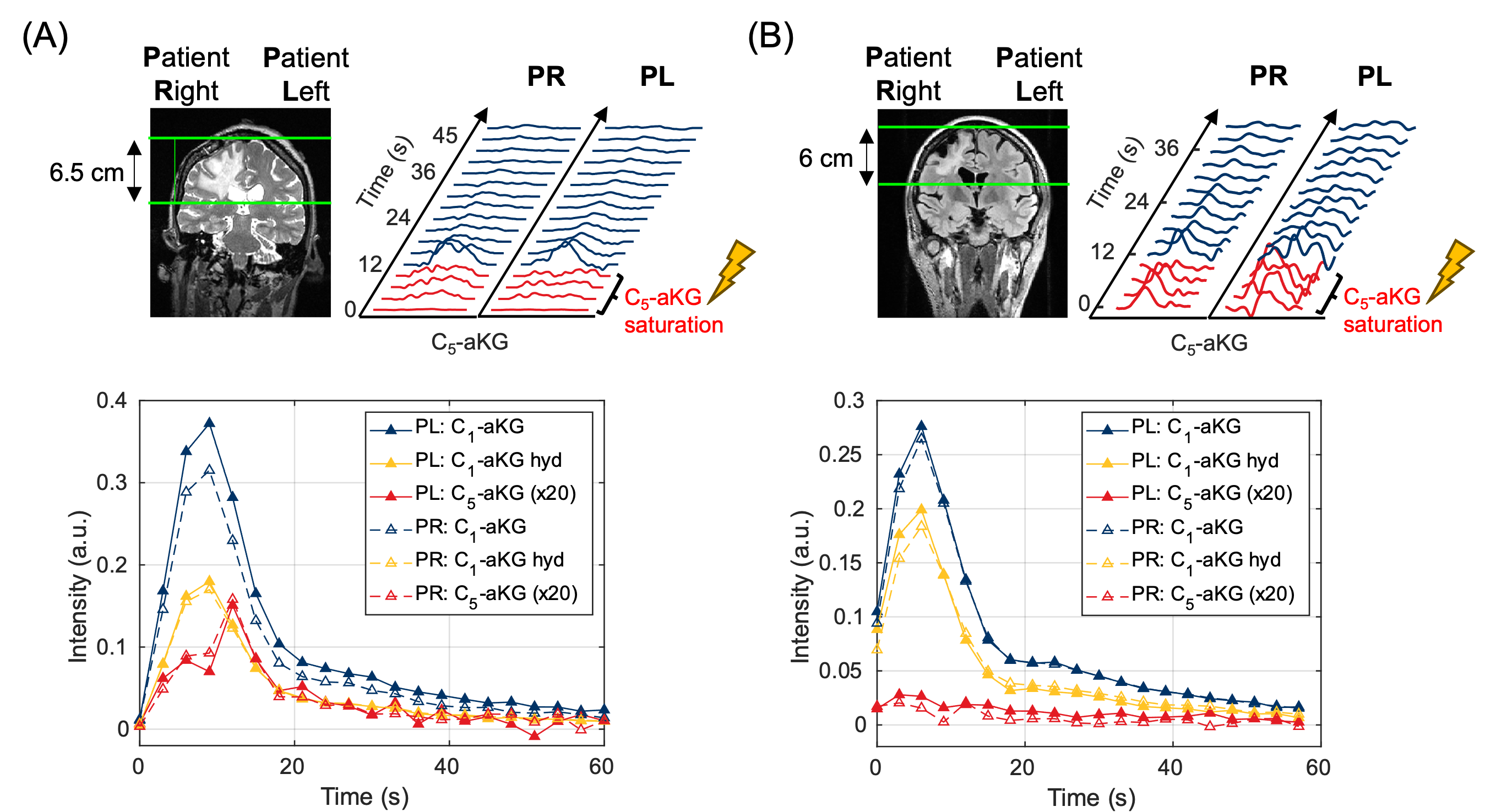
Mean [1-13C]aKG concentrations and polarization levels were 85.7 ± 4.9 mM and 29.6 ± 5.6 %, respectively (Figure 2A). The T1 relaxation time constants of 13C1-aKG, 13C1-aKG hydrate, and 13C5-aKG were consistent over 12 studies (Figure 2B-C). The HP 13C MRS data in the healthy human brain shows peaks at 13C1-aKG (0 Hz), 13C1-aKG hydrate (251 Hz), and 13C5-aKG (352 Hz) acquired after HP [1-13C]aKG injection (Figure 3A). The 13C1-glutamate signal, which was absent in the solution-state spectra, was detected. The frequency-selective saturation pulse reduced the 13C5-aKG signal (Figure 3B) by ~50 % compared to control experiments (Figure 3E) to better separate 13C1-2HG from 13C5-aKG signal if IDH-mutant tumor is present. Data was also acquired from glioma patients using the 13C5-aKG saturation method. Figure 4 shows two results from two patient studies. By reconstructing muti-channel data from coil elements on patient’s right or left sides enabled spatial selectivity7 for comparing HP 13C aKG signal intensities between tumor and contralateral brain. In both datasets, signal dynamics in the 2HG region showed no significant difference between these tumors and contralateral side, indicating little to no 2HG production. It is important to note the IDH status of 3 patients was either unknown or wild-type. In the other 3 patients, the tumors were confirmed as IDH-mutant previously, but were treated by surgical resection and/or chemotherapy/IDH inhibitors, which would alter tumor metabolism and affect 2HG production. The HP aKG results were also consistent with the absence of 2HG signal in single-voxel 1H spectroscopic data in all patients. Future HP aKG glioma studies are ongoing.Discussion and Conclusion
This study investigated first in-human brain HP [1-13C]aKG MR molecular imaging. We demonstrated feasibility and the ability to detect glutamate production from aKG and its potential to serve as a biomarker of IDH mutational status in glioma. Based on these findings, HP [1-13C]aKG metabolic imaging may be valuable clinically to detect 2HG in IDH-mutant tumors and monitor levels following therapy.Acknowledgements
This research was supported by the NIH (P01CA118816, P41EB013598) and the UCSF NICO project.References
1.Greuter L, Guzman R, Soleman J. Pediatric and Adult Low-Grade Gliomas: Where Do the Differences Lie? Children. 2021;8(11):1075. doi:10.3390/children81110752.
2. Chaumeil MM, Larson PEZ, Yoshihara HAI, et al. Non-invasive in vivo assessment of IDH1 mutational status in glioma. Nat Commun. 2013;4(1):2429. doi:10.1038/ncomms34293.
3. Hong D, Batsios G, Viswanath P, et al. Acquisition and quantification pipeline for in vivo hyperpolarized 13C MR spectroscopy. Magnetic Resonance in Med. 2022;87(4):1673-1687. doi:10.1002/mrm.290814.
4. Hong D, Kim Y, Mushti C, et al. Monitoring Response to a Clinically Relevant IDH Inhibitor in Glioma – Hyperpolarized 13C Magnetic Resonance Spectroscopy Approaches. Neuro-Oncology Advances. 2023;vdad143. doi:10.1093/noajnl/vdad1435.
5. Olesen JL, Ianus A, Østergaard L, Shemesh N, Jespersen SN. Tensor denoising of multidimensional MRI data. Magnetic Resonance in Med. 2023;89(3):1160-1172. doi:10.1002/mrm.294786.
6. Zhu Z, Zhu X, Ohliger MA, et al. Coil combination methods for multi-channel hyperpolarized 13C imaging data from human studies. Journal of Magnetic Resonance. 2019;301:73-79. doi:10.1016/j.jmr.2019.01.0157.
7. Ma J, Pinho MC, Harrison CE, et al. Dynamic 13C MR spectroscopy as an alternative to imaging for assessing cerebral metabolism using hyperpolarized pyruvate in humans. Magnetic Resonance in Med. 2022;87(3):1136-1149. doi:10.1002/mrm.29049
Figures