0213
In vivo detection and imaging of aminopeptidase activities related to renin-angiotensin system using newly designed hyperpolarized MR probes1Chemistry and biotechnology, The University of Tokyo, Tokyo, Japan, 2National Institutes for Quantum Science and Technology, Chiba, Japan, 3National Institutes of Health, Bethesda, MD, United States
Synopsis
Keywords: Hyperpolarized MR (Non-Gas), Molecular Imaging, Molecular design
Motivation: Detection of aminopeptidase (AP) activities related to renin-angiotensin system (RAS) can lead to diagnosis of various diseases. Magnetic resonance imaging utilizing suitable hyperpolarized molecular probes can non-invasively detect in vivo AP activities. However, there have been no hyperpolarized molecular probes for APA, APB, and leucine AP.
Goal(s): We aimed to design and develop new hyperpolarized molecular probes for the detection and imaging of RAS related AP activities in vivo.
Approach: Based on the previously reported APN probe scaffold, three new hyperpolarized probes were designed.
Results: Using the developed probes, target AP activities were successfully detected and visualized in vivo.
Impact: This study exhibits a framework that artificially designed hyperpolarized molecular probes can detect in vivo aminopeptidase activities, which is assumed impossible with isotope labeled natural substrates and broadens the possibility of hyperpolarized MR diagnosis based on AP activities.
Introduction
The renin-angiotensin system (RAS) is an important metabolic pathway primarily involved in blood pressure regulation1, and abnormalities in this system cause hypertension, tumor growth, and inflammation2. Angiotensin II (Ang II), a bioactive octapeptide and a key player in the RAS, is metabolized by aminopeptidase A (APA) to produce Ang III, which is further metabolized by APN or APB to produce Ang IV, a ligand for AT4 (also known as leucine AP (LAP)) (Fig. 1) . As described above, RAS-related APs play an important role in regulating the amount of each hormone peptide, and detection of these activities can lead to detection of various diseases such as hypertension and tumors.Nuclear magnetic resonance/magnetic resonance imaging (NMR/MRI) combined with dynamic nuclear polarization (DNP) is a powerful method for real-time detection of target enzymatic activity in vivo3. In general, for the detection of enzymatic activity by DNP-NMR/MRI, 13C- or 15N-labeled natural substrates are mostly used as DNP-NMR molecular probes4,5. However, the molecular weight of Ang II, the natural substrate of RAS-related APs, exceeds 1000, which is assumed to have a very short hyperpolarization lifetime. In this study, we artificially designed and developed DNP-NMR molecular probes that can detect RAS-related AP activities in vivo. Previously, we have developed a DNP-NMR molecular probe, Ala-[1-13C]Gly-d2-NMe2, which can detect APN activity in vivo6. Based on this molecular probe scaffold, we designed and developed new DNP-NMR molecular probes for other RAS-related AP (APA, APB, and LAP) activities (Fig. 2). The developed molecular probes were then successfully used to detect and image RAS-related AP activities in vivo.
Methods
Hyperpolarized 13C MRI: Hyperpolarized 13 C MRI experiments were conducted on 3T MRI scanners (MR Solutions Inc. and Bruker Biospin). Representative conditions are as follows. 18 μL of probe solution (ca. 2.2 M (APA probe), 3.2 M (APB probe), or 3.5 M (LAP probe)) containing 30 mM AH111501 was hyperpolarized using a SpinAligner DNP polarizer (Polarize IVS). A hyperpolarized sample was rapidly dissolved in the dissolution buffer (3.2 mL of DPBS containing 0.68 mM ethylenediaminetetraacetic acid). 200 µL of the hyperpolarized solution was injected intravenously into the healthy mice. The image processing was performed using Image J software.Results
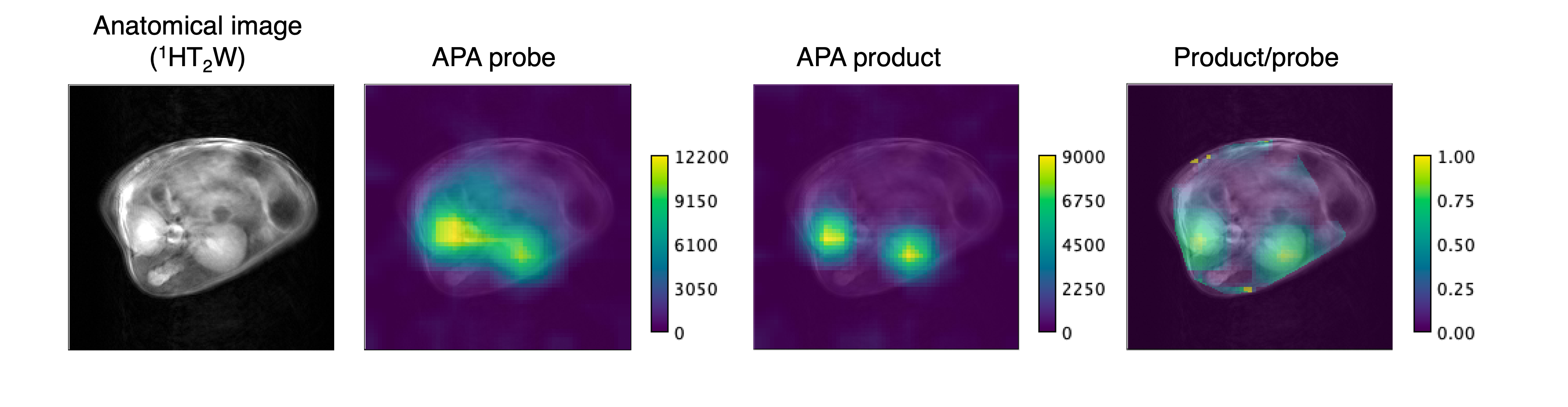
Since AP hydrolyzes peptides specifically at the N-terminal amino acid residue, DNP-NMR probes for each AP activity was developed by replacing the N-terminal Ala residue of Ala-[1-13C]Gly-d2-NMe2 with an amino acid residue recognized preferably by APA, APB, or LAP (Fig. 3). All the developed molecular probes satisfied the necessary properties for DNP-NMR molecular probes: long hyperpolarization lifetime (T1 = 31 ± 0.7 s (Glu), 26 ± 0.8 s (Arg), 28 ± 0.5 s (Leu) at 3 T), large chemical shift change (2.7 ppm), and high water solubility (more than 2 M). After intravenous administration of each probe to healthy mice, we observed the production of a new metabolite peak (Fig. 4A). Moreover, the AUC ratio of each probe in the presence/absence of the inhibitor for each target AP was calculated ex vivo (in mouse kidney homogenate) or in vivo. The results showed a decrease in the AUC ratio in the presence of the inhibitor (Fig. 4B), suggesting that each probe can detect the target AP activity under hyperpolarized state. Finally, we visualized the metabolism of Glu-[1-13C]Gly-d2-NMe2 around the kidney of healthy mice by chemical shift imaging. Considering that the metabolism of the probe correlates with APA activity, the results suggest that this probe can be used for site-specific APA activity imaging, which has never been achieved before.Discussion
The newly developed molecular probes Xaa-[1-13C]Gly-d2-NMe2 (Xaa = Glu, Arg, and Leu) exhibited properties required for in vivo DNP-NMR experiments, including long polarization lifetime, large chemical shift change, high water solubility, and non-toxicity. The probes were successfully utilized for in vivo detection of the target AP activity. Considering that the natural substrate Ang II is likely not available as a DNP-NMR molecular probe for AP activity detection due to its large molecular weight, the importance of being able to develop DNP-NMR molecular probes through appropriate molecular design is emphasized. The metabolic imaging results of Glu-[1-13C]Gly-d2-NMe2 suggest the possibility of real-time imaging of APA activity in vivo. APA is known to be involved in important diseases such as tumor angiogenesis and Alzheimer's disease7,8, and this probe could be used to diagnose these diseases.Conclusion
In this study, we designed and developed novel DNP-NMR molecular probes for real-time detection and imaging of RAS-related AP activities in vivo. Further studies are currently underway for disease diagnosis and monitoring of therapeutic responses.Acknowledgements
This research was supported by MEXT Q-LEAP [Grant Number JPMXS0120330644 (to Y.T., and S.S.)], JST FOREST Program [Grant Number JPMJFR225G (to Y.T.)]. This study was also supported by intramural research program at NCI/NIH.
References
1. V. J. Dzau, Circulating versus local renin-angiotensin system in cardiovascular homeostasis. Circulation, 1988;77 (6 Pt 2):pp. I4-I13
2. S. Ishikane, et al. The role of angiotensin II in cancer metastasis: Potential of renin-angiotensin system blockade as a treatment for cancer metastasis. Biochem. Pharmacol., 2018;151:96‐103.
3. J. H. Ardenkjær-Larsen, et al. Increase in signal-to-noise ratio of > 10,000 times in liquid-state NMR. Proc. Natl. Acad. Sci. USA, 2003;100:10158-10163.
4. Y. Kondo, et al.Design of Nuclear Magnetic Resonance Molecular Probes for Hyperpolarized Bioimaging. Angew. Chem. Int. Ed., 2021;60:14779-14799.
5. K. R. Keshari et al., Chemistry and biochemistry of 13C hyperpolarized magnetic resonance using dynamic nuclear polarization. Chem. Soc. Rev., 2014;43:1627-1659.
6. Y. Saito, et al. Structure-guided design enables development of a hyperpolarized molecular probe for the detection of aminopeptidase N activity in vivo. Sci. Adv., 2022;8:eabj2667.
7. A. Valverde et al. Aminopeptidase A contributes to biochemical, anatomical and cognitive defects in Alzheimer’s disease (AD) mouse model and is increased at early stage in sporadic AD brain. Acta Neuropathol., 2021;141:823-839.
8. S. Marchiò et al. Aminopeptidase A is a functional target in angiogenic blood vessels. Cancer Cell, 2004;5(2):151-162.
Figures
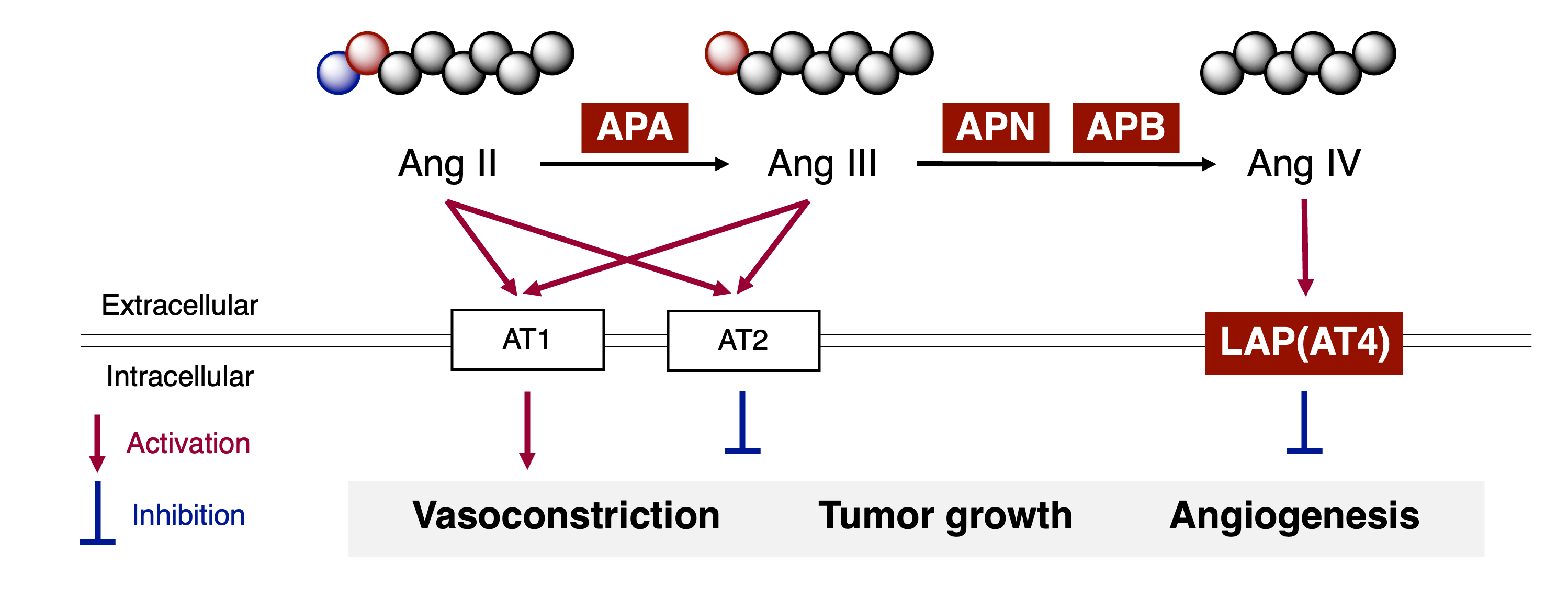
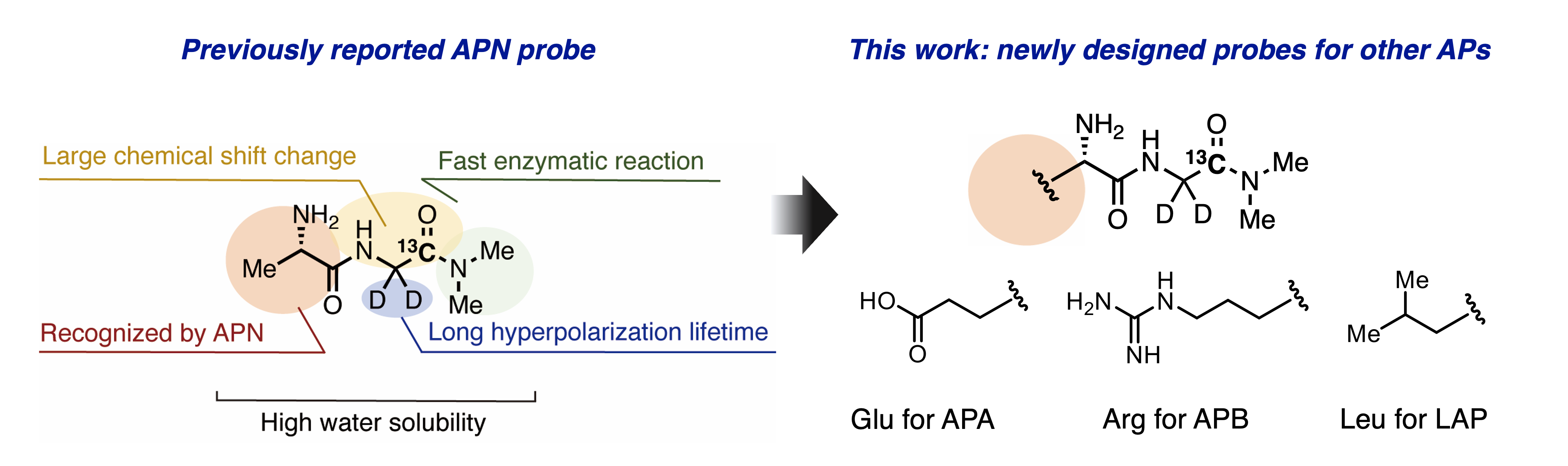
Figure 2. Design of DNP-NMR molecular probes for RAS-related AP activities.
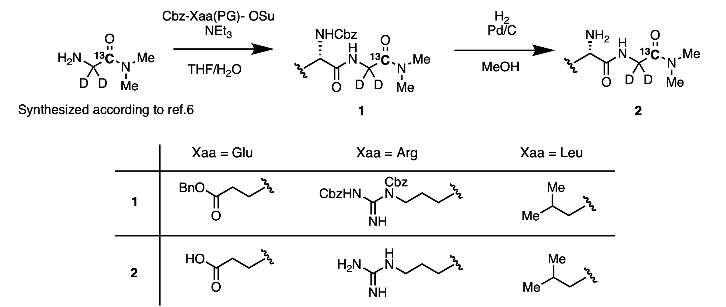
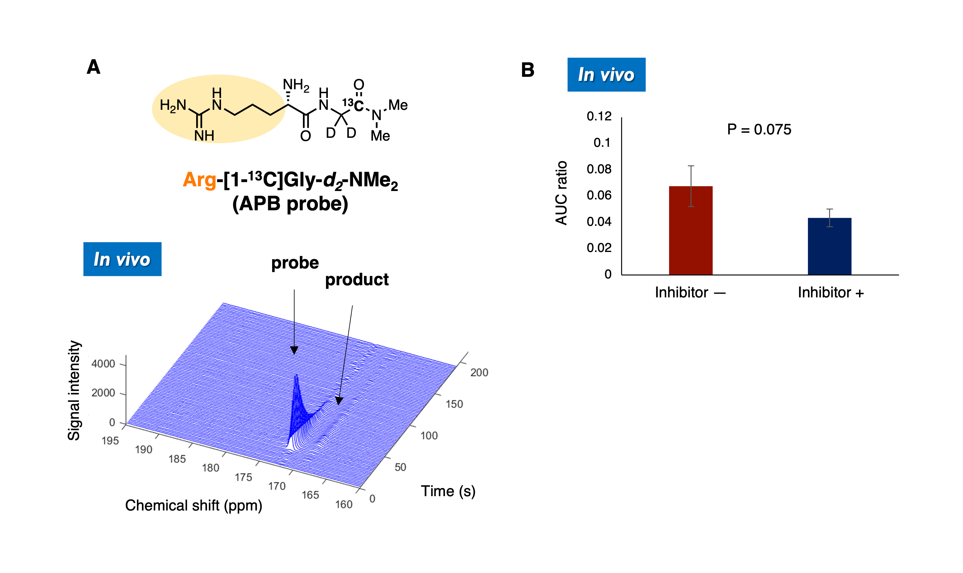
Figure 4. Hyperpolarized experiments of Arg-[1-13C]Gly-d2-NMe2 in vivo with or without the inhibitor (n = 3). (A) 18 µL of probe solution containing 30 mM AH111501 was hyperpolarized by SpinAligner. 13C MR spectra were acquired from the body, following intravenous injection of 200 µL of hyperpolarized solution. Conditions; Dissolution buffer: 3.2 mL DPBS containing 0.68 mM EDTA, TR :1 s, flip angle: 10°, 3 T. (B) In the experiments with the inhibitor, a mouse was pretreated with bestatin (10 mg/kg) 1 h before the measurement. AUC, area under the curve. Error bars indicate the SD.