0206
Effects of multi-vendor gradient non-linearity correction on breast tumor ADC measurements in the ACRIN 6698 trial1Radiology and Biomedical Imaging, University of California San Francisco, San Francisco, CA, United States, 2Radiology, Stanford University, Stanford, CA, United States, 3Radiology, University of Michigan, Ann Arbor, MI, United States, 4Radiology, University of Minnesota, Minneapolis, MN, United States, 5Radiology, University of Washington, Seattle, WA, United States, 6Quantum Leap Healthcare, San Francisco, CA, United States, 7University of Michigan, Ann Arbor, MI, United States
Synopsis
Keywords: Breast, Diffusion/other diffusion imaging techniques, gradient nonlinearity correction, breast cancer, ADC
Motivation: MRI scanner gradient non-linearity (GNL) is a known source of variability and spatially-dependent bias in quantitative ADC measurements derived from diffusion-weighted imaging (DWI).
Goal(s): Evaluate the effects of GNL correction on DWI from the ACRIN 6698 multi-center trial, which investigated ADC as a marker breast cancer response in patients receiving neoadjuvant therapy.
Approach: Retrospective GNL correction was performed on the ACRIN 6698 DWI data
Results: Percent decrease in mean tumor ADC post-GNC ranged from 0.5%-11% for different MRI gradient sets, illustrating that GNL can confer significant bias to ADC measurements and should be corrected in multi-center clinical DWI trials.
Impact: Implementation of gradient non-linearity correction to reduce bias and variability of tumor ADC measurements from DWI data acquired in multi-center, multi-platform oncology trials may improve the quantitative utility of ADC for characterizing response to treatment.
Introduction
Diffusion-weighted imaging (DWI) can provide quantitative information on tissue water mobility and microstructure in the form of calculated metrics such as the apparent diffusion coefficient (ADC). The American College of Radiology (ACRIN) 6698 multi-center trial investigated apparent diffusion coefficient (ADC) for prediction of breast cancer response to neoadjuvant therapy1. MRI scanner gradients have inherent non-linearities that can be a significant source of spatially-dependent bias in DWI-measured ADC2 and of inter-platform variability in multi-center clinical trials3. Previous studies have demonstrated improved ADC accuracy for off-center anatomy after correction for spatial non-uniformity of diffusion weighting (DW) induced by system-specific gradient nonlinearity (GNL) on a single vendor platform4 and in multi-center data5,6. This study evaluated the effects of retrospective GNL correction (GNC) on ADC measurements and response prediction in the ACRIN 6698 study.Methods
This retrospective study was performed using the publicly available DWI data from the 242 patients included in the final data analysis for the ACRIN 6698 trial1 (ClinicalTrials.gov: NCT01564368), hosted on TCIA7. ACRIN 6698 was a sub-study of the I-SPY 2 TRIAL, a multi-center neoadjuvant clinical trial for women with high-risk, stage II/III breast cancer. At all ten sites, bilateral breast single-shot EPI DWI data were acquired with 4 b-values (b=0, 100, 600, 800 s/mm2) for each patient prior to treatment (T0), at early treatment (T1, 3 weeks after the start of treatment), mid-treatment (T2, 12 weeks after the start of treatment), post-treatment (T3, pre-surgery). Patient DWI data were acquired 1.5T and 3T MRI scanners manufactured by the three major vendors (GE, Philips, Siemens). The same system was used for longitudinal DWI scans of an individual patient. The ten MRI scanners had seven unique gradient configurations. A vendor-agnostic GNC program5,6 utilizing vendor-supplied spherical harmonics for gradient characterization was used to generate corrected DWI and ADC maps. Tumor regions of interest (ROIs) utilized in the original 6698 analysis were applied to both uncorrected and GNC ADC maps. The uncorrected and GNC tumor ADCs were evaluated for mean, standard deviation, and percentage change resulting from GNC for each MRI scanner gradient system. Tumor ROI positions were characterized by a single 3D centroid (center-of-gravity) position relative to magnet isocenter and an ROI extent along each axis to assess the relationship of distance from isocenter to GNL bias. Performance for predicting pathologic complete response (pCR) was assessed for uncorrected and GNC ADCs by using the area under the receiver operating characteristic curve (AUC) for the overall cohort.Results
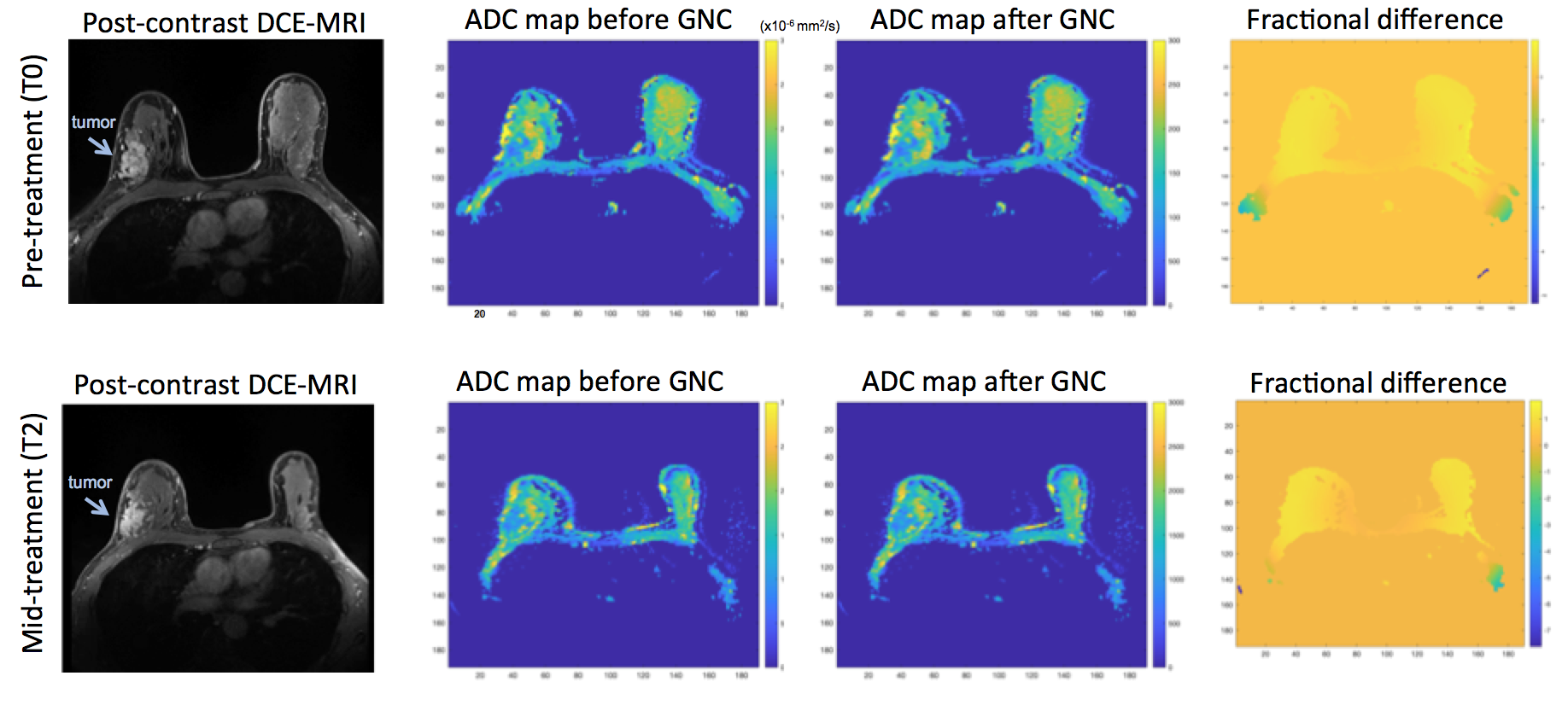
A total of 865 DWI exams from the 242 patients were included in this analysis. Table 1. summarizes the gradient configurations and field strengths for the 10 MRI scanners. Figure 1. demonstrates the relationship between tumor ADC bias and distance of tumor ROI from magnet isocenter for the 7 unique scanner gradient configurations, with increasing distance resulting in increasing bias. Representative uncorrected and GNC ADC images from a patient (Figure 2.) exhibit increasing fractional bias at greater distances from isocenter. Uncorrected and GNL-corrected mean tumor ADC and standard deviation for the full cohort at T0 (Table 2.) show GNC generally reduced the tumor ADC, and the difference ranged from 11.04% to 0.05% across all the gradient systems across vendor platforms. GNC also decreased the standard deviation of tumor ADC distribution to a similar degree across all systems. Tumor ADC values and percent ADC changes between T0 and later time points showed finite impact from GNC, however these changes did not significantly affect the AUCs at any treatment time point (Table 3.).Discussion
This retrospective study applying gradient nonlinearity correction to multi-site DWI data acquired on a variety of MRI scanners manufactured by different vendors demonstrated the spatially dependent effect of gradient correction on the tumor ADC measurements. The observed bias ranges aligned with previous published phantom and patient results5,6. The minor effect of GNC on the predictive power of ADC is not unexpected in a longitudinal study such as ACRIN 6698, in which all patients were scanned on the same MRI scanner for all imaging time points. Despite acquiring longitudinal measurements on the same scanner, differences in patient positioning can lead to GNL affecting observed ADC change if GNC is not performed. The range of ADC bias for different gradient configurations illustrates that GNL can confer appreciable bias to ADC measurements and supports the use of GNL correction in multi-center clinical trials using DWI, particularly when same system scanning cannot be ensured for longitudinal studies.Conclusion
The study demonstrated feasibility of retrospective GNC for correction of ADC bias in a multi-platform imaging trial and the effects on tumor ADC values.Acknowledgements
National Institutes of Health Grants: NIH R01 CA190299, U01 CA225427, R01 CA132870.
References
1. Partridge SC, Zhang Z, Newitt DC, Gibbs JE, Chenevert TL, Rosen MA, Bolan PJ, Marques HS, Romanoff J, Cimino L, Joe BN, Umphrey HR, Ojeda-Fournier H, Dogan B, Oh K, Abe H, Drukteinis JS, Esserman LJ, Hylton NM, Team AT, Investigators IST. Diffusion-weighted MRI Findings Predict Pathologic Response in Neoadjuvant Treatment of Breast Cancer: The ACRIN 6698 Multicenter Trial. Radiology. 2018;289(3):618-27.
2. Bammer R, Markl M, Barnett A, et al. Analysis and generalized correction of the effect of spatial gradient field distortions in diffusion-weighted imaging. Magn Reson Med. 2003; 50:560–569.
3. Malyarenko DI, Newitt D, Wilmes LJ, Tudorica A, Helmer KG, Arlinghaus LR, JacobsMA, Jajamovich G, Taouli B, Yankeelov TE, Huang W, Chenevert TL. Demonstration of nonlinearity bias in the measurement of the apparent diffusion coefficient in multicenter trials. Magn Reson Med. 2016;75(3):1312–1323.
4.Newitt DC, Tan ET, Wilmes LJ, et al, Gradient Nonlinearity Correction to Improve Apparent Diffusion Coefficient Accuracy and Standardization in the American College of Radiology Imaging Network 6698 Breast Cancer Trial, J Magn Reson Imaging. 2015 Oct; 42(4): 908–919.
5. Malyarenko DI, Newitt DC, Amouzandeh G, Wilmes LJ, Tan ET, Marinelli L, Devaraj A, Peeters JM, Giri S, Vom Endt A, Hylton NM, Partridge SC, Chenevert TL. Retrospective Correction of ADC for Gradient Nonlinearity Errors in Multicenter Breast DWI Trials: ACRIN6698 Multiplatform Feasibility Study. Tomography. 2020 Jun;6(2):86-92.
6. Malyarenko DI, Wilmes LJ, Arlinghaus LR, JacobsMA, HuangW, Helmer KG, Taouli B, Yankeelov TE, Newitt D, Chenevert TL. QIN DAWGvalidation of gradient nonlinearity bias correction workflow for quantitative diffusion-weighted imaging in multicenter trials. Tomography. 2016;2:396-405.
7. Clark K, Vendt B, Smith K, Freymann J, Kirby J, Koppel P, Moore S, Phillips S, Maffitt D, Pringle M, Tarbox L, Prior F. The Cancer Imaging Archive (TCIA): maintaining and operating a public information repository. J Digit Imaging. 2013 Dec;26(6):1045-57. doi: 10.1007/s10278-013-9622-7. PMID: 23884657; PMCID: PMC3824915.
Figures
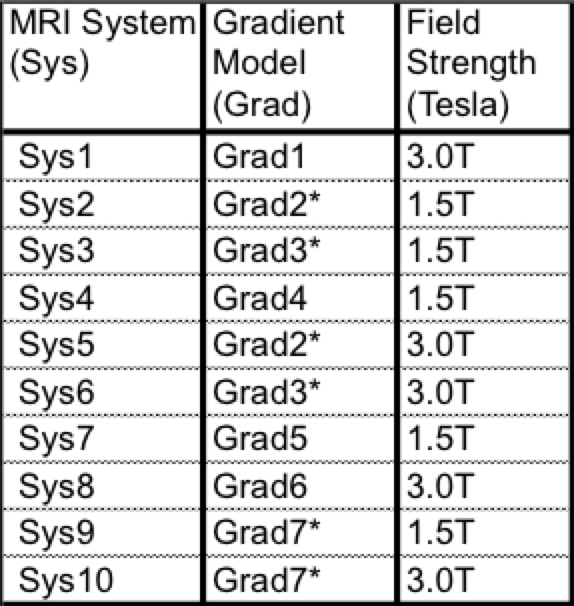
Table 1. List of gradient configurations and field strengths for all MRI scanner systems in the ACRIN 6698 trial. Asterisks indicate gradient configurations that are in more than one MRI scanner.
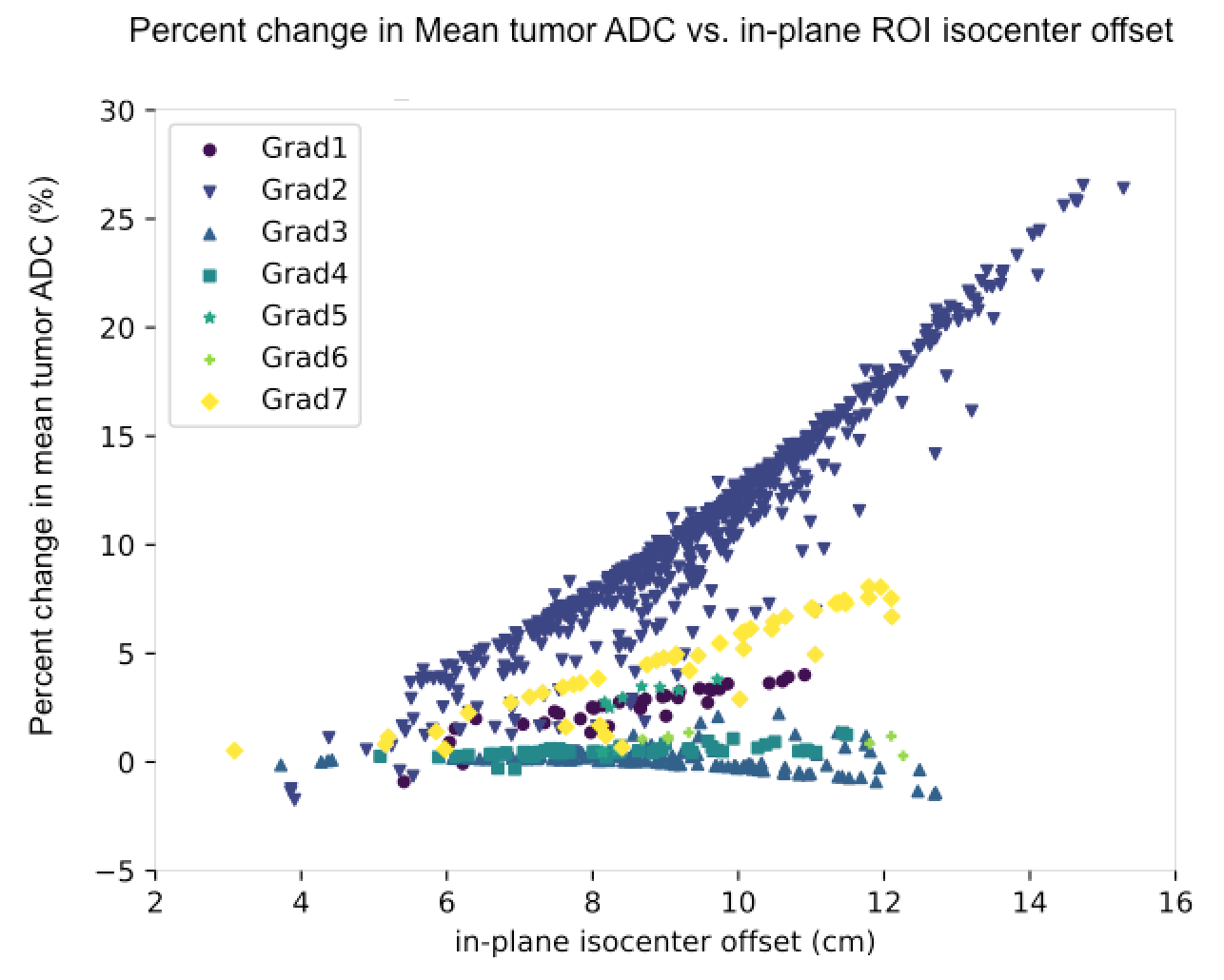
Figure 1. Percent difference in mean tumor ADC (with and without GNC) for all patient DWI scans (n=865), plotted as a function of distance from scanner iso-center for the seven gradient configurations of the ten MRI scanners in the ACRIN 6698 trial. All datapoints for: 7 gradient systems, exams from visits T0, T1, T2, T3, combined 1.5T and 3T
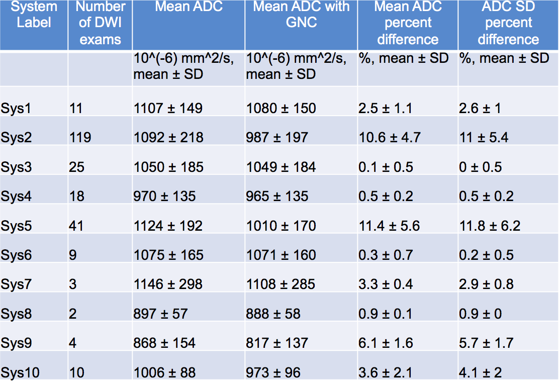
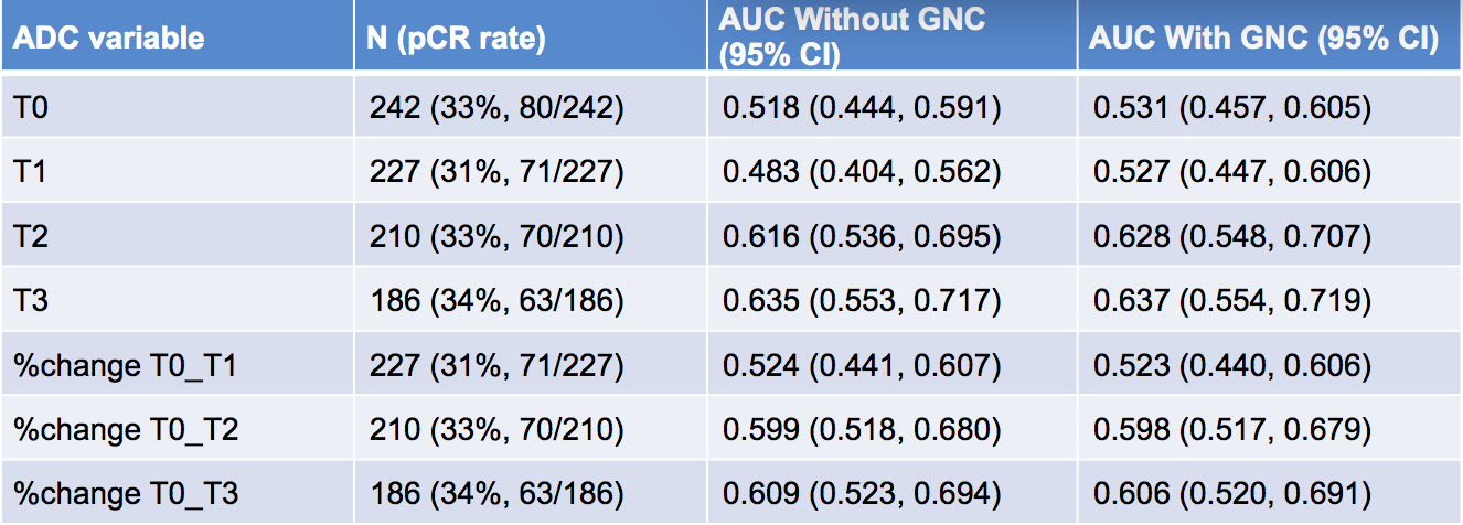
Table 3. Effect of gradient non-linearity correction on AUCs for ADC prediction of pathologic complete response (pCR), for mean tumor ADC values and percent change in mean tumor ADC between pre-treatment and the three treatment time points.