0205
Amide-proton transfer weighted imaging combined with time-dependent diffusion weighted imaging for evaluation of breast tumors1The First Affiliated Hospital of Zhengzhou University, Zhengzhou, China, 2Philips Healthcare, Beijing, China
Synopsis
Keywords: Breast, Breast
Motivation: Breast cancer is a complex and heterogeneous disease, and its different subtypes show very different biological characteristics, which will also affect the choice of treatment.
Goal(s): The objective of this study was to explore the value of APTWI combined with td-MRI in evalution of breast tumors.
Approach: The performance of APTWI was compared to conventional td-MRI in differentiation between benign and malignant breast tumors.
Results: APTWI overperform td-MRI in differentiation between benign and malignant breast tumors. And ADC values derived from td-MRI at different gradient oscillation frequencies show potential diagnostic values in predicting different risk factors in breast cancer.
Impact: These may provide new ideals for the diagnosis, treatment and prognosis evaluation of breast cancer.
Introduction
Breast cancer is a complex and heterogeneous disease, and its different subtypes show very different biological characteristics, which will also affect the choice of treatment. Therefore, the biological assessment of breast cancer is crucial. Breast magnetic resonance imaging (MRI) is an important tool in the diagnosis of breast cancer in clinical practice. Amide-proton transfer weighted imaging (APTWI) enables the evaluation of free proteins and peptides in tissues based on the chemical exchange between amide protons and water particles without using exogenous contrast agents [1]. Besides, time-dependent diffusion MRI (td-MRI) technology has great potential in assessing cellular microstructure by linking the diffusion-time dependence of water diffusion in living cells to specific microstructural parameters. Applications of APTWI and td-MRI in the field of breast lesions is gradually increasing. For example, Klomp et al[2] and Zaric et al[3] have assessed the reproducibility of APTWI in the breast at 7.0T MRI, and Xu et al[4]. and Wu et al[5]. have investigated the performance of td-MRI in evaluation of breast tumors. This study aims to explore the value of APTWI combined with td-MRI in evalution of breast tumors.Materials and Methods
This prospective study collected 200 patients with suspected breast tumors from the First Affiliated Hospital of Zhengzhou University from March to August 2023. All patients underwent T1-weighted imaging, T2-weighted imaging, conventional diffusion weighted imaging, APTWI, td-MRI, and dynamic contrast enhanced MRI scans. The APTWI imaging parameters were as follows: 3D turbo spin echo acquisition; TR(5400ms)/TE(7.8ms); flip angle=90°; number of signal average=1; slice thickness=6 mm; field of view=130×130mm2; voxel size=2.0×2.0×7mm3; matrix size 64×65; total scan time=4min57s. The td-MRI imaging parameters were as follows (PGSE/OGSE17Hz/OGSE33Hz): TR(4000ms)/TE(145ms); flip angle=90; number of signal average=1; slice thickness=6 mm; field of view=192×192mm2; voxel size = 2.53×2.58×5 mm3; matrix size 76×74; b-value[s/mm2] = 0/250/500/750/1000/1400/1800 (PGSE); 0/250/500/750/1000 (OGSE17Hz); 0/100/200/300 (OGSE33Hz); bandwidth[pixel/Hz] = 37.2; total scan time 4min24s/4min12s/2min08s. The asymmetric magnetization transfer rate at the frequency offset of 3.5 ppm [MTRasym(3.5ppm)] by APTWI and apparent diffusion coefficients (ADC) from td-MRI with different diffusion times (ADCPGSE, ADC17Hz and ADC33Hz) for breast tumors were measured respectively. The independent samples t test was used to compared the MTRasym(3.5ppm) and ADC values between benign and malignant breast tumors, between breast cancer with low- and high-histological grading, as well as between breast cancer with positive and negative expression of ER, PR, Her-2, and Ki-67, respectively. The receiver operating characteristic (ROC) curve was used to access the diagnostic performance of MTRasym and ADC values. P < 0.05 indicated that the difference was statistically significant.Results and Discussion:
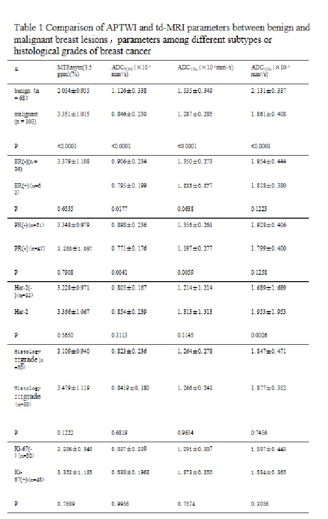
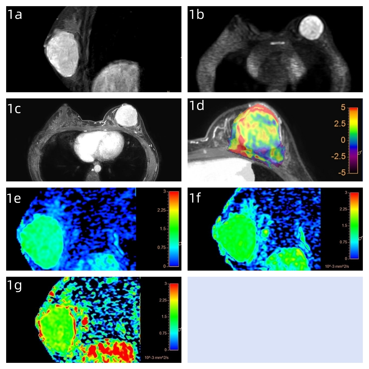
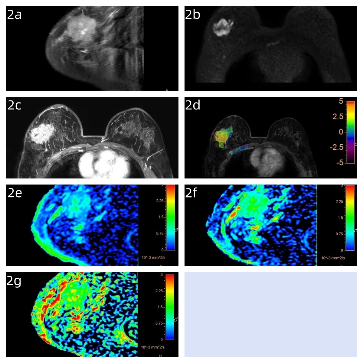
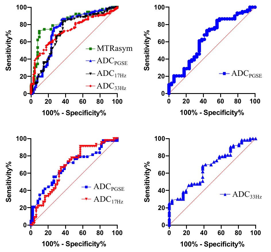
171 patients with 171 lesions were included and divided into malignant (n=103) and benign lesion group (n=68) according to the histopathological results. Figures 1 and 2 show representative images of patients with benign and malignant lesions.MTRasym values of malignant lesions were significantly higher than those of benign lesions (3.351±1.015% vs 2.054±0.955%). ADC values of malignant breast lesions were all lower than those of benign breast lesions (ADCPGSE: 0.846±0.230 vs 1.126±0.338 × 10⁻³ mm2/s, ADC17Hz: 1.287±0.285 vs 1.535±0.349 × 10⁻³ mm2/s, ADC33Hz: 1.861±0.408 vs. 2.131±0.337 × 10⁻³ mm2/s), and the differences were statistically significant. For both of benign and malignant breast lesions ADC33Hz > ADC17Hz > ADCPGSE. (Table 1)
Differences in MTRasym(3.5ppm) and ADC values between low- and high-grade tumors were without statistical significance. MTRasym(3.5ppm) also showed no significant difference between breast tumors with positive and negative expression of ER, PR, Her-2 and Ki-67, respectively. The ADCPGSE values of ER(+) tumors were significantly lower than those of ER(-) tumors. The ADCPGSE and ADC17Hz values of PR(+) tumors were significantly lower than those of PR(-) tumors. The ADC33Hz values of Her-2(+) were significantly lower than those of Her-2(-) tumors. (Table 1)
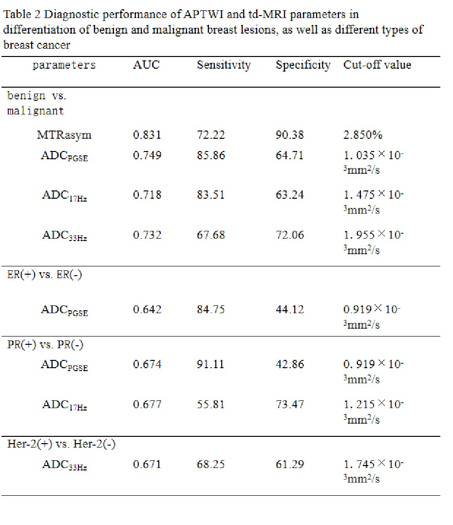
For differentiation between benign and malignant breast lesions, MTRasym(3.5ppm) showed higher AUC (0.831) and specificity (90.38) than those of the ADCs, the ADCPGSE showed relatively high AUC (0.749) and sensitivity (85.86) among the ADCs, and the ADC33Hz showed a fair good specificity of 72,06. For ER expression status discrimination, the ADCPGSE showed an AUC of 0.642, a sensitivity of 84.75, and a specificity of 44.12. For PR expression status discrimination, ADC17Hz showed higher AUC (0.677) and specificity (73.47) than ADCPGSE. For Her-2 expression status discrimination, the ADC33Hz showed an AUC of 0.671, the sensitivity of 68.25, and the specificity of 61.29.(Table 2)
Conclusion
APTWI showed higher diagnostic efficacy than td-MRI in discrimination between benign and malignant breast tumors. While the ADC values obtained by td-MRI at different gradient oscillation frequencies show better diagnostic performance in identifying different gene expressions in breast cancer.Key words
amide proton transfer imaging, time-dependent diffusion weighted imaging, breast tumor, benign and malignant, risk factorAcknowledgements
No acknowledgment.References
[1]Stejskal EO, Tanner JE. Spin diffusion measurements: spin echoes in the presence of a time-dependent field gradient. J Chem Phys. 1965;42:288–292.[2]Klomp DW, Dula AN, Arlinghaus LR, et al. Amide proton transfer imaging of the human breast at 7T: Development and reproducibility. NMR Biomed 2013;26:1271-1277.
[3]Zaric O, Farr A, Poblador Rodriguez E, et al. 7T CEST MRI: A potential imaging tool for the assessment of tumor grade and cel proliferation in breast cancer. Magn Reson Imaging 2019;59:77-87.
[4] Xu J, Jiang X, Li H, et al. Magnetic resonance imaging of mean cell size in human breast tumors[J]. Magnetic resonance in medicine, 2020, 83(6): 2002-2014.
[5]Ba R, Wang X, Zhang Z, et al. Diffusion-time dependent diffusion MRI: effect of diffusion-time on microstructural mapping and prediction of prognostic features in breast cancer[J]. European Radiology, 2023: 1-12.
Figures