0199
Water/fat separated Prostate Diffusion-Weighted Imaging using Dixon-encoded multi-shot EPI with structured low-rank reconstruction1C.J. Gorter MRI Center, Department of Radiology, LUMC, Leiden, Netherlands, 2Centre for Medical Imaging, University College London, London, United Kingdom, 3Philips, Best, Netherlands, 4Philips Research Hamburg, Hamburg, Germany
Synopsis
Keywords: Prostate, Prostate
Motivation: Conventional single-shot EPI (ssh-EPI) is fast, but often causes geometric distortions, especially near the rectum.
Goal(s): In this research, a Dixon-msh-EPI technique is validated for prostate Diffusion-weighted Imaging (DWI).
Approach: Dixon-msh-EPI is proposed to reduce such distortions, while jointly separating water/fat components by a special structured low-rank reconstruction that also corrects shot-to-shot phase variations.
Results: Experiments were conducted on 7 healthy male volunteers using a 3T scanner comparing DW-ssh-EPI and Dixon-msh-EPI performance. The Dixon-msh showed significantly reduced geometric distortion and effective fat signal suppression, as scored by two readers. While both methods provided comparable image quality, Dixon-msh-EPI demonstrated improved motion-correction and geometric accuracy.
Impact: Dixon-msh-EPI offers improved prostate DWI by significantly reducing geometric distortions and enhancing across b-value registration. This technique could lead to more accurate diagnoses and has the potential to refine clinical MRI protocols, emphasizing precision in prostate diffusion imaging.
Introduction
Early detection and accurate characterization of prostate cancer are critical for effective treatment and thus improved patient outcomes1,2. Diffusion-weighted imaging (DWI) with ADC mapping could provide a non-invasive insight into the microstructure of prostate tissue3, aiding in the diagnostic process. While single-shot EPI (ssh-EPI) is generally preferred for its speed in clinical prostate DWI, it is severely limited in image quality due to potential geometric distortions and T2* blurring3. Especially in the prostate, air/tissue susceptibility can vary significantly due to its close location to the rectum. Multi-shot EPI3 (msh-EPI) can potentially address all these issues and provide higher spatial resolution, although it faces challenges with motion-induced shot-to-shot phase variations4. Moreover, in common with all EPI-based acquisitions, unsuppressed fat is a confounding factor due to the spatial displacements of fat signals, resulting from the chemical-shift effect5,6.To address these issues, we propose to use a Dixon-msh EPI acquisition combined with a structured low-rank reconstruction to simultaneously address shot-to-shot phase variations and the fat-suppression problem7. The DW Dixon-ms-EPI has notable advantages, including (1) effective phase-corrected averaging without fat-suppression pulse and measuring 2D-navigators4; (2) correction for residual geometric distortions using estimated B0 map for free; (3) improved fat suppression; and (4) better "fat-based" image registration across b-values. In this study, we also evaluate Dixon-msh-EPI against standard fat-suppressed ssh-EPI for prostate DWI.
Methods
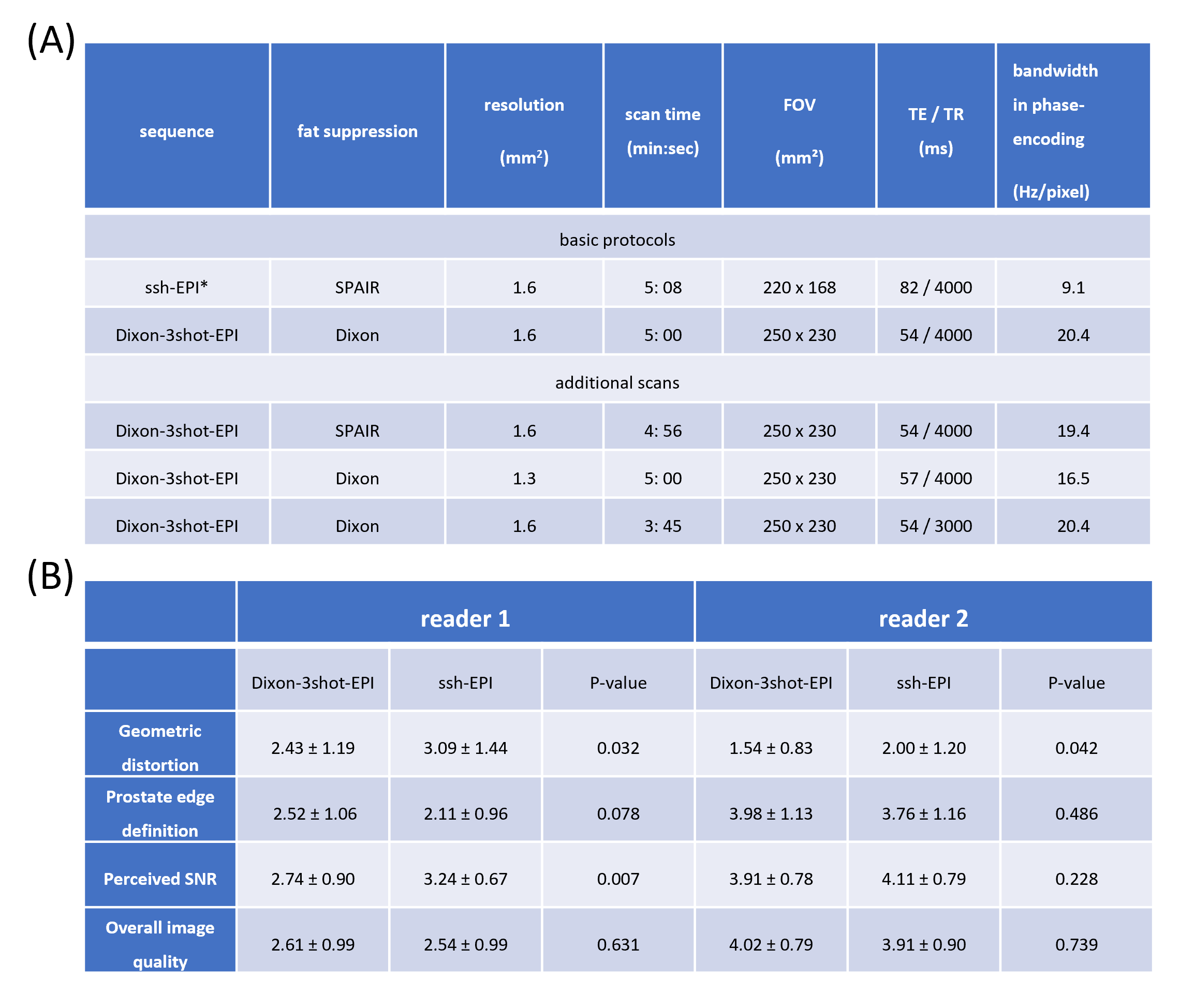
Dixon combined with msh-EPI (Dixon-ms-EPI) using structured low-rank reconstruction for DWI allows navigation-free joint estimation of water, fat and B0 components. By applying two Hankel-matrix completions8, the data redundancy along multi-shot/Dixon dimensions can be used to guide the reconstruction:$$\left\{P_w,P_f\right\}=\underset{\hat{x}_w,\hat{x}_f\in\mathbb{C}^{Q{\times}N{\times}L}}{\operatorname{argmin}}\|AP-d\|_2^2+\lambda_1\left\|H\left(FP_w\right)\right\|_*+\lambda_2\left\|H\left(F P_f\right)\right\|_*,$$where $$$P=\left[P_w,P_f\right]^T=\left[\rho_{w,1,1},\ldots,\rho_{w,N,1},\ldots,\rho_{w,1,L},\ldots,\rho_{w,N,L},\rho_{f,1,1},\ldots,\rho_{f,N,1},\ldots,\rho_{f,1,L},\ldots,\rho_{f,N,L}\right]^T$$$represents the water/fat complex-valued images with $$$L$$$ shots and $$$N$$$ Dixon points, and $$$A$$$ is the system matrix that enforces data consistency, $$$H$$$ represents the Hankel-matrix transformation. There are also some advantages of doing Dixon for prostate DWI: after the reconstruction, residual geometric distortions can be further corrected using the Dixon B0 fieldmap, with conjugate-phase reconstruction (CPR)9 for free. Such B0 map is estimated during reconstruction from the b=0 s/mm2 data 5–7. Separated fat signals can also be used for macroscopic motion tracking in DW Dixon-msh-EPI (Rigid + diffeomorphic field registrations were used in previous work10), and fat can be suppressed over the complete multi-peak fat spectrum6,11.Experiments were conducted using a 3T scanner on 7 healthy male volunteers who provided informed consent. They underwent a standard prostate 2D-T2w-TSE scan, followed by two baseline DWI scans and a few additional scans. Detailed scan parameters for these sequences are presented in Table 1 (A). Reconstruction utilizing SENSE for the ssh-EPI was compared to the structured low-rank for the Dixon 3-shot EPI. CPR was not used in this comparison.
The comparative analysis involved evaluating ADC values, which were obtained using a mono-exponential model in ROIs placed in the transition zone areas while being free of susceptibility-related artifacts. Statistical significance was tested using a paired Wilcoxon test. Two expert readers assessed image quality from both DWI protocols using a 5-point Likert scale12. Criteria included geometric distortion, prostate edge clarity, perceived SNR, and overall image quality.
Results
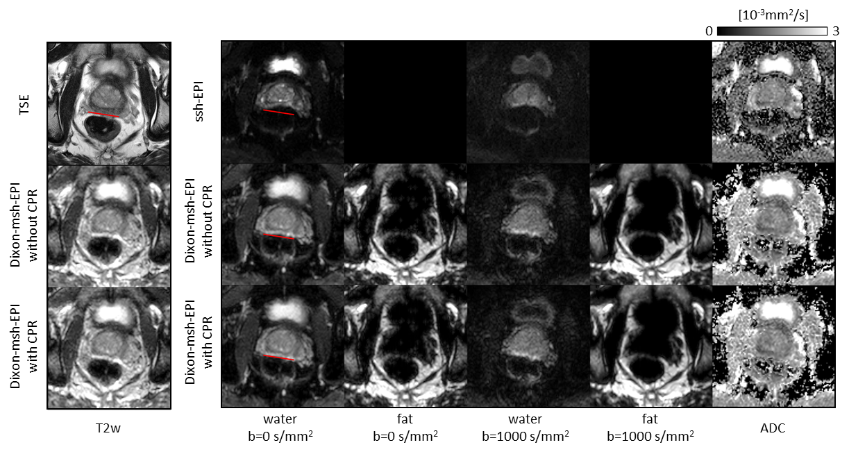
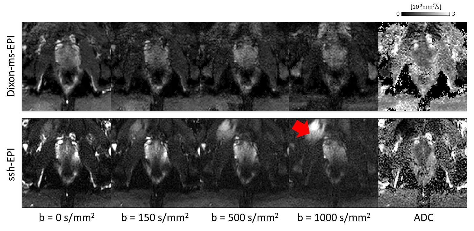
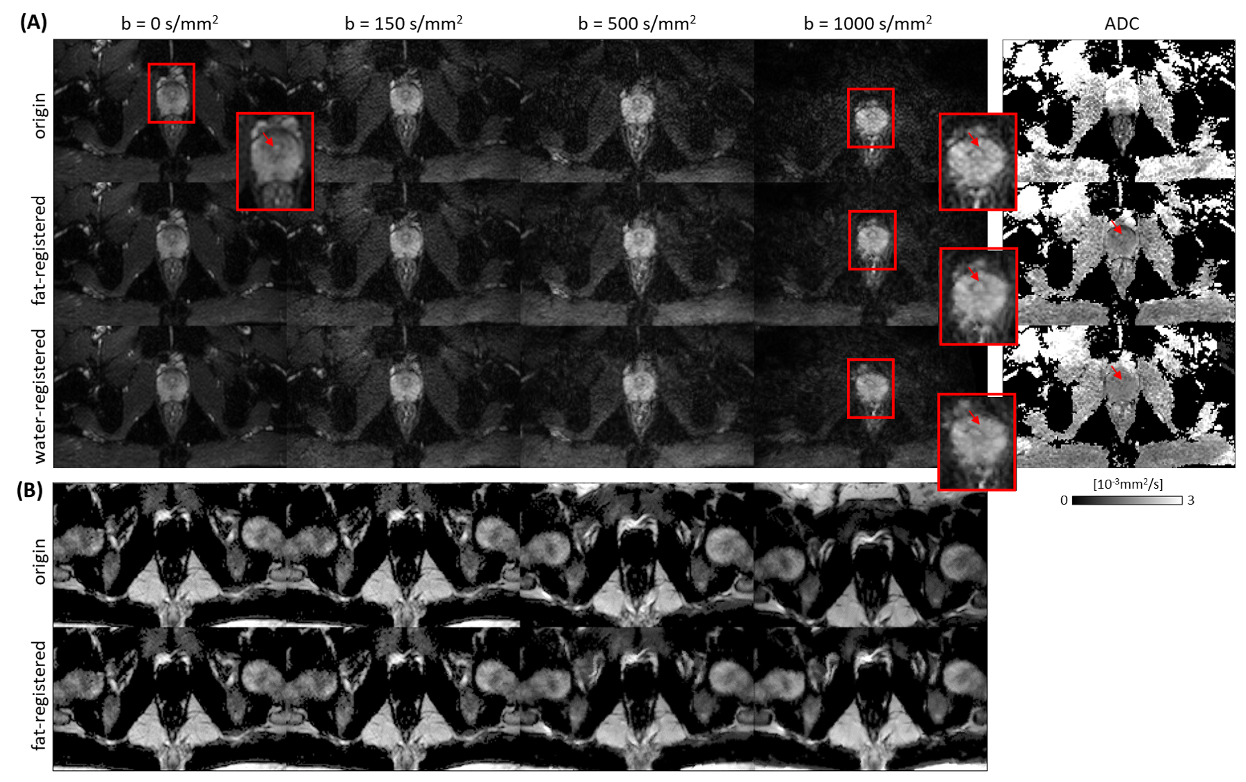
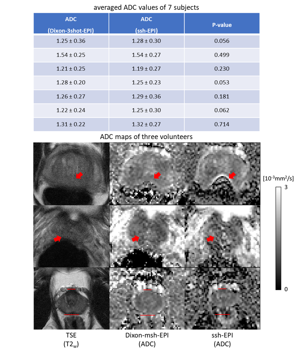
Dixon-ms-EPI showed less geometric distortion than ssh-EPI as scored by both readers; however, only reader 2 perceived the SNR of ssh-EPI to be significantly higher than Dixon-ms-EPI, as shown in Table 1(B). Figure 1 shows a slice illustrating the reduced geometric distortion of Dixon-msh-EPI versus ssh-EPI, compared to the T2w-TSE reference image. It is also evident that the CPR + B0 map can further correct the remaining geometric distortions. In Figure 2, Dixon ssh-EPI effectively suppressed fat signals, unlike fat-suppressed SPAIR13 ssh-EPI, which failed in this chosen case. In Figure 3, the prostate scan exhibited motion-induced deformities in the water-registered images, which can be much better registered using fat images. Figure 4 shows comparable ADC values between Dixon-ssh-EPI and ssh-EPI (top table), but ssh-EPI had certain regions with noticeable ADC mapping degradation (bottom).Discussion and conclusion
This study confirmed that Dixon-msh-EPI outperforms ssh-EPI with regard to reduced geometric distortions, whereas other factors of overall image quality were comparable. Note that scan time was kept constant. The inherent ability of Dixon-msh-EPI to access fat information can aid in motion correction by applying more robust fat-based registration. Dixon-msh-EPI also allowed a further reduction in geometric distortion by exploiting the jointly estimated B0 map, and showed its effectiveness in fat suppression. Therefore, it presents a valuable alternative to fat-suppressed ssh-EPI. In addition, eliminating the SPAIR pulse saved 130 ms per shot, which improved scan efficiency. Notably, this initial study was based on healthy volunteers, further clinical studies with patients are needed to understand its diagnostic efficacy.Acknowledgements
The authors would like to acknowledge NWO-TTW (HTSM-17104).References
1. Hoeks CMA, Barentsz JO, Hambrock T, et al. Prostate cancer: Multiparametric MR imaging for detection, localization, and staging. Radiology. 2011;261(1).
2. Giganti F, Kirkham A, Kasivisvanathan V, et al. Understanding PI-QUAL for prostate MRI quality: a practical primer for radiologists. Insights Imaging. 2021;12(1).
3. Tamada T, Kido A, Ueda Y, et al. Comparison of single-shot EPI and multi-shot EPI in prostate DWI at 3.0 T. Sci Rep. 2022;12(1):1-10.
4. Jeong HK, Gore JC, Anderson AW. High-resolution human diffusion tensor imaging using 2-D navigated multishot SENSE EPI at 7 T. Magn Reson Med. 2013;69(3):793-802. doi:10.1002/mrm.24320 5. Dong Y, Riedel M, Koolstra K, van Osch MJP, Börnert P. Water/fat separation for self-navigated diffusion-weighted multishot echo-planar imaging. NMR Biomed. 2023;36(1):e4822.
6. Dong Y, Koolstra K, Riedel M, van Osch MJP, Börnert P. Regularized joint water–fat separation with B0 map estimation in image space for 2D-navigated interleaved EPI based diffusion MRI. Magn Reson Med. 2021;86(6):3034-3051.
7. Dong Y, Koolstra K, Li Z, Riedel M, Osch MJP van, Börnert P. Structured low-rank reconstruction for navigator-free water/fat separated multi-shot diffusion-weighted EPI. Magn Reson Med. Published online September 27, 2023.
8. Mani M, Jacob M, Kelley D, Magnotta V. Multi-shot sensitivity-encoded diffusion data recovery using structured low-rank matrix completion (MUSSELS). Magn Reson Med. 2017;78(2):494-507.
9. Man LC, Pauly JM, Macovski A. Multifrequency interpolation for fast off-resonance correction. Magn Reson Med. 1997;37(5):785-792.
10. Garyfallidis E, Brett M, Amirbekian B, et al. Dipy, a library for the analysis of diffusion MRI data. Front Neuroinform. 2014;8(FEB).
11. Ren J, Sherry AD, Malloy CR. 1H MRS of intramyocellular lipids in soleus muscle at 7 T: Spectral simplification by using long echo times without water suppression. Magn Reson Med. 2010;64(3):662-671.
12. Stocker D, Manoliu A, Becker AS, et al. Image Quality and Geometric Distortion of Modern Diffusion-Weighted Imaging Sequences in Magnetic Resonance Imaging of the Prostate. Invest Radiol. 2018;53(4):200-206.
13. Udayasankar UK, Martin D, Lauenstein T, et al. Role of spectral presaturation attenuated inversion-recovery fat-suppressed T2-weighted MR imaging in active inflammatory bowel disease. Journal of Magnetic Resonance Imaging. 2008;28(5):1133-1140.
Figures