0196
Free-breathing Motion-Corrected Liver DWI at 3T with Optimized Fat-suppression Scheme1Radiology, Fujian Medical University Union Hospital, Fuzhou, China, 2Fujian Medical University Union Hospital, Fuzhou, China, 3MR Application Development, Siemens Shenzhen magnetic Resonance Ltd., Shenzhen, China, Shenzhen, China, 4MR application, Siemens Healthineers Ltd,Guangzhou,China, Guangzhou, China, 5MR Application Predevelopment, Siemens Healthineers AG, Erlangen, Germany, Erlangen, Germany
Synopsis
Keywords: Liver, Diffusion/other diffusion imaging techniques
Motivation: A free-breathing motion-insensitive and high-quality DWI is desired for abdominal DWI in clinical practice.
Goal(s): This study is to propose and evaluate a novel ss-EPI DWI sequence incorporating motion correction , complex averaging , and a combination of a reparametrized sinc fatsat pulse with an optimized water excitation pulse for abdominal DWI.
Approach: Four different DWI sequecnes were performed on volunteers. The overall image qualities and the SNR on the liver papernchyma were evaluated and compared.
Results: The SNRs of the liver and the image qualities were significantly higher with the proposed Free-breathing motion-corrected DWI ( MOCO-DWI ) sequence compared to other EPI DWI sequences.
Impact: The free-breathing MOCO-DWI sequences with complex-averaging and new fat suppression scheme is recommended for high-quality liver DWI in clinical routine.
Objectives
In liver DWI, single-shot echo-planar imaging (ss-EPI) sequences are commonly employed due to their rapid acquisition and less sensitivity to respiratory and cardiac motion artifacts. Free-breathing without respiratory triggering is commonly used in clinical practice due to its high SNR-time efficiency. However, the motion occurring between different b values or averages can compromise the quality of the resulting DWI images and ADC maps, which can lead to image blurring, less reliable ADC maps, and consequently reduced lesion detection capability. Breath-holding (BH) and respiratory/navigator-triggered acquisition etc., have been utilized to improve ADC measurement reproducibility, signal-to-noise ratio (SNR) and image quality in liver DWI. However, the utility of breath-holding will decrease patient comfort, while respiratory/navigator-triggering will prolong the measurement time. Thus this study aimed to propose and evaluate a free-breathing motion-corrected (MOCO) DWI sequence for liver application at 3T. The sequence incorporates motion correction(1), complex averaging (2), and a new fat suppression scheme with combination of a reparametrized sinc fatsat pulse (3) with an optimized water excitation pulse.Methods
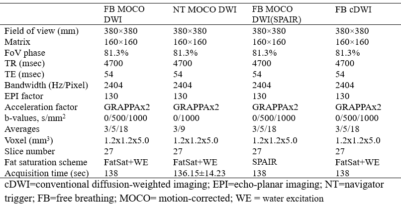
Forty-two volunteers underwent three motion-corrected single-shot echo-planar imaging (ss-EPI) DWI research application sequences and a conventional ss-EPI DWI sequence on a 3T MRI system ( MAGNETOM Prisma, Siemens Healthineers, Erlangen, Germany). Among the three MOCO DWI protocols, two of them were performed with free breathing, one of them with navigator-triggered (NT). While for the two free-breathing MOCO DWI, one of them was configured with the new fat suppression scheme, while the other was with the conventional spectral attenuated inversion recovery (SPAIR) pulse. The conventional DWI (cDWI) was configured wiht SPAIR pulse and performed with free-breathing. Detailed scan parameters were showed in table 1. Signal-to-noise ratio (SNR) of the liver parenchyma was measured at b = 1000 s/mm2. Over image qualities and the sharpness of the liver contour were independently evaluated by three radiologists using 5-point Likert scalesResults
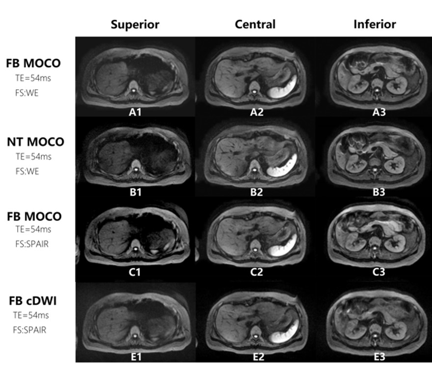
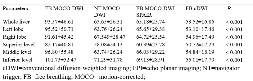
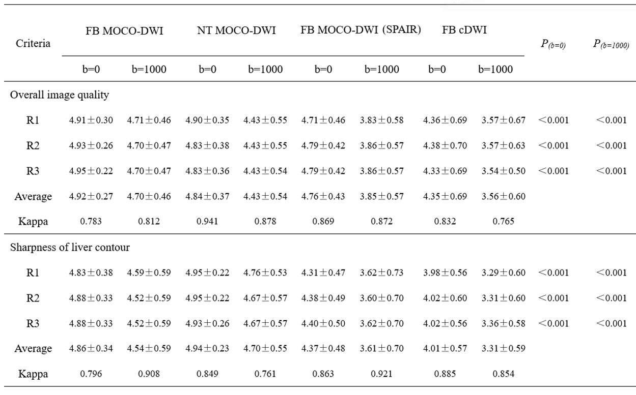
The SNRs (table 2) of the liver were significantly higher for free-breathing MOCO-DWI with new fat- suppression scheme compared to other EPI DWI sequences at b = 1000 s/mm2 (p < 0.05). The qualitative results (table 3) demonstrated markedly superior image quality ratings for FB MOCO-DWI and NT MOCO-DWI compared to the other EPI DWI sequences at b = 1000 s/mm2 (p < 0.05)Conclusions
The proposed EPI DWI sequence, which includes motion correction, complex averaging, and a noval fat suppression scheme utilizing spectral fat saturation and binomial water excitation pulses, was proven to be clinically viable for liver MRI. The FB MOCO-DWI sequence, with its enhanced SNR and outstanding image quality, is highly recommended for routine clinical liver DW imaging at 3T.Acknowledgements
The authors thank the members of Magnetic Resonance team at Fujian Medical University Union Hospital and Siemens Healthineers for technical support.References
1. Jolly, M. P. , Guetter, C. , & Guehring, J. "Cardiac Segmentation in MR Cine Data Using Inverse Consistent Deformable Registration". Proceedings of the 2010 IEEE International Symposium on Biomedical Imaging: From Nano to Macro, Rotterdam, The Netherlands, 14-17 April, 2010. IEEE.
2. Kordbacheh H, Seethamraju RT, Harisinghani MG, et al. Image quality and diagnostic accuracy of complex-averaged high b value images in diffusion-weighted MRI of prostate cancer. Abdom Radiol (NY). 2019 Jun;44(6):2244-2253.
3. Chebrolu V V , Frick M , Kollasch P , et al. "Fat Saturation in Musculoskeletal MRI using Spectral Heterogeneity Adaptive RF Pulses (SHARP)." Proceedings Joint Annual Meeting ISMRM-ESMRMB 2018.
Figures