0176
Quantitative Placental R2* Mapping on Rhesus Macaques with Thrombotic and Inflammatory Injury Model1Department of Medical Physics, University of Wisconsin-Madison, Madison, WI, United States, 2Wisconsin National Primate Research Center, University of Wisconsin-Madison, Madison, WI, United States, 3Department of Obstetrics and Gynecology, University of Wisconsin-Madison, Madison, WI, United States, 4Department of Radiology, University of Wisconsin-Madison, Madison, WI, United States, 5Department of Biomedical Engineering, University of Wisconsin-Madison, Madison, WI, United States, 6Department of Comparative Biosciences, University of Wisconsin-Madison, Madison, WI, United States
Synopsis
Keywords: Placenta, Quantitative Imaging, R2* mapping, Biomarkers, Contrast Agents, Ferumoxytol
Motivation: Pregnancy complications are often associated with placenta dysfunction, which lacks non-invasive assessment in early pregnancy.
Goal(s): Evaluating placental R2* as a non-invasive and quantitative biomarker to identify and monitor pregnancy complications.
Approach: We introduce a novel thrombotic and inflammatory placental injury model of rhesus macaques and report mean values, histograms, and spatial distribution of placental R2* based on blood oxygenation level dependent (BOLD) MR imaging at different gestational ages before and after iron nanoparticle (ferumoxytol) administration.
Results: Higher overtime and post-contrast increase in mean values and more heterogeneous spatial patterns of R2* are observed in placentas with induced injury.
Impact: A refined nonhuman primate model for thrombosis and inflammation is introduced for investigating placental pathology. Larger R2* increases over time and immediately after ferumoxytol injections suggest the potential for R2* to be an indicator of pathology with or without contrast.
Introduction
Uteroplacental inflammation and blood flow deficiency can lead to or exacerbate pregnancy complications such as fetal growth restriction and preeclampsia. Non-invasive diagnostic tools that identify placental pathophysiologic conditions early in pregnancy would enable early interventions and improved pregnancy outcomes. Recent studies have shown that Ferumoxytol, a superparamagnetic iron oxide nanoparticle originally designed for anemia treatments and used off-label as an MRI contrast agent, does not have negative impacts on the maternal-fetal interface (MFI) or placental/fetal health1, has been found to be useful in human imaging of placenta accreta2-4, and has been suggested as an alternative for diagnosis of PE during pregancy5. Since phagocytosis of ferumoxytol by macrophages is a part of the inflammatory response6, we hypothesize that ferumoxytol uptake can be used to detect and possibly quantify placental inflammation. R2* (=1/T2*) has been introduced as a potential marker of placental dysfunction as the blood oxygenation level-dependent (BOLD) signal correlates with placental oxygenation7-10. It also increases regionally with the concentration of ferumoxytol. In this pilot study, we introduce a rhesus macaque (RM) model designed to induce placental inflammation and thrombosis and compare their R2* results for mean values, histograms, and spatial distribution across 5 gestation time points with controls.Methods
Subjects and Interventions12 RM with singleton pregnancy were injected with 100 μg of monocyte chemoattractant protein-1 (MCP-1) (4RM), 0.5 ml of Tisseel (3RM), 3x0.5 ml of Tisseel at 3 separate locations (2RM), or saline (3RM) as controls, all into the anterior placental disc under ultrasound guidance at ~101 days of gestational age (GA). MCP-1 is a central effector of inflammatory responses and was selected for its potential to direct migration of macrophages and induce placental inflammation. Tisseel is an FDA-approved fibrin sealant used surgically to control bleeding and is used here to induce thrombosis in the intervillous space.
MRI Acquisition and Processing
All subjects were imaged in right-lateral position on a 3.0-T clinical scanner (GE Healthcare) at GA ~100, 114, 115, 144, and 145 days, hereby referred to as Scans 1-5. Intervention injections were performed approximately 1 day after Scan 1 and BOLD MR data was acquired with two 3D multi-echo spoiled gradient echo sequences (Sequence A: first TE=1.5ms;ΔTE=2.4ms,#TEs=12;scan time=8min17sec; Sequence B: first TE=1.3ms;ΔTE=1.1ms,#TEs=8;scan time=6min4sec) that fully covered the placenta. Ferumoxytol (4mg/kg) was administered between the two sequences at Scan 2 and Scan 4. R2* maps and water images were generated using the method proposed by Zhu11. Placental segmentation was performed with ITK-SNAP12 based on structural images and water images. Mean R2*, R2* histograms, and spatial distribution of R2* were analyzed. The placenta discs were inspected by a pathologist after Caesarian delivery at ~155 days of gestation.
Results
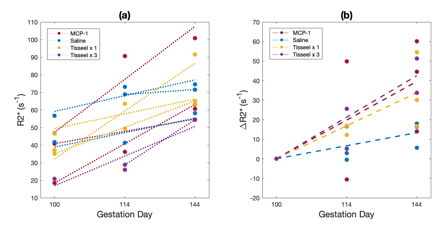
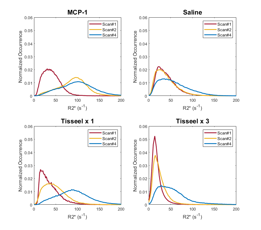
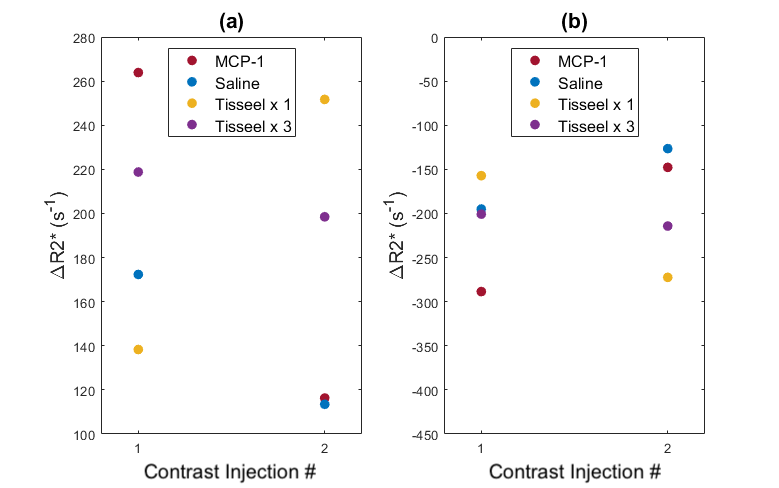
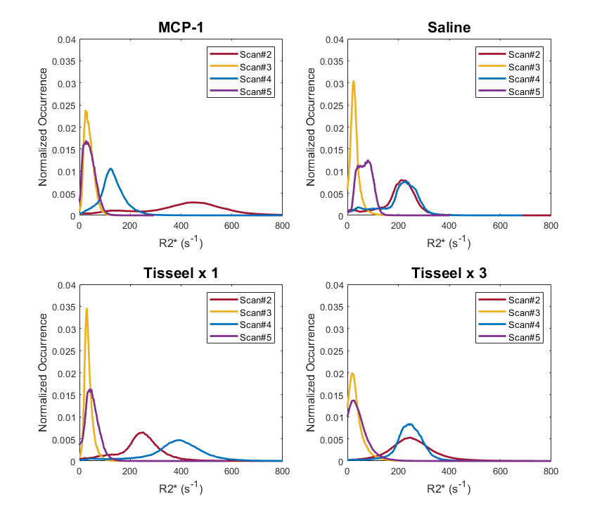
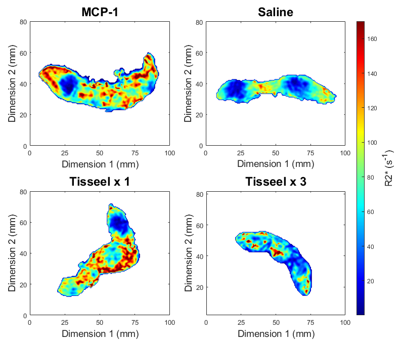
The delivered placental discs of control subjects were pathologically unremarkable. Small fibrin thrombi were observed within the placental parenchyma in the Tisseel-injected cases, with greater tissue ischemia and blood pooling beneath decidua appearing in the cases with 3 Tisseel injection sites. Ischemic lesions were observed without significant inflammation in subjects injected with MCP-1. Mean placental R2* values before ferumoxytol injection of all groups increased with time throughout gestation, with more significant increases observed in all treatment groups than in the control group (Figure 1), consistent with observations in other studies in the presence of pathologies13,14.Pre-contrast R2* histograms become broader and shift towards higher R2* means with gestation. This trend is amplified in the histograms of the MCP-1- and Tisseel-injected subjects (Figure 2). When compared between immediately after and one day following Ferumoxytol injections, the treatment groups show larger increases in mean R2* values and subsequent larger decreases 1 day after contrast injections (Figure 3), which is also observed in the R2* histograms (Figure 4). The last pre-contrast scans during Scan 4 show more heterogeneous R2* distributions in the placenta with more regions of higher R2* values in the treatment groups (Figure 5).
Discussion and Conclusion
The study shows an overall increase in R2* over gestation, with more pronounced increases in mean values and more heterogeneous spatial patterns of R2* observed in MCP-1- and Tisseel-injected placenta, in which remarkable pathology was observed. The increase of mean R2* with increased gestational age shows the same trend as observed in several other studies that report similar increases in R2* (or equivalent decreases in T2*3,4) and indicates a decrease in oxygenation of placental blood with gestation. The amplified increases in R2* over time and right after ferumoxytol injections in pathological placentas suggest the potential usage of R2* as a marker for inflammatory and/or ischemic conditions with or without ferumoxytol injections.Acknowledgements
We gratefully acknowledge GE Healthcare for research support of UW-Madison, and funding support from NIH-NICHD (R01HD103443).
References
1. Nguyen, S. M., Wiepz, G. J., Schotzko, M., Simmons, H. A., Mejia, A., Ludwig, K. D., Zhu, A., Brunner, K., Hernando, D., Reeder, S. B., Wieben, O., Johnson, K., Shah, D., & Golos, T. G. (2020). Impact of ferumoxytol magnetic resonance imaging on the rhesus macaque maternal-fetal interface. Biology of reproduction, 102(2), 434–444. https://doi.org/10.1093/biolre/ioz181Figures