0172
An AI-Based Solution for MR Image Analysis of the Female Reproductive System1Voxelwise Imaging Technology Inc, Vancouver, BC, Canada, 2Prenuvo, Vancouver, BC, Canada
Synopsis
Keywords: Uterus, Reproductive, Female Reproductive System, Machine Learning, Artificial Intelligence
Motivation: To quantify female reproductive anatomy in MR imaging.
Goal(s): To develop an AI-based solution to segment the regions of interest (RoIs) for the uterine zone, ovaries, pelvic fluid, and detect benign uterine conditions.
Approach: A deep learning based method is applied on a large representative population of 9334 sagittal T2-weighted female pelvis scans to extract normative menstrual cycle- and aging-curves for various RoIs.
Results: Our proposed normative curves define the standard menstrual cycle and aging trends. RoI segmentation, fibroid, and cyst detection models achieve average foreground dice, specificity and accuracy scores of 83.9%, 95.2% and 94.37%, respectively.
Impact: Proposing a robust, precise AI solution for analyzing female reproductive organs on MR imaging, including uterine zones, ovaries, pelvic fluid, and fibroids/cysts. Using this, we define standard aging and menstrual cycle curves for women.
Introduction:
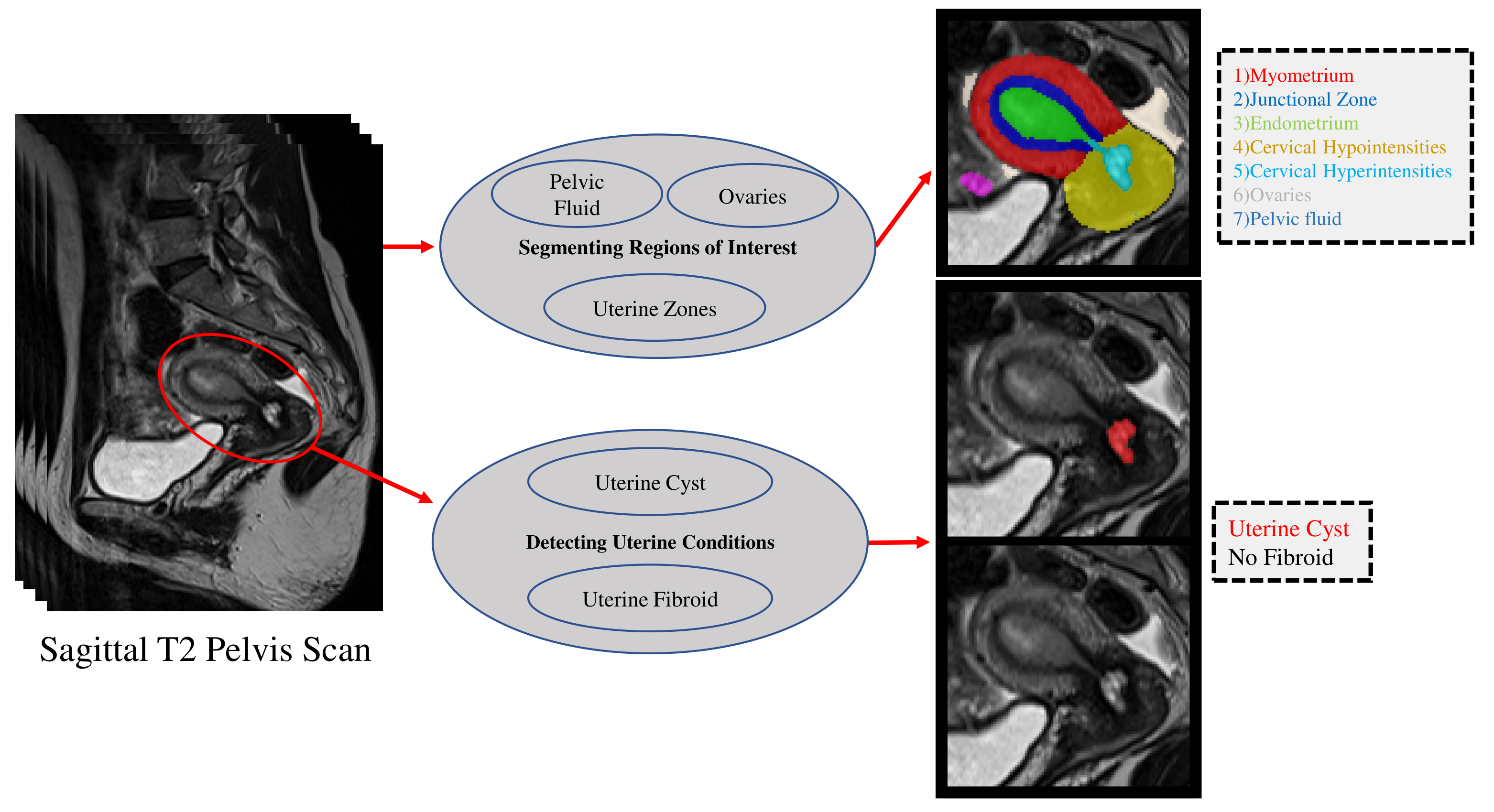
MR images of the female pelvis provide detailed high resolution images of the reproductive organs which facilitates in-depth analysis of the reproductive system. Nevertheless, ultrasonography stands as the widely accepted imaging modality for assessing female reproductive organs within the medical community and is favored by patients for its ease of access and affordability1, although some studies demonstrate MRI's superiority in diagnosing certain conditions1,2. Consequently, a significant number of existing AI-driven tools for gynecological analysis have been built for ultrasound modality3. Among the limited publications on developing AI-driven tools for female pelvis MRI analysis, most of them focus on malignant conditions or placenta monitoring4,5,6,7, while a few seek this potential for the general female population8. In this work, we proposed a comprehensive AI-driven tool to segment regions of interest (RoIs) for the female reproductive system, namely uterine zones, ovaries, pelvic fluid, and to detect uterine fibroid and cyst conditions (Figure 1). Compared to8, the proposed solution is more accurate, robust, and efficient by using significantly larger training datasets with more precise ground-truth labels sourced from multiple MRI machines and improving the modeling, e.g., training a unified RoI segmentation network and formulating condition diagnosis as an instance detection task. We have further proposed normative menstrual cycle and aging graphs for a large population of sagittal T2-weighted sequences from a representative sample (N=9334) of adults ranging from age 22 to 70 years.Methods:
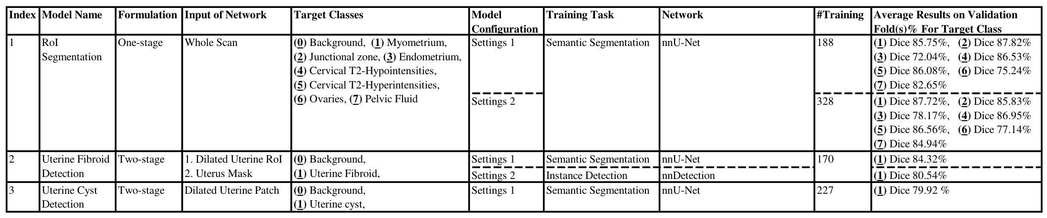
Data is acquired from sagittal T2-weighted pelvic sequences obtained as part of a comprehensive preventive whole-body MRI screening protocol. nnU-Net9 and nnDetection10 frameworks are used for semantic segmentation and instance detection tasks, respectively. Ground-truth segmentation masks are annotated using ITK-SNAP11. One-stage methods take the entire sagittal scan as input, while the two-stage approach restricts the input to a cropped section of the scan. Further details regarding the components of the proposed solution, training sets, and results on validation folds are summarized in Table 1.Results and Discussions:
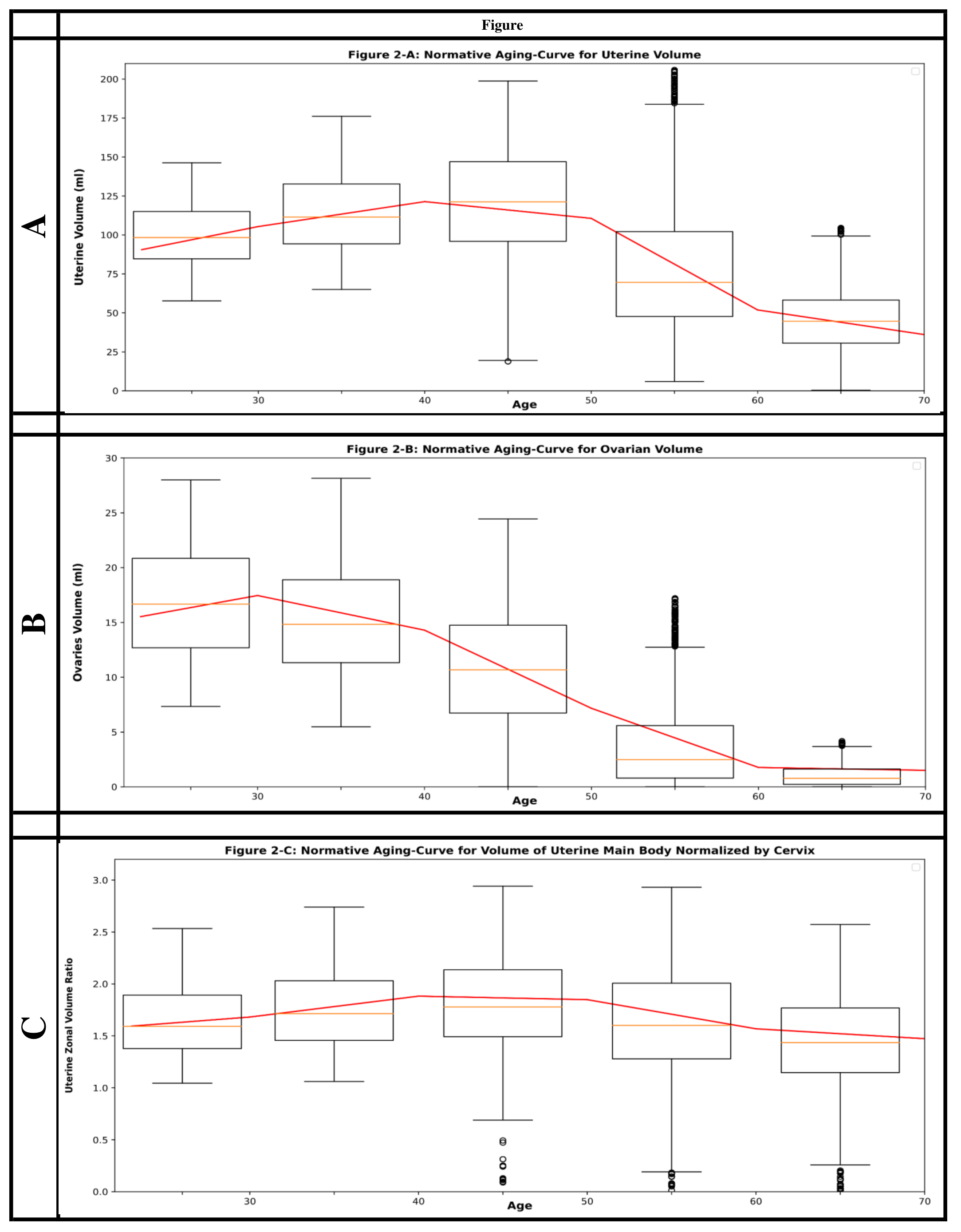
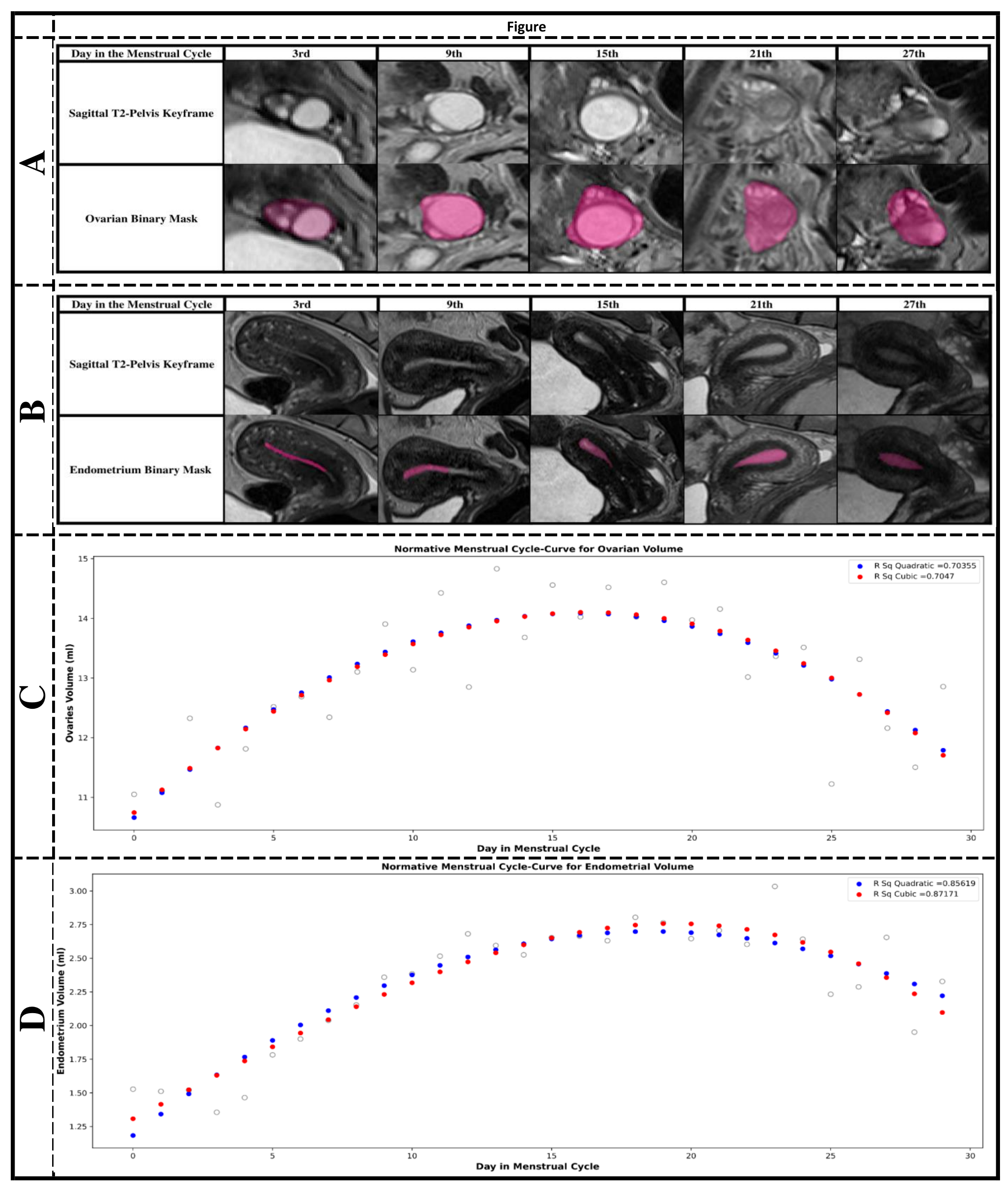
In this section, we discuss the elements and results of our proposed solution, considering results on large populations (Figure 2 & 3), hold-out sets (Table 2), and validation folds (Table 1).- RoI segmentation model segments the uterine zones, ovaries, and pelvic fluid. This enables us to do 2D and 3D measurements over the regions of interest, e.g, measuring the endometrial and junctional zone thickness and quantifying volume of the uterus and ovaries. It further helps with obtaining the input RoIs for the two-stage methods and localizing the conditions. Two settings for training the model are introduced in Table 1. By obtaining the inference results of the model trained with settings 1 (Table 1) over a large population (N=9334), we extracted the normative aging-curves for various RoIs (Figure 2). For the subset of menstrual patients with reported regular menstrual cycles (N=3029), we obtained normative menstrual cycle-curves for ovaries and endometrium (Figure 3). To further enhance the model’s robustness for rare reproductive organ appearances, we queried our radiology report database to extract patients with gynecological conditions. Then, the training dataset was expanded from 188 to 328 samples. These additional samples were manually annotated and consist of cases for which the prior model with settings 1 had subpar performance. The expanded dataset was used to retrain the RoI segmentation model with settings 2.
- Uterine cyst model was trained on 227 cases delineated with various types of uterine cysts including Nabothian- and adenomyosis related-cyst in our radiology reports. Compared with the Nabothian cyst model in8, this model detects cysts in various parts of the uterus which may help with diagnosing more uterine conditions including adenomyosis.
- Uterine fibroid models are obtained by training nnU-Net9 and nnDetection10 on 170 cases reported with uterine fibroid in our report database. Our nnDetection based fibroid model, as noted in Table 2, significantly improves fibroid classification specificity score (95.2% v.s. 76.19%) compared to the nnU-Net based model, while maintaining a competitive sensitivity. We attribute the performance gain to incorporating instance level information in addition to semantic labels. Both models outperform the solution in8 as we use a single foreground class with precise ground-truth masks and larger training set. In rare cases, both models miss degenerated or exophytic fibroids. Also, nnDetection predicts the bounding box that fits the fibroid, leading to errors in volume quantification and lower dice scores (Table 1).
Conclusion:
This study contributes to the advancement of AI-driven tools for MRI analysis in the field of gynecology, enhancing the accuracy and diagnostic capabilities for a broader range of female reproductive conditions. The findings support the potential of MRI as a valuable diagnostic modality in female reproductive health.Acknowledgements
We appreciate the support and collaboration of the MRI Technologists, Backend, and Patient Care teams at Prenuvo in the patient care and data acquisition process.References
1. Hubert, J. and Bergin, D., 2008. Imaging the female pelvis: when should MRI be considered?. Applied radiology, 37(1).
2. Palmér, M., Åkesson, Å., Marcickiewicz, J., Blank, E., Hogström, L., Torle, M., Mateoiu, C., Dahm-Kähler, P. and Leonhardt, H., 2023. Accuracy of transvaginal ultrasound versus MRI in the PreOperative Diagnostics of low-grade Endometrial Cancer (PODEC) study: a prospective multicentre study. Clinical radiology, 78(1), pp.70-79.
3. Chen, Z., Wang, Z., Du, M. and Liu, Z., 2022. Artificial intelligence in the assessment of female reproductive function using ultrasound: A review. Journal of Ultrasound in Medicine, 41(6), pp.1343-1353.
4. Shrestha, P., Poudyal, B., Yadollahi, S., Wright, D.E., Gregory, A.V., Warner, J.D., Korfiatis, P., Green, I.C., Rassier, S.L., Mariani, A. and Kim, B., 2022. A systematic review on the use of artificial intelligence in gynecologic imaging–Background, state of the art, and future directions. Gynecologic Oncology.
5. Urushibara, A., Saida, T., Mori, K., Ishiguro, T., Inoue, K., Masumoto, T., Satoh, T. and Nakajima, T., 2022. The efficacy of deep learning models in the diagnosis of endometrial cancer using MRI: a comparison with radiologists. BMC Medical Imaging, 22(1), pp.1-14.
6. Twickler, D.M., Do, Q.N., Xi, Y., Shahedi, M., Dormer, J., TT, A.D., Lewis, M.A., Spong, C.Y., Dashe, J.S., Madhuranthakam, A. and Fei, B., 2020. 228: Automated segmentation of the human placenta and uterus with MR imaging using artificial intelligence (AI). American Journal of Obstetrics & Gynecology, 222(1), pp.S158-S159.
7. Lu, M., Wang, T., Zhu, H. and Li, M., 2023, October. HACL-Net: Hierarchical Attention and Contrastive Learning Network for MRI-Based Placenta Accreta Spectrum Diagnosis. In International Conference on Medical Image Computing and Computer-Assisted Intervention (pp. 304-314). Cham: Springer Nature Switzerland.
8. Khaghani, J., Basar S., Chodakiewitz Y., London S., Attariwal, R., and Hashemi, S., 2023. A Deep Learning-Based Tool for Analyzing the Female Reproductive System in MR images. International Society for Magnetic Resonance in Medicine. International Society for Magnetic Resonance in Medicine (ISMRM), June 03-08 2023, Toronto.
9. Isensee, F., Jaeger, P.F., Kohl, S.A., Petersen, J. and Maier-Hein, K.H., 2021. nnU-Net: a self-configuring method for deep learning-based biomedical image segmentation. Nature methods, 18(2), pp.203-211.
10. Baumgartner, M., Jäger, P.F., Isensee, F. and Maier-Hein, K.H., 2021. nnDetection: a self-configuring method for medical object detection. In Medical Image Computing and Computer Assisted Intervention–MICCAI 2021: 24th International Conference, Strasbourg, France, September 27–October 1, 2021, Proceedings, Part V 24 (pp. 530-539). Springer International Publishing.
11. Yushkevich, P.A., Piven, J., Hazlett, H.C., Smith, R.G., Ho, S., Gee, J.C. and Gerig, G., 2006. User-guided 3D active contour segmentation of anatomical structures: significantly improved efficiency and reliability. Neuroimage, 31(3), pp.1116-1128.
12. Kelsey, T.W., Dodwell, S.K., Wilkinson, A.G., Greve, T., Andersen, C.Y., Anderson, R.A. and Wallace, W.H.B., 2013. Ovarian volume throughout life: a validated normative model. PloS one, 8(9), p.e71465.
Figures