0171
Magnetic resonance imaging-based radiomic for predicting infiltration levels of CD68+ tumor-associated macrophages in Glioblastomas1Lanzhou University Second Hospital, Lanzhou, China
Synopsis
Keywords: Preclinical Image Analysis, Nervous system
Motivation: Predicting Tumor-Associated Macrophages (TAMs) levels using preoperative non-invasive imaging can influence patients with Glioblastoma (Gb) treatment decision-making and evaluate prognosis.
Goal(s): This study aimed to combine imaging and radiomics features of preoperative for predicting CD68 + macrophage infiltration.
Approach: Retrospective collection 143 patients with Gb. Divided patients into high CD68+TAMs(≥14.8%)and low CD68+TAMs (<14.8%) groups. The radiomics features extraction were based on CE-T1WI and T2WI. Multi-parameter stepwise regression was used to create the models.
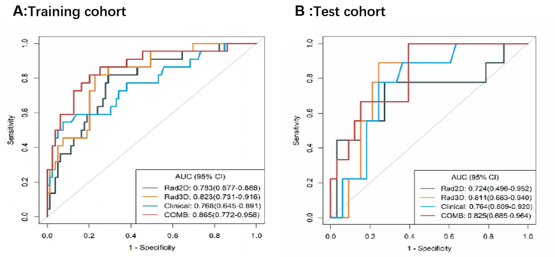
Results: The combined model, with ADCmin and radiomics features, had the best performance revealing AUCs of 0.865 and 0.825 for the training and testing sets, respectively.
Impact: To provide imaging biomarkers for the evaluation of the TAMs infiltration of Gb by using machine learning combined with MR imaging parameters, reveal the relationship between images features and TAMs, and construct an evaluation model to predict macrophage before surgery.
Introduction
Glioblastoma (Gb) is the most common primary malignant brain tumor, with an incidence of 3.26 per 100,000, accounting for 50.1% of all central nervous system (CNS) malignant tumors. The median survival time is only 8 months[1,2]. Gb exhibits strong invasiveness, and its tumor cells grow rapidly and invade the surrounding healthy brain tissue, resulting in short-term in-situ recurrence after surgery. The micro-dynamic changes of Gb drive its heterogeneity, which plays an important role in treating drug resistance[3]. Therefore, exploring tumor heterogeneity to Gb therapy may open novel perspectives for immunotherapy or targeted therapy.Tumor-Associated Macrophages (TAMs) in the immune microenvironment of Gb are the most important biomarkers for evaluating malignancy, degree of invasion, and prognosis of patients[4], and their increased levels are associated with poor prognosis[5]. TAMs are also a major part of cancer immunotherapy, producing high levels of immunosuppressive cytokines. To down-regulate the T-cell response and lead to high tumorigenicity[6]. Therefore, treating TAMs is a promising treatment for glioma [7]. Currently, tumor macrophages have become the main focus of immuno-oncology drugs [8, 9]. Particularly, CD68, CSF-1 are important targets for evaluating the total TAM population in the TME.To quantify macrophage, CD68 immunohistochemistry has been widely reported as a specific marker of TAMs in tumors[10-13]. Imaging provides unique information for predictive survival and prognosis independent of pathological and clinical data in managing patients with Gb. Magnetic resonance imaging (MRI) can be used as a simple method to monitor the macroscopic characterization and microstructure of gliomas[14]. Furthermore, MRI-based radiomics provide complementary information on morphological and texture features, which has potential clinical value as a noninvasive imaging biomarker in TAMs evaluation.Hence, the this study aimed to provide reliable imaging biomarkers for the evaluation of the TAMs infiltration extent of glioblastoma by using machine learning combined with MRI imaging parameters.Methods
This retrospective study involved 143 patients (training set: n = 101; test set: n = 42) with Gb . 143 surgical specimens from patients with Gb and labeled macrophages with immunohistochemical staining on paraffin sections of CD68, a phenotypic marker of macrophages.The optimal cut-off value (14.8%) was based on the minimum p-value formed by the Kaplan-Meier survival analysis and log-rank tests which divided patients into high CD68+TAMs(≥14.8%)and low CD68+TAMs (<14.8%) groups. The radiomics features extraction were based on CE-T1WI and T2WI. Multi-parameter stepwise regression was used to create the clinical, radiomics, and combined models, which were each evaluated using the ROC curve .Results
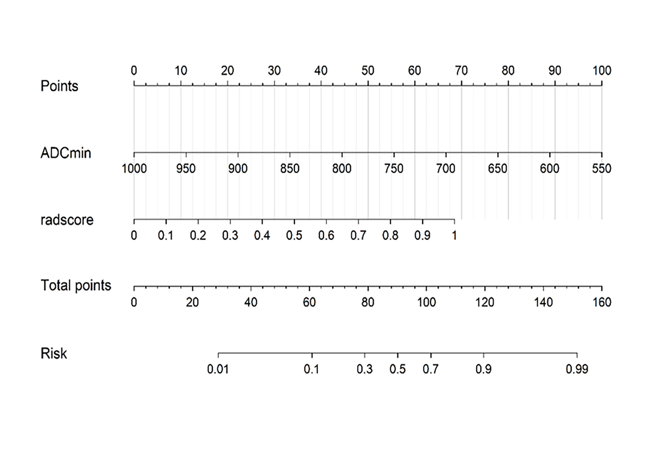
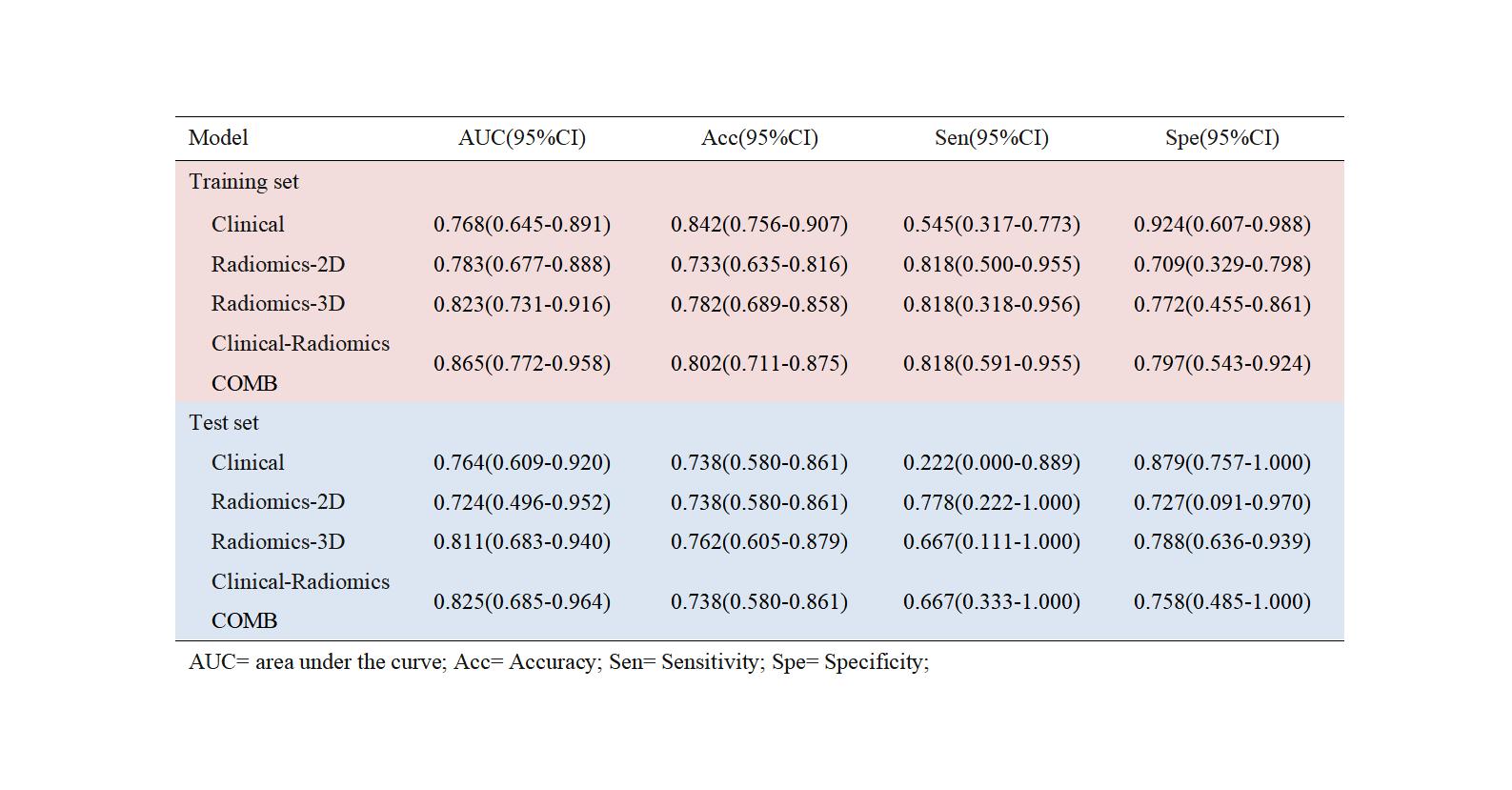
A clinical model based on the ADCmin revealed area under curve (AUCs) of 0.768 and 0.764 for the training and testing sets, respectively. The 2D Radiomics model revealed AUCs of 0.783 and 0.724 for the training and testing sets, respectively. The 3D Radiomics model revealed AUCs of 0.823 and 0.811 for the training and testing sets, respectively. The combined model, with ADCmin and radiomics features, had the best performance revealing AUCs of 0.865 and 0.825 for the training and testing sets, respectively. The calibration curve of the combined model nomogram(Fig.1) showed good agreement between the estimated and actual probabilities. The efficacy of the three models in predicting CD68+TAMs is shown in Table 1 and Fig.2.Discussion
We evaluated 9 microscopic visual fields based on CD68 immunohistochemistry and obtained an average value. The average expression of CD68 + macrophage in 143 patients with Gb was 9.39%. Survival analysis of CD68 + TAMs expression revealed that the survival time of patients with Gb with higher CD68 expression was significantly shorter than that of patients with Gb with lower CD68 expression.The clinical model constructed by a single ADCmin value has a good prediction effect (AUC=0. 768). The MR imaging DWI technique was shown to provide insight into the microstructure, and the ADC map can reflect the degree of macrophages to a certain extent. Based on the clinical model, we further established the relationship between biological characteristics and key radiomics features of TAMs. Previous studies have shown that the radiomics model can accurately predict the incidence of glioma biological behavior[15]. Our study, based on preoperative CE-T1WI and T2WI sequences, extracted radiomics features in the tumor core region .Finally, we combined imaging features with all radiomics features, which performed better than clinical and radiomics models alone. The AUC of the training set was 0.865, and the accuracy, sensitivity, and specificity values were 0.802, 0.818, and 0.797, respectively.Conclusions
In conclusion, We extracted radiomic features from preoperative MRI as non-invasive markers of CD68 +TAM infiltration. The prediction model established by combining preoperative image features and radiomics features performed well in predicting Gb macrophage infiltration.Acknowledgements
This work was supported by National Natural Science Foundation of China (grant number 82071872, 82371914 ); Science and Technology Program Funding Project of Gansu Province (grant number 21YF5FA123);China International Medical Foundation (grant number Z-2014-07-2101).References
1. Ostrom QT, Price M, Neff C, Cioffi G, Waite KA, Kruchko C, et al. CBTRUS Statistical Report: Primary Brain and Other Central Nervous System Tumors Diagnosed in the United States in 2015-2019. Neuro Oncol 2022;24(Suppl 5):v1-v95. 2. Louis DN, Perry A, Wesseling P, Brat DJ, Cree IA, Figarella-Branger D, et al. The 2021 WHO Classification of Tumors of the Central Nervous System: a summary. Neuro Oncol 2021;23(8):1231-1251. 3. Neftel C, Laffy J, Filbin MG, Hara T, Shore ME, Rahme GJ, et al. An Integrative Model of Cellular States, Plasticity, and Genetics for Glioblastoma. Cell 2019;178(4):835-849.e21. 4. Wang LJ, Xue Y, Lou Y. Tumor-associated macrophages related signature in glioma. Aging (Albany NY) 2022;14(6):2720-2735. 5. Tian Y, Ke Y, Ma Y. High expression of stromal signatures correlated with macrophage infiltration, angiogenesis and poor prognosis in glioma microenvironment. PeerJ 2020;8:e9038. 6. Hussain SF, Yang D, Suki D, Aldape K, Grimm E, Heimberger AB. The role of human glioma-infiltrating microglia/macrophages in mediating antitumor immune responses. Neuro Oncol 2006;8(3):261-79. 7. Ruffell B, Coussens LM. Macrophages and therapeutic resistance in cancer. Cancer Cell 2015;27(4):462-72. 8. Song CH, Kim N, Nam RH, Choi SI, Jang JY, Kim JW, et al. Combination treatment with 17beta-estradiol and anti-PD-L1 suppresses MC38 tumor growth by reducing PD-L1 expression and enhancing M1 macrophage population in MC38 colon tumor model. Cancer Lett 2022;543:215780. 9. Tang Q, Yang S, He G, Zheng H, Zhang S, Liu J, et al. Tumor-derived exosomes in the cancer immune microenvironment and cancer immunotherapy. Cancer Lett 2022;548:215823.10. Wang XL, Jiang JT, Wu CP. Prognostic significance of tumor-associated macrophage infiltration in gastric cancer: a meta-analysis. Genet Mol Res 2016;15(4).11. Wang L, Zhang C, Zhang Z, Han B, Shen Z, Li L, et al. Specific clinical and immune features of CD68 in glioma via 1,024 samples. Cancer Manag Res 2018;10:6409-6419.12. Zhang Y, Xie Y, He L, Tang J, He Q, Cao Q, et al. 1p/19q co-deletion status is associated with distinct tumor-associated macrophage infiltration in IDH mutated lower-grade gliomas. Cell Oncol (Dordr) 2021;44(1):193-204.13. Zeiner PS, Preusse C, Golebiewska A, Zinke J, Iriondo A, Muller A, et al. Distribution and prognostic impact of microglia/macrophage subpopulations in gliomas. Brain Pathol 2019;29(4):513-529.14. Xue C, Zhou Q, Xi H, Zhou J. Radiomics: A review of current applications and possibilities in the assessment of tumor microenvironment. Diagn Interv Imaging 2023;104(3):113-122.15. Zhang X, Lu H, Tian Q, Feng N, Yin L, Xu X, et al. A radiomics nomogram based on multiparametric MRI might stratify glioblastoma patients according to survival. Eur Radiol 2019;29(10):5528-5538.Figures