0169
Multifunctional Nanocomposite for Enhanced Diagnosis and Therapeutic Intervention in Breast Cancer via T1-T2 dual-enhanced and NIR II imaging1Department of Radiology, Jinan University, Guangzhou, China, 2School of Biomedical Engineering, Guangzhou Medical University, Guangzhou, China, 3School of Materials Science and Engineering, Sun Yat-sen University, Guangzhou, China, 4Department of Radiology, The Sixth Affiliated Hospital (Qingyuan People's Hospital), Guangzhou Medical University, Guangzhou, China
Synopsis
Keywords: Small Animals, Cancer, cancer immunotherapy, gas-photothermal therapy, magnetic resonance imaging, stimulator of interferon genes pathways, triple-negative breast cancer
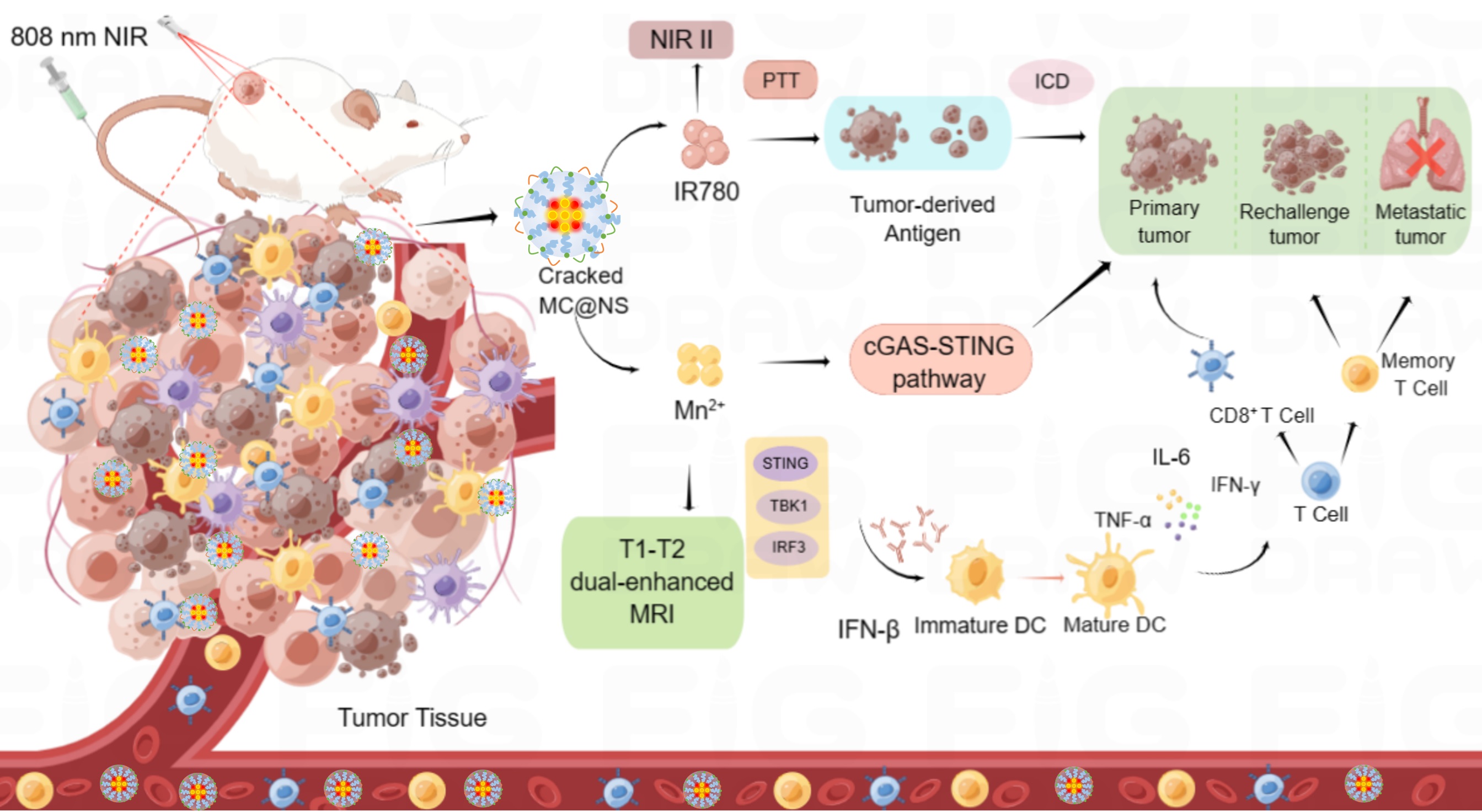
Motivation: Breast cancer exhibits high incidence and mortality rates. We reports a functional nanosystem composed of Pluronic F127, manganese chloride (MnCl2), and IR780 dye. The nanosystem possesses multimodal imaging capabilities using near-infrared II (NIR-II) and T1-T2 dual-enhanced MRI, guiding photothermal therapy, and enhancing the STING pathway to combat triple-negative breast cancer.
Goal(s): Effectively suppress TNBC growth and metastasis, with good biocompatibility..
Approach: MC@NS nanosystem could diagnose the tumor and confirm the time window of treatment via NIR II/MRI dual-modal imaging, and effectively suppress tumor growth thought phototherapy, STING pathway and anti-tumor immunity.
Results: It effectively suppress tumor growth thought phototherapy, STING pathway and immunotherapy.
Impact: This nanosystem, through T1-T2 dual-enhanced MRI and NIR II multimodal imaging, enabled tumor diagnosis and guided photothermal therapy, effectively suppressing tumor growth by combining photothermal therapy and STING pathway to enhance immunogenic cell death, providing a novel theranostic strategy.
Breast cancer has become the highest incidence tumor in the world, and it is an urgent to develop new diagnostic and therapeutic approaches to treat breast cancer[1, 2]. Our study reports a functional nanosystem that can diagnose tumors and confirm the optimal time-window for laser radiation via T1-T2 dual-enhanced MRI and NIR II multimodal imaging, and effectively suppress tumor growth and metastasis through phototherapy, STING pathway and anti-tumor immunity.
METHODS:
Preparation and characterization of MnCO-based nanosystem (MC@NS)
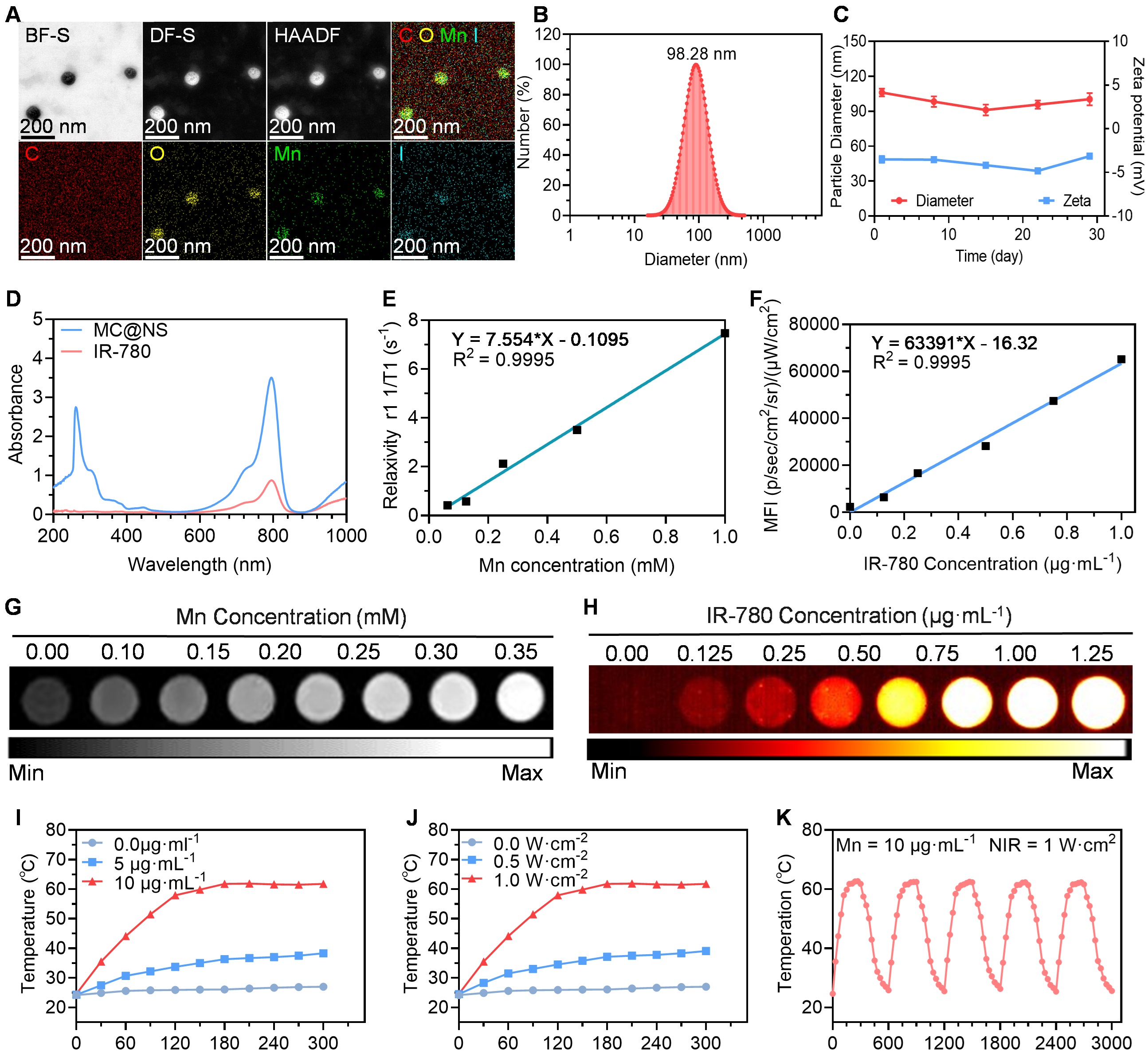
Firstly, the MnCl2-based nanosystem (MC@NS) was obtained though ultrasonic self-assembly of pluronic F127, MnCl2 and IR780 dyes. The site, morphology and the zeta potential of MC@NS was characterized by transmission electron microscopy (TEM) and dynamic light scattering (DLS)[3]. Secondly, the encapsulation of IR780 within the material was confirmed through ultraviolet spectroscopy. Subsequently, the imaging property of MC@NS assessed by a second near-infrared imager and a 9.4 T MRI system respectively[4]. To evaluate the photothermal effect, MC@NS at different concentrations and laser powers were exposed to continuous laser for 5 minutes[5].
Biocompatibility, phototherapy and STING pathway in vitro
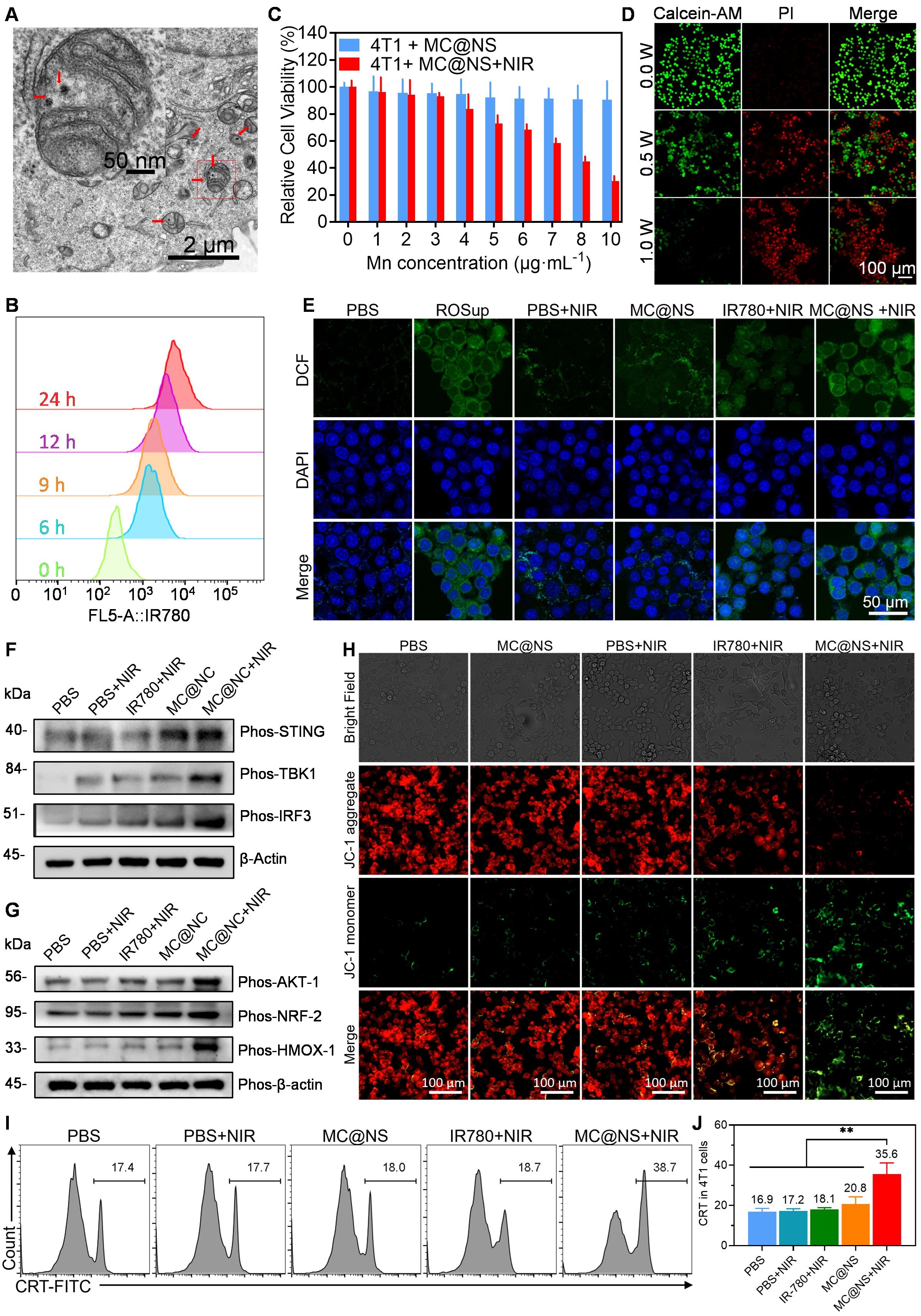
The uptake level of MC@NS at various time points in 4T1 cells was assessed by flow cytometry (FCM). The photothermal effect at the cellular level was performed by CCK-8 assay and calcien-AM with propidium iodide staining. The ROS level with different treatments was measured by DCF staining. The intracellular expression levels of the STING pathway and AKT signaling pathway were measured by western blot[6]. Furthermore, the JC-1 dye, which indicates mitochondrial membrane potential, was stained and observed under a fluorescence microscope. CRT was stained and analyzed through flow FCM.
MRI-NIRF dual-modality imaging in vivo
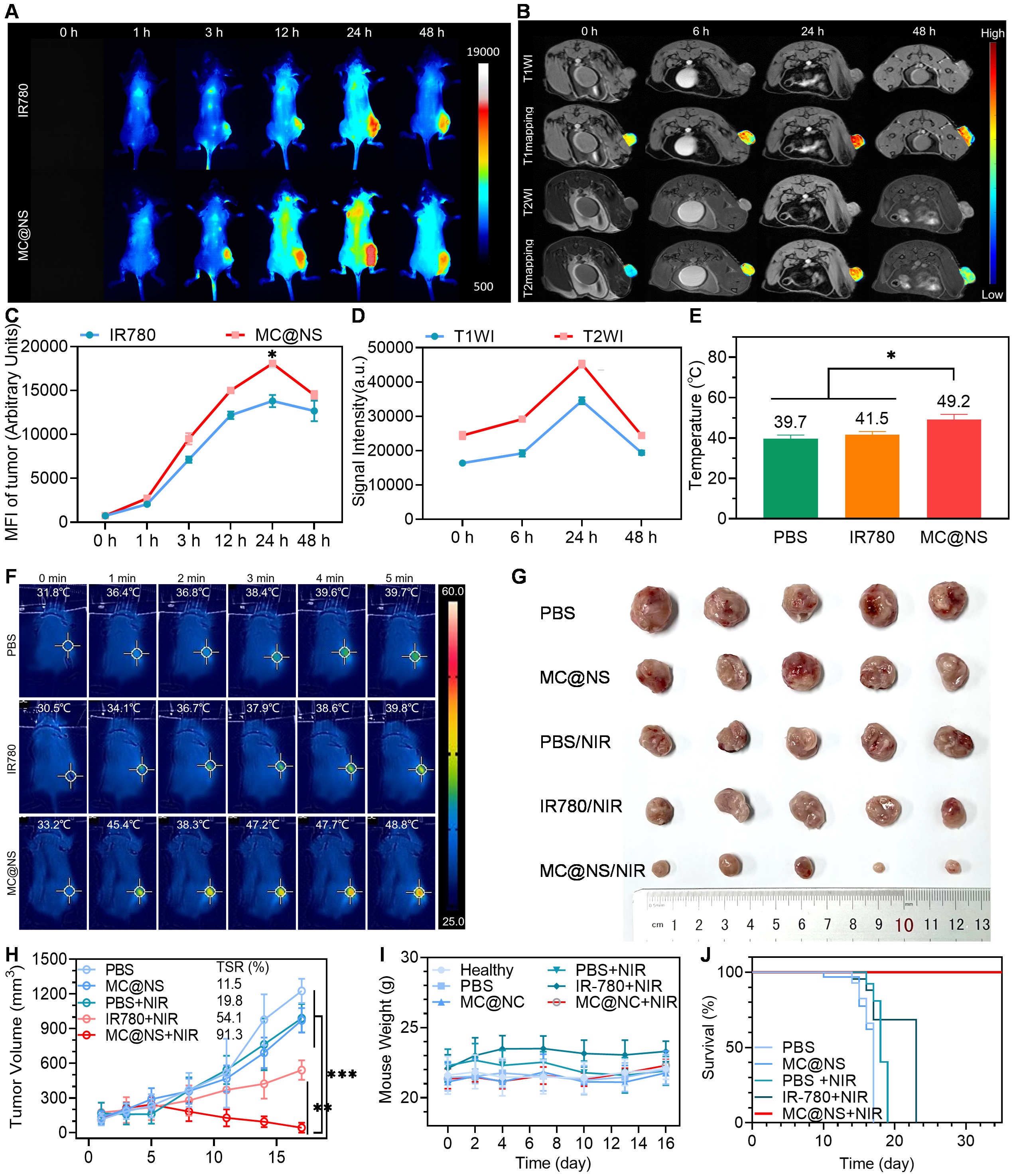
Breast cancer model was established via Balb/c mouse (female, 6 weeks old) injected 4T1 cells. When the tumor volume reached to 800 mm3 approximately, the tumor-bearing mice were randomly divided into three groups (free IR780 and MC@NS, n=3/group) and underwent NIRF II imaging. MR imaging in vivo (n=3) was performed on a 9.4 T MR system (Bruker, Gremany). The T1-weighted images, T2-weighted images and mapping of tumors were obtained (0 h, 6 h, 24 h, 48 h). Parameters of T1WI, T2WI, T1 mapping and T2 mapping were as follow successively: TR/TE: 300/3.0 ms, TR/TE: 2000/20 ms, TR/TE: 393.5-400.0-800.0-1500.0-3000.0-5500/12.0 ms and TR/TE: 2200/7.5-15.0-22.5-30.0-37.5-45.0-52.5-60.0-67.5-75-82.5-90 ms, FOV: 60 mm, slice thickness: 0.7 mm.
Anti-tumor Immunity synergistic therapy in vivo
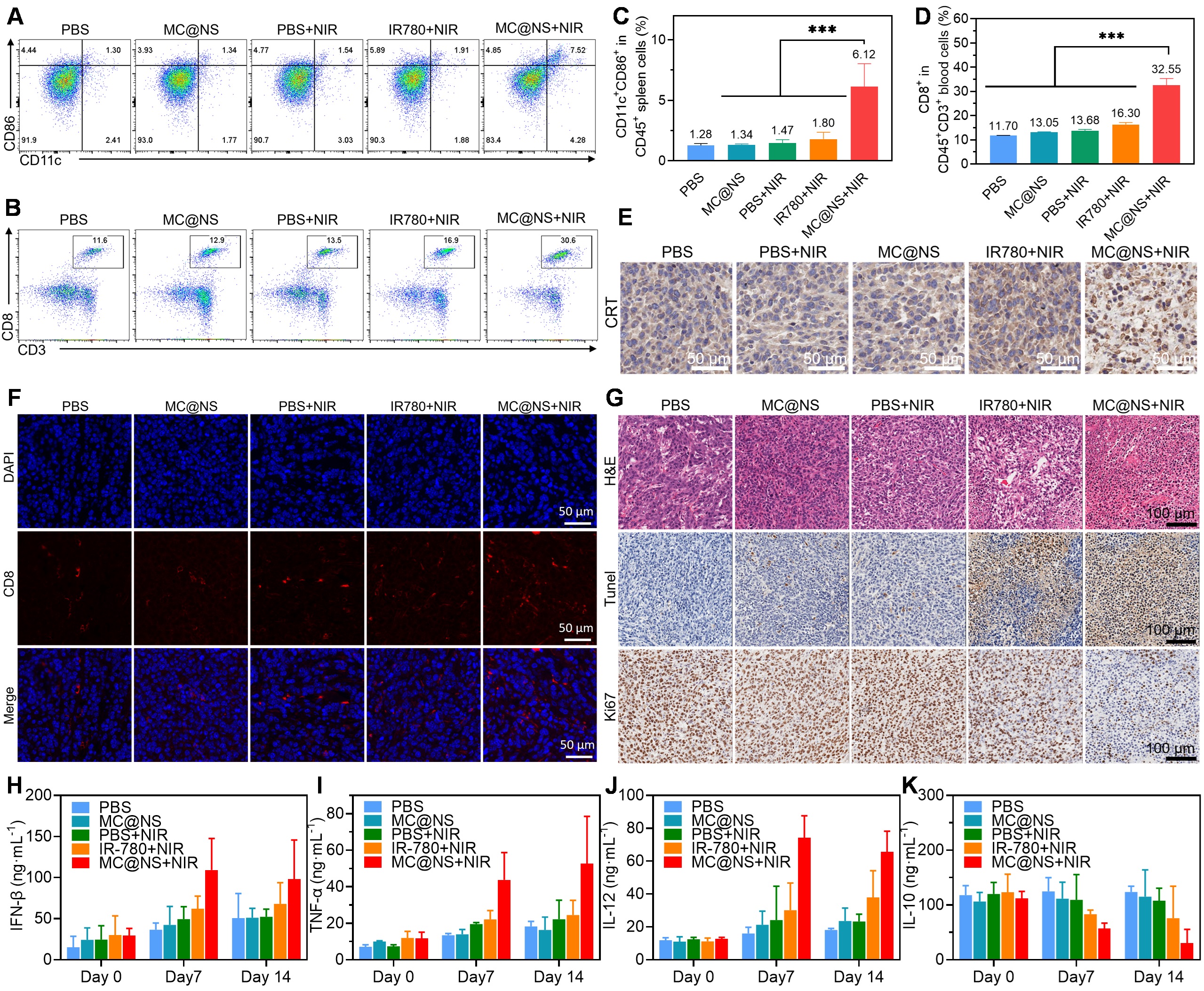
Firstly, the mice were classified as five groups randomly (n=5) as the tumors had reached approximately 300 mm3: ①PBS, ②MC@NS, ③PBS+NIR, ④IR780+NIR, ⑤MC@NS+ NIR. The tumor volume and mouse body weight were measured every two days. Secondly, dendritic cells (CD45+CD11c+CD80+CD86+), cytotoxic T cells (CD45+CD3+CD8+) in spleens or blood of mice with different treatments on the 7th days. The level of IFN-β, TNF-α, IL-12 and IL-10 were measured by enzyme-linked immunosorbent assay. And the tumors and vital organs were collected on the 14th days and stained with CRT, CD8, hematoxylin-eosin, Ki67 and TUNEL.
All statistical analyses were performed using the software GraphPad Prism 8.0 (*p < 0.05, **p < 0.01).
RESULTS
Firstly, MC@NS was confirmed to have a core-shell morphology via the TEM, and elemental composition was verified through mapping. Dynamic light scattering (DLS) revealed a particle size of approximately 98 nm and a stable zeta potential (Figure 1A-C). Secondly, the ultraviolet spectroscopy further confirms the encapsulation of IR780 within the nanosystem (Figure 1D). Next, it has excellent cellular uptake capacity and photothermic effect in 4T1 cells (Figure 2A&B, in vitro Figure 1I-K, 2C&D and in vivo Figure 3D). Based on MC@NS phototherapy can generate increased levels of reactive oxygen species (ROS) (Figure 2E) and activate the STING pathway (Figure 2F). Interestingly, remarkable NIR II and T1-T2 dual-enhanced MRI imaging potential of MC@NS were demonstrated in vitro (Figure 1E-H) and in vivo (Figure 3A&B), which appropriately guides laser-induced photothermal therapy. Based on MC@NS phototherapy, the temperature of mouse tumors can be raised to approximately 48℃ (Figure 3E&F), effectively suppressing tumor growth (Figure 3G-J). Together, all of these activated immunotherapy due to MC@NS-based phototherapy and STING pathway[7, 8], and the antitumor immune cells (DCs, CD8+ CLT cells) were upregulated along (Figure 4A-D, 4F&G)[9]. Meanwhile, The level of anti-tumor cytokines (IFN-β, TNF-α, IL-12) was increased and promo-tumor cytokines (IL-10) were decreased (Figure 4H-K)[10].
CONCLUSION
We developed a multifunctional MC@NS nanosystem, which could diagnose the tumor and confirm the time window of treatment via NIR II/MRI dual-modal imaging, and effectively suppress tumor growth thought phototherapy, STING pathway and immunotherapy, providing a promising strategy for cancer theranostic applications.
Acknowledgements
This work was supported by the National Natural Science Foundation of China (81971574, 82271938, 82371908,), the Natural Science Foundation of Guangdong Province (2021A1515011350), the GuangDong Basic and Applied Basic Research Foundation (2021A1515220060, 2022A1515110792), the Science and Technology Project of Guangzhou (202102010025) , the Special Fund for the Construction of High-level Key Clinical Specialty (Medical Imaging) in Guangzhou, Guangzhou Key Laboratory of Molecular Imaging and Clinical Translational Medicine (202201020376).References
[1]. Rebecca L. Siegel MPH Kimberly D. Miller MPH Nikita Sandeep Wagle MBBS, M.P.A.J., Cancer statistics, 2023. CA: A Cancer Journal for Clinicians, 2023: p. 17-48.
[2]. Sung, H., et al., Global Cancer Statistics 2020: GLOBOCAN Estimates of Incidence and Mortality Worldwide for 36 Cancers in 185 Countries. CA: a cancer journal for clinicians, 2021. 71(3): p. 209-249.
[3]. Wu, D., et al., Mesoporous Polydopamine Carrying Manganese Carbonyl Responds to Tumor Microenvironment for Multimodal Imaging‐Guided Cancer Therapy. Advanced functional materials, 2019. 29(16): p. 1900095-n/a.
[4]. Guan, X., et al., Iron oxide-based enzyme mimic nanosystem for dual-modality imaging guided chemical phototherapy and anti-tumor immunity against immune cold triple-negative breast cancer. Chemical engineering journal (Lausanne, Switzerland : 1996), 2021. 425: p. 130579.
[5]. Peng, J., et al., Photosensitizer Micelles Together with IDO Inhibitor Enhance Cancer Photothermal Therapy and Immunotherapy. Adv Sci (Weinh), 2018. 5(5): p. 1700891.
[6]. Sun, X., et al., Amplifying STING activation by cyclic dinucleotide-manganese particles for local and systemic cancer metalloimmunotherapy. Nature nanotechnology, 2021. 16(11): p. 1260-1270.
[7]. Chu, K.F. and D.E. Dupuy, Thermal ablation of tumours: biological mechanisms and advances in therapy. Nature reviews. Cancer, 2014. 14(3): p. 199-208.
[8]. Shengjun Xu, S.L.Q.S., Self-Activated Cascade-Responsive Sorafenib and USP22 shRNA Co-Delivery System for Synergetic Hepatocellular Carcinoma Therapy. Advanced Science, 2021. 2003042(8): p. 1-15.
[9]. Yan, J., et al., Engineering Radiosensitizer-Based Metal-Phenolic Networks Potentiate STING Pathway Activation for Advanced Radiotherapy. Adv Mater, 2022. 34(10): p. e2105783.
[10]. Li, X., et al., Cancer immunotherapy based on image-guided STING activation by nucleotide nanocomplex-decorated ultrasound microbubbles. Nat Nanotechnol, 2022. 17(8): p. 891-899.
Figures