0166
Multi-Modal Protein-Engineered Theranostic Fibers: Drug encapsulation, Imaging, and enhanced 19F MRS1Department of Chemical and Biomolecular Engineering, NYU Tandon, Brooklyn, NY, United States, 2New York University, Brooklyn, NY, United States, 3Department of Radiology, NYU Grossman School of Medicine, New York, NY, United States, 4Flatiron Institute - Simons Foundation, New York City, NY, United States, 5Computer Science Department, NYU Courant, New York City, NY, United States, 6Center for Genomics and Systems Biology, NYU, New York, NY, United States, 7Chemistry, NYU, New York, NY, United States, 8Department of Biomaterials, NYU College of Dentistry, New York, NY, United States
Synopsis
Keywords: Probes & Targets, Multimodal, theranostic, temperature probe, fluorine
Motivation: Theranostic materials allows for simultaneous therapeutic and diagnostic disease intervention.
Goal(s): We aimed to engineer a protein-based theranostic with multiple imaging modalities.
Approach: Using noncannonical amino acid incorporation of trifluoroleucine (TFL), we synthesize fluorinated coiled-coil, Q2TFL, imageable by 1H MRI and high-frequency ultrasound, and sensitive to 19F MRS.
Results: Q2TFL demonstrates reduced signal contrast in 1H MRI, echogenic signals under high-frequency ultrasound, and enhances sensitivity in linear ratiometric 19F MRS. This allows for thermoresponsiveness and potential protein conformation analysis. Q2TFL serves as a promising platform for versatile and effective theranostic agents, bringing together therapeutic and diagnostic modalities in a compact and efficient manner.
Impact: Q2TFL, a fluorinated protein fiber, enables drug delivery and offers unique multimodal imaging. It acts as a temperature probe and protein structure monitor, paving the way for innovative theranostic biomaterials.
Introduction
Theranostic agents represent a growing field in biomedicine that help to overcome limitations in biomaterials providing therapy and diagnosis of diseases 1. These materials help to monitor the development of disease after therapeutic treatment as well as provide a simultaneous diagnosis and treatment of a disease1. To create an ideal theranostic biomaterial, without compromising drug encapsulation, diagnostic imaging must be optimized for improved detection2. One such method to improve this specificity is the incorporation of fluorine into biomaterials3. Since fluorine is largely absent from organisms, yet exists in 100% natural abundance, it is useful as a contrast agent due to its specific signal in 19F magnetic resonance spectroscopy (MRS) 4. We develop a protein-based fluorinated self-assembling fiber, Q2TFL as a theranostic agent capable of 19F MRS using noncanonical amino acid incorporation of trifluoroleucine (TFL). We demonstrate that Q2TFL has increased sensitivity for 19F MRS, and increased thermostability compared to previous constructs and can encapsulate the hydrophobic small molecule, curcumin (CCM), which provides further stabilization. Furthermore, we show that Q2TFL may be used in vivo as a visible fiber assembly via 1H magnetic resonance imaging (MRI) and high-frequency ultrasound as well as a sensitive biomaterial using 19F MRS. Interestingly, we show that Q2TFL possesses a ratiometric 19F MRS signal proportional to its protein structure and environmental temperature indicating its potential as a multifunctional in vivo probe.Methods
Protein biosynthesis and characterization. Q2TFL and QTFL were expressed as described previously 5 via NCAA incorporation of pqE30/Q and pQE60/Q2 plasmids using electrically competent LAM1000 E. coli cells6. Protein was purified using affinity chromatography on a cobalt-charged HiTrap IMAC FF 5 mL column. Q2TFL and QTFL protein were assessed for secondary structure via circular dichroism and attenuated total reflectance-fourier transform infrared spectroscopy. Transmission electron microscopy was used to confirm nanofiber assembly and matrix-assisted laser desorption/ionization-time of flight mass spectrometry was used to assess percentage of TFL incorporation. Protein binding was assessed using fluorescence and confocal microscopy.19F NMR. 19F detection was studied using a Bruker AVIII-500 (11.7 T) NMR instrument equipped with a broadband BB(F)O CryoProbe using one-pulse sequence was used to acquire the 19F signal with a spectral width 113,636.4 Hz corresponding to 241.5 ppm, 0.577 s acquisition time, and 256 scans.
Phantom and In Vivo MRI. MRI and MRS were performed on a Biospec 70/30 micro-MRI system (Bruker – Billerica MA, USA) equipped with zero helium boil-off 300 mm horizontal bore 7-T superconducting magnet (300 MHz) based on ultra-shield refrigerated magnet technology. The magnet was interfaced to an actively shielded gradient coil insert (Bruker BGA-12S-HP; OD=198-mm, ID=114-mm, 660-mT/m gradient strength, 130-μs rise time) and powered by high-performance gradient amplifier (IECO, Helsinki – Finland) operating at 300A/500V. The MRI/MRSR setup utilized in this study involved in-house design of two distinct radiofrequency resonators through mutual inductance enabling dual 1H/19F resonances for scanning a mouse body.
Results and Discussion
Q2TFL possessed 95% incorporation of TFL and exhibited strong α-helical character and nanofiber assembly which was strengthened by its ability to bind small hydrophobic molecule, CCM. Signal-to-noise ratios of triplet peaks by 19F NMR demonstrated a proportional relationship to protein TFL content. Q2TFL was assessed for temperature dependence by altering the environmental temperature in NMR where Q2TFL exhibited an increase in SNR with an increase in temperature (Figure 1a-c). A linear temperature dependence between the ratios of NMR peak SNRs ( SNRT) demonstrating the potential to use Q2TFL as an environmental temperature and protein conformation probe (Figure 1d-f). Finally, Q2TFL was assessed in vivo using ultrasound (US)-guided intra-articular injection in C57Bl6 mice. Q2TFL demonstrated the ability for bimodal mapping through echogenicity for high-frequency US visualization, and T2-darkening MRI contrast relative to the surround tissue, while also being traceable by 19F MRS in vivo (Figure 2).Conclusion
Q2TFL forms fibers on the nano- to mesoscale and possesses improved thermostability and SNR compared to our previously fluorinated fiber construct ref), QTFL, demonstrating its ability for 19F MRS detection, and is capable of CCM encapsulation highlighting its therapeutic potential. Importantly, processing of TFL triplet behavior in Q2TFL allows potentially for additional function as a temperature probe and monitoring relative protein structure of the agent. Finally, we demonstrate the ability of Q2TFL to provide multimodal contrast both in 1H MRI and high frequency ultrasound with sensitive traceability by 19F MRS in vivo. These results provide important criteria towards fluorination of coiled-coils for supramolecular assembly and design towards 19F MRS theranostic agents; and provide a foundation for future in vivo investigations in this area and ultimate extension to 18F PET imaging.Acknowledgements
This work was supported by NSF-DMREF under Award Number DMR 1728858. ATR-FTIR experiments were performed at the NYU Chemistry Department Shared Instrument Facility. The facility is supported by the National Science Foundation (NSF) Chemistry Research Instrumentation and Facilities Program (CHE-0840277) and Materials Research Science and Engineering Center (MRSEC) Program (DMR-1420073). Part of this work was performed at the NYU Grossman School Medicine Preclinical Imaging Laboratory, a shared resource partially supported by the NIH/SIG 1S10OD018337-01, the Laura and Isaac Perlmutter Cancer Center Support Grant, NIH/NCI 5P30CA016087, and the NIBIB Biomedical Technology Resource Center Grant NIH P41 EB017183. This work was partially supported by the NYU Shifrin Myers Breast Cancer Discovery Fund (SMBCDF).
References
1. Jeyamogan, S.; Khan, N. A.; Siddiqui, R. Application and Importance of Theranostics in the Diagnosis and Treatment of Cancer. Archives of Medical Research 2021, 52 (2), 131-142. DOI: https://doi.org/10.1016/j.arcmed.2020.10.016.
2. Eggeling, C. Advances in bioimaging—challenges and potentials. Journal of Physics D: Applied Physics 2018, 51 (4), 040201. DOI: 10.1088/1361-6463/aaa259.
3. Monkovic, J. M.; Gibson, H.; Sun, J. W.; Montclare, J. K. Fluorinated Protein and Peptide Materials for Biomedical Applications. In Pharmaceuticals, 2022; Vol. 15.
4. Marsh, E. N. G.; Suzuki, Y. Using 19F NMR to Probe Biological Interactions of Proteins and Peptides. ACS Chemical Biology 2014, 9 (6), 1242-1250. DOI: 10.1021/cb500111u.
5. More, H. T.; Zhang, K. S.; Srivastava, N.; Frezzo, J. A.; Montclare, J. K. Influence of fluorination on protein-engineered coiled-coil fibers. Biomacromolecules 2015, 16 (4), 1210-1217. DOI: 10.1021/bm5019062 From NLM.
6. Montclare, J. K.; Son, S.; Clark, G. A.; Kumar, K.; Tirrell, D. A. Biosynthesis and Stability of Coiled-Coil Peptides Containing (2S,4R)-5,5,5-Trifluoroleucine and (2S,4S)-5,5,5-Trifluoroleucine. ChemBioChem 2009, 10 (1), 84-86, https://doi.org/10.1002/cbic.200800164. DOI: https://doi.org/10.1002/cbic.200800164 (acccessed 2022/10/25).
Figures
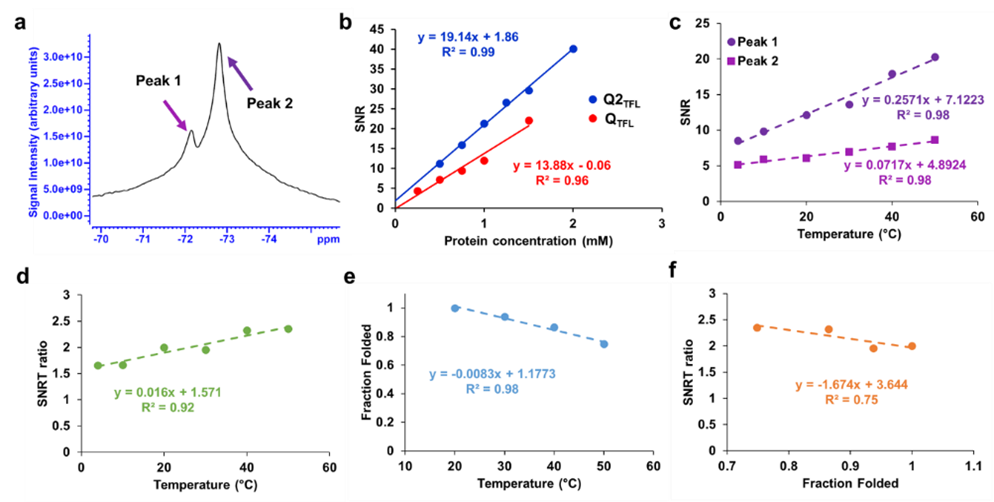
Figure 1 a. NMR spectrum at 500 MHz of Q2TFL at 1.5 mM showing two peaks b. SNR of Q2TFL and QTFL as a function of protein concentration c. Temperature dependence of SNR from independent peaks. d. Linear correlation of temperature with SNRT ratio showing ability to predict temperature. e. Linear correlation of temperature with average (n=3) fraction folded of Q2TFL f. Linear correlation of average fraction folded with SNRT ratio showing ability to predict relative structure.
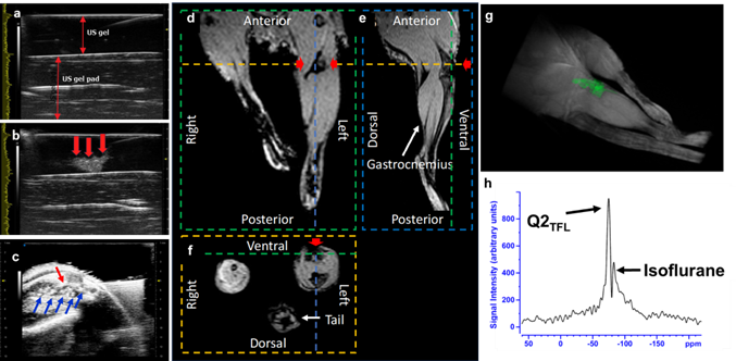
Figure 2. a. The B-mode image of the ultrasound gel b. and after pipetting the Q2TFL (red arrows) into the US gel. c. Ultrasound guided syringe (red arrows) injection imaging of Q2TFL (blue arrows) into left hind leg. d-f. The 3D 1H MRI dataset covering the lower region of the mouse’s body showing hypointense Q2TFL fiber signal (red arrows). g. 3D rendering showcasing the distribution of the Q2TFL fibers h. 19F MR spectroscopy performed in vivo after injection of Q2TFL.