0161
OPM-MEG/ULF-MRI Hybrid System: towards acquisition of both MR image and neural magnetic field1Department of Electrical Engineering, Kyoto University, Kyoto, Japan, 2Hamamatsu Photonics K.K., Hamamatsu, Japan
Synopsis
Keywords: Hybrid & Novel Systems Technology, Low-Field MRI
Motivation: To overcome the limitations of signal co-registration in MEG, we constructed MEG/MRI hybrid system.
Goal(s): To demonstrate feasibility of this system. High-sensitivity OPMs in a reasonable magnetic shield with MRI system.
Approach: Phantom experiments. Phantom includes triangle coil and MEG measures its magnetic field. MRI scanned this phantom with 3D-SE sequence.
Results: We recognized signal peak generated by 100 nAm current dipole moment in amplitude spectrum density visually. We also confirmed the structure of the phantom in 3D MR images.
Impact: We made MEG/MRI hybrid system for the purpose of accurate signal co-registration between them. MEG employed scalar-mode OPM, and its noise level was 367 fT/rHz. We scanned phantom using 7-mT MRI scanner and confirmed its structure.
INTRODUCTION
On the magnetoencephalography (MEG), there is limitation related to co-registration between anatomical images and calculated position of signal source of neural magnetic field1–5. In general, it is difficult to obtain MEG and MR images simultaneously, because MEG can be acquired in magnetic shield room and MR scanners employ high-field static magnetic field. In this study, we tried to realize acquisition of both data with the MEG/MRI hybrid system employing ultra-low-field (ULF) MRI scanners and scalar-mode optically pumped magnetometer (OPM)6 at the same experimental system.On the ULF-MRI scanner, Mathieu Sarracanie et al. reported that 6.5-mT MRI system was able to visualize brain image7 and ULF-MRI scanners whose static magnetic field is several mT have potential to provide anatomical images enough to recognize brain regions. On the other hand, the scalar-mode OPM can measure small magnetic field as the Larmor frequency change of vaporized alkali metal. There are reports that can measure magnetocardiography without magnetic shields6. However, the system integration of OPM-MEG and ULF-MRI have not been reported. Therefore, we constructed this hybrid system and demonstrated its feasibility through measurements of magnetic field generated by triangle coil and obtain MR images.
METHODS
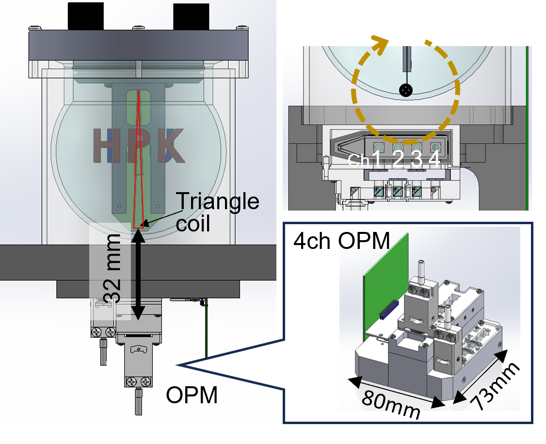
Shields: This system is surrounded by electromagnetic shield made of aluminum and EMS panels on all six side. The EMS panels can attenuate the geomagnetic field to roughly one fifth. Compared to the normal magnetic shield, EMS panels has low cost, even though the attenuation rate is lower.MEG: To investigate the sensitivity of scalar-mode, we measured sinusoidal magnetic field in the MEG/MRI hybrid system when the static magnetic field of MRI was off. The triangle coil illustrated in Figure 1 is the source of measurement target and we employed home-made 4-ch scalar-OPM module. The magnitudes of current dipole moment generated in the triangle coil were 10, 20 ,50 100, 250, and 1000 nAm, respectively. We also employed the gradiometers between Ch.2 and Ch.3. The signal frequency was 10 Hz and on data acquisition, the sampling rate was 180 Sa/s.
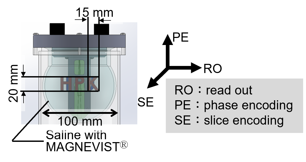
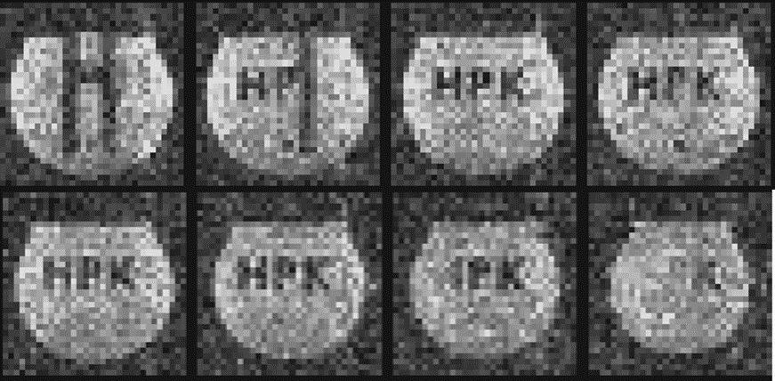
MRI: The homemade MRI system has biplanar coil sets including 7.0 mT static magnetic field and gradient systems. We scanned the phantom illustrated in Figure 2 employing 3D-SE sequence. On the scanning parameters, TR/TE, flip angle, bandwidth, and number of excitation (NEX) were 300 ms/ 25 ms, 90 degree, 3.2 kHz and 8, respectively.
The voxel size was 3 mm isocubic and its matrix size was 32x32x32.
RESULTS
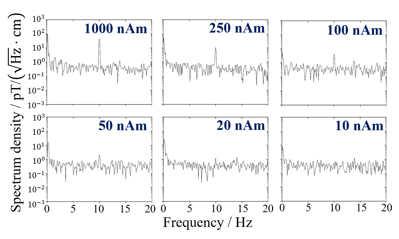
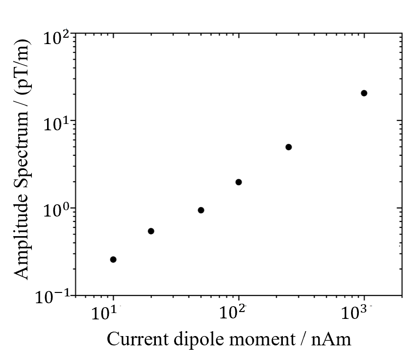
MEG: We showed the amplitude spectrum density of measurements in each current dipole moments in Figure 3. The peak at 10 Hz could be observed obliviously in case of 100, 250, and 1000 nAm. We also plotted the relationship between current dipole moment and response of gradiometer in Figure 4. The noise level was 367 fT/rHz/cm.MRI: We showed MR image of the phantom along the slice encoding in Figure 5. Even though the signal-to-noise ratio (SNR) of MR image is low due to the ultra-low static magnetic field, the structure of the phantom can be confirmed visually. Due to the external magnetic noise, there is bright points in MR images at the same position.
DISCUSSION
Our MEG system was able to measure tens nAm current dipole moment at 32 mm far from the signal source in presence of MRI system. Even though improvement of sensitivity is preferable in preparation for measurement of neural magnetic field, we were able to demonstrate the feasibility of scalar-mode OPM with simple magnetic shield and as magnetometer or gradiometer in MRG/MRI hybrid system.The MRI system can visualize the tomographic image of the phantom using spin-echo sequence. We were able to confirm the structure of it visually despite lower SNR than medical MR images, which expected to contribute to signal co-registration in the anatomical brain region. On the other hand, there is some unexpected bright points in MR images. The position of them is stable, which means that there is a noise at the same frequency within the frequency bandwidth.
CONCLUSION
We demonstrated the feasibility of the MEG/MRI hybrid system employing scalar-mode OPM and 7-mT ultra-low-field MRI scanner. This system enables us to register signal position obtained by the inverse problem of MEG to the anatomical MR images precisely. As the future medical application example, we expect that this system will continue to preoperative identification of epilepsy focal location. To achieve various medical applications, we plan to improve sensitivity of scalar-OPM enough to detect tens fT magnetic fields and SNR of MR images by identifying the noise sources and suppressing them.Acknowledgements
This work was partially supported by a Grant-in-Aid for Research (21H03807) from the Ministry of Education, Culture, Sports, Science, and Technology (MEXT), Japan.References
1. Chella F, Marzetti L, Stenroos M, et al. The impact of improved MEG–MRI co-co-registration on MEG connectivity analysis. Neuroimage. 2019;197(April):354-367. doi:10.1016/j.neuroimage.2019.04.0612. Whalen C, Maclin EL, Fabiani M, Gratton G. Validation of a method for coregistering scalp recording locations with 3D structural MR images. Hum Brain Mapp. 2008;29(11):1288-1301. doi:10.1002/hbm.20465
3. Adjamian P, Barnes GR, Hillebrand A, et al. Co-co-registration of magnetoencephalography with magnetic resonance imaging using bite-bar-based fiducials and surface-matching. Clin Neurophysiol. 2004;115(3):691-698. doi:10.1016/j.clinph.2003.10.023
4. Henson RN, Mattout J, Singh KD, Barnes GR, Hillebrand A, Friston K. Population-level inferences for distributed MEG source localization under multiple constraints: Application to face-evoked fields. Neuroimage. 2007;38(3):422-438. doi:10.1016/j.neuroimage.2007.07.026
5. Hillebrand A, Barnes GR. Practical constraints on estimation of source extent with MEG beamformers. Neuroimage. 2011;54(4):2732-2740. doi:10.1016/j.neuroimage.2010.10.036
6. Limes ME, Foley EL, Kornack TW, et al. Portable Magnetometry for Detection of Biomagnetism in Ambient Environments. Phys Rev Appl. 2020;14(1):11002. doi:10.1103/PhysRevApplied.14.011002
7. Sarracanie M, Lapierre CD, Salameh N, Waddington DEJ, Witzel T, Rosen MS. Low-Cost High-Performance MRI. Sci Rep. 2015;5:1-9. doi:10.1038/srep15177
Figures