0159
Cervical Spine MRI on a RF Shielding-Free 0.05T MRI Scanner1Laboratory of Biomedical Imaging and Signal Processing, The University of Hong Kong, Hong Kong, China, 2Department of Electrical and Electronic Engineering, The University of Hong Kong, Hong Kong, China
Synopsis
Keywords: Low-Field MRI, Low-Field MRI
Motivation: To develop low-cost and patient-friendly MRI scanners to address global healthcare disparities.
Goal(s): To demonstrate cervical spine (C-spine) MRI on a low-cost and RF shielding-free 0.05T MRI scanner.
Approach: Typical imaging protocols were implemented on a newly developed 0.05T MRI scanner. The scanner is compact, RF shielding-free, and acoustically quiet during scanning. Further, a deep learning electromagnetic interference (EMI) elimination method and a data-driven reconstruction strategy were designed.
Results: The deep learning EMI elimination method effectively removed EMI noise, and the data driven reconstruction method suppressed image noise and artifacts while increasing spatial resolution, leading to significantly improved image quality.
Impact: We demonstrate high-quality C-spine MRI on a low-cost and shielding-free 0.05T MRI scanner through exploiting computing power and extensive high-field MRI data. These developments will lead to a new generation of affordable, patient-centric, and computing-powered MRI scanners.
Introduction
MRI is one of the most important medical imaging modalities in modern healthcare1. However, after five decades of development, MRI accessibility remains low and highly inhomogeneous worldwide2, mainly due to the cost-prohibitive nature and specialized infrastructural requirements associated with existing high-field (1.5T or 3T) superconducting MRI scanners. Consequently, there has been a growing interest and urgency to address the unmet clinical needs and global healthcare disparities by developing ultra-low-field (ULF) MRI scanners that operate below 0.1T for low-cost or/and point-of-care imaging applications3-9. Recent ULF MRI technology developments in system engineering10-13 as well as computing14-16 have yielded promising results especially in intensive care, yet they have been confined to brain imaging.Cervical spine (C-spine) MRI serves as a key diagnostic tool to visualize cervical spine structures and identify neck abnormalities. However, C-spine MRI at ULF is challenging due to the extremely low SNR associated with the substantial field reduction. In this study, using a newly developed 0.05T MRI scanner, together with a novel deep learning EMI elimination method17, we have conducted preliminary feasibility study of RF shielding-free 0.05T C-spine MRI. Further, we have demonstrated that 0.05T C-spine image quality can be significantly advanced through a new deep learning 3D reconstruction strategy18.
Methods
RF Shielding-free 0.05T MRI ScannerC-spine MRI experiments were conducted on a home-built shielding-free 0.05T MRI scanner. Using a similar design to our recently developed 0.055T brain MRI scanner13, it operates using a standard AC power outlet and is low cost to build. A C-spine coil with diameter of 20cm was used. Additionally, ten EMI sensing coils with diameter of 5cm were placed around scanner and inside electronic cabinet and used to detect EMI signals from both external environment and internal scanner electronics during scanning13,19.
Data Acquisition
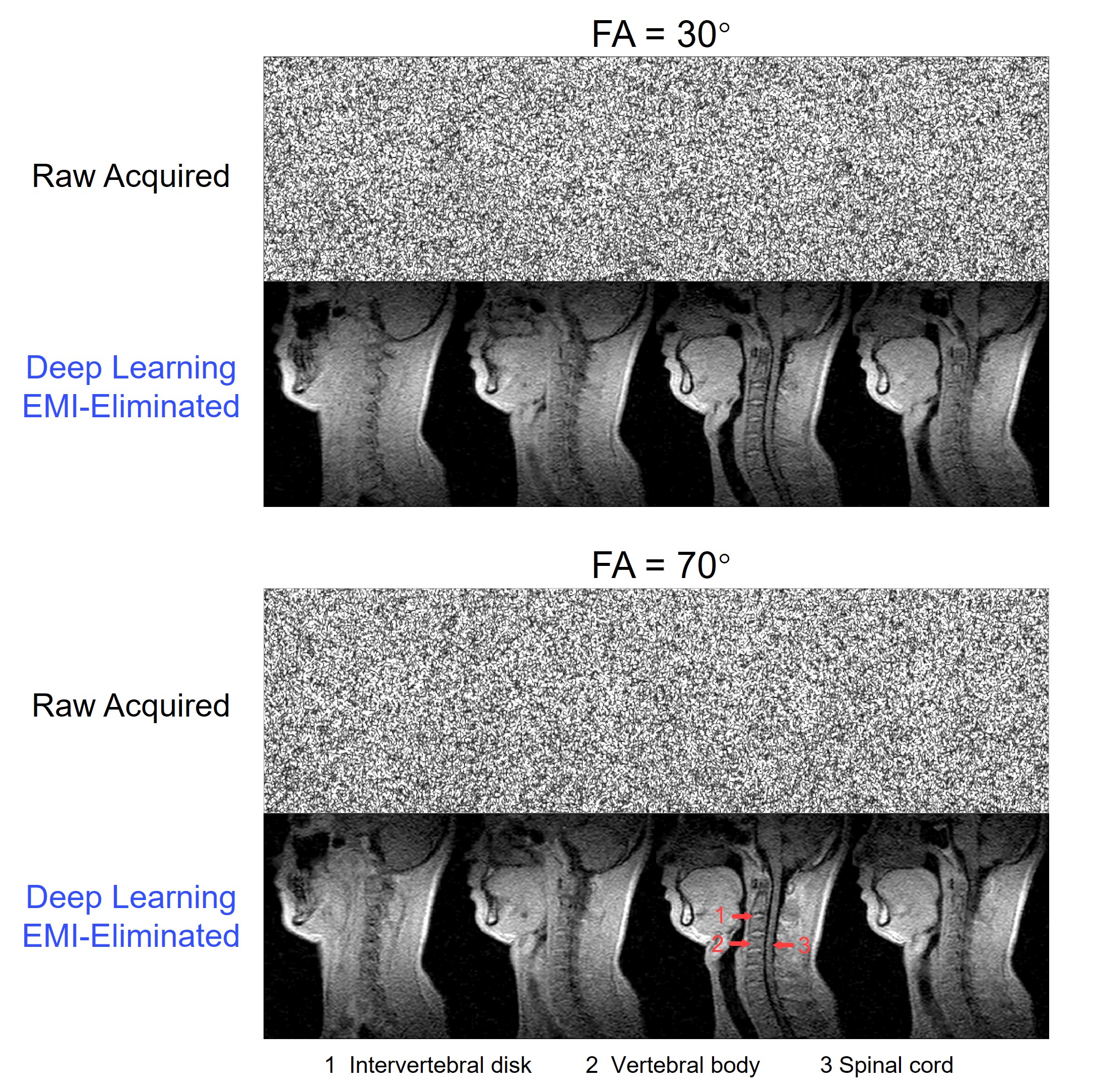
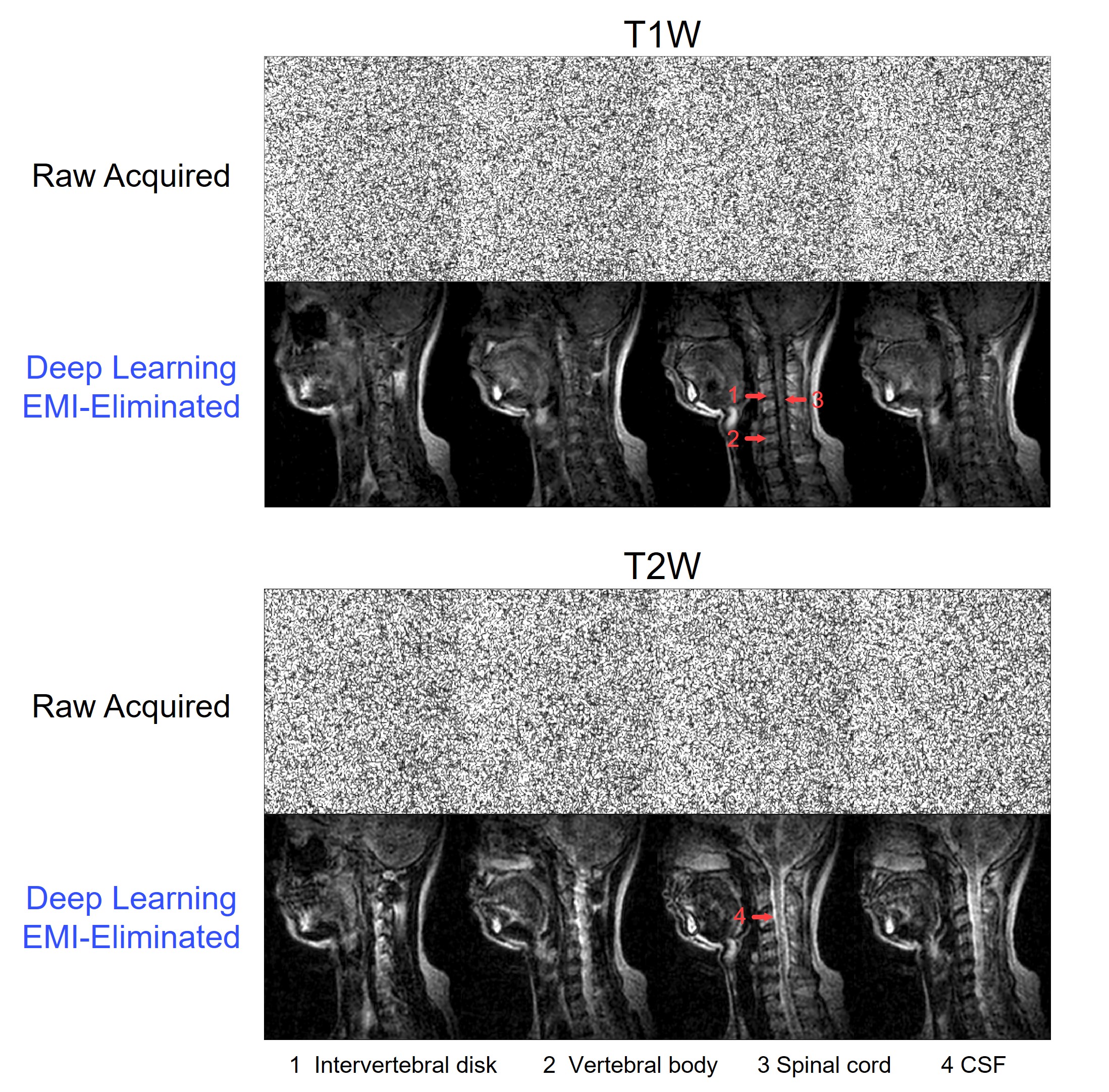
T1W and T2W C-spine datasets were acquired using cartesian 3D GRE (T1W: TR/TE = 48ms/5.7ms, and FA = 30° or 70°) and 3D FSE (T1W: TR/TE/ETL = 210ms/76ms/9; T2W: TR/TE/ETL = 2300ms/136ms/25) sequences. Data acquisition adopted k-space partial Fourier sampling in frequency encoding direction for 3D GRE sequence, while in one or two phase encoding directions for 3D FSE sequences. For each protocol, scan time was kept at around 8 mins, and the sequence parameters were optimized for image SNR and contrasts. All datasets were acquired from normal adult subjects.
Deep Learning EMI Elimination
After each scan, EMI elimination was performed using a novel deep learning method17, term Deep-DSP, recently developed by our group. Unlike existing methods, Deep-DSP directly predicts EMI-free MR signals from signals simultaneously acquired by MRI receive coil and EMI sensing coils, enabling effective EMI elimination by exploiting electromagnetic coupling among coils as well as typical MR signal characteristics.
Deep Learning Image Reconstruction
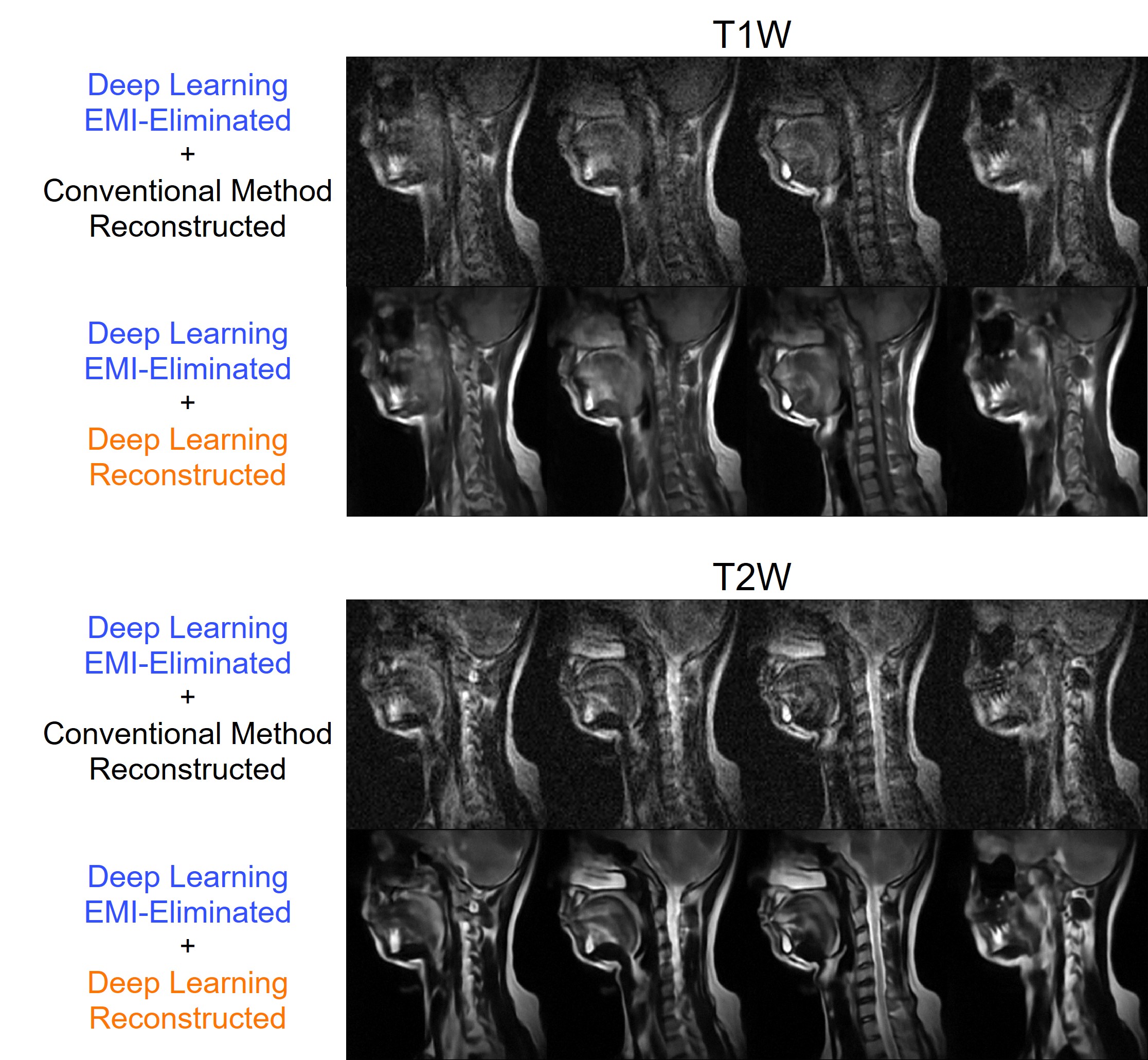
To further advance the image quality, image reconstruction was performed through our newly developed deep learning 3D reconstruction strategy18. This new data-driven strategy integrates partial Fourier image reconstruction and super-resolution, leading to substantial suppression of artifacts and noise while increasing spatial resolution.
Results
Figs. 1 and 2 present typical T1W and T2W C-spine imaging results acquired with 3D GRE and 3D FSE sequences. In the absence of RF shielding-cage, C-spine images were completely immersed within strong EMI noise. However, Deep-DSP largely removed the EMI noise. Intervertebral disk and body, together with spinal cord and CSF inside spinal canal, can be identified on the EMI-eliminated images. Fig. 3 shows the C-spine T1W and T2W images reconstructed using conventional Fourier method vs. our deep learning reconstruction method. Our deep learning image reconstruction and super-resolution method substantially advanced image quality, leading to improved visualization of fine anatomical structures.Discussion and Conclusions
ULF MRI offers several advantages, including open scanning environment, low acoustic noise levels during scanning, low sensitivity to metallic implants, less image susceptibility artifacts at air/tissue interfaces, and extremely low RF specific absorption rate. However, ULF MRI is inherently challenged by the low SNR due to the substantial field reduction. In this study, we succeed in demonstrating the potential of high-quality 0.05T C-spine MRI through our new deep learning EMI elimination and 3D reconstruction methods. With these advances, we anticipate a new class of affordable, simple and computing-powered MRI scanners, addressing unmet clinical needs in various healthcare applications globally.Further studies can be carried out in two directions. First, current protocols will be further optimized to improve robustness to motion artifacts (e.g., by using radial sampling). Second, RF shielding-free 0.05T C-spine MRI of a large cohort of patients will be conducted, with direct comparisons with clinical 1.5T or 3T results, to demonstrate the feasibility of 0.05T C-spine MRI in producing diagnostically useful information.
Acknowledgements
This work was supported in part by Hong Kong Research Grant Council (R7003-19F, HKU17112120, HKU17127121, HKU17127022 and HKU17127523 to E.X.W).References
[1] Fuchs VR, Sox Jr HC. Physicians’ views of the relative importance of thirty medical innovations. Health Aff 2001;20(5):30-42.
[2] Magnetic resonance imaging (MRI) units (indicator). OECD; 2022.
[3] Cho A. MRI for all. Science 2023;379(6634):748-751.
[4] Webb A, Obungoloch J. Five steps to make MRI scanners more affordable to the world. Nature 2023;615(7952):391-393.
[5] Naddaf M. One MRI for 4.7 million people: the battle to treat Syria's earthquake survivors. Nature 2023;615(7951):193-194.
[6] Kimberly WT, Sorby-Adams AJ, Webb AG, Wu EX, Beekman R, Bowry R, Schiff SJ, de Havenon A, Shen FX, Sze G, Schaefer P, Iglesias JE, Rosen MS, Sheth KN. Brain imaging with portable low-field MRI. Nat Rev Bioeng 2023;1(9):617-630.
[7] Hennig J. An evolution of low-field strength MRI. Magma 2023;36(3):335-346.
[8] Salameh N, Lurie DJ, Ipek Ö, Cooley CZ, Campbell-Washburn AE. Exploring the foothills: benefits below 1 Tesla? Magma 2023;36(3):329-333.
[9] Wald LL, McDaniel PC, Witzel T, Stockmann JP, Cooley CZ. Low‐cost and portable MRI. J Magn Reson Imaging 2020;52(3):686-696.
[10] Cooley CZ, McDaniel PC, Stockmann JP, Srinivas SA, Cauley SF, Śliwiak M, Sappo CR, Vaughn CF, Guerin B, Rosen MS, Lev MH, Wald LL. A portable scanner for magnetic resonance imaging of the brain. Nat Biomed Eng 2021;5(3):229-239.
[11] O’Reilly T, Teeuwisse WM, de Gans D, Koolstra K, Webb AG. In vivo 3D brain and extremity MRI at 50 mT using a permanent magnet Halbach array. Magn Reson Med 2021;85(1):495-505.
[12] He Y, He W, Tan L, Chen F, Meng F, Feng H, Xu Z. Use of 2.1 MHz MRI scanner for brain imaging and its preliminary results in stroke. J Magn Reson 2020;319:106829.
[13] Liu Y, Leong ATL, Zhao Y, Xiao L, Mak HKF, Tsang A, Lau GKK, Leung GKK, Wu EX. A Low-cost and shielding-free ultra-low-field brain MRI scanner. Nat Commun 2021;12(1):1-14.
[14] de Leeuw den Bouter ML, Ippolito G, O'Reilly TPA, Remis RF, van Gijzen MB, Webb AG. Deep learning-based single image super-resolution for low-field MR brain images. Sci Rep 2022;12(1):6362.
[15] Lau V, Xiao L, Zhao Y, Su S, Ding Y, Man C, Wang X, Tsang A, Lau GKK, Leung GKK, Leong ATL, Wu EX. Pushing the limits of low-cost ultra-low-field MRI by dual-acquisition deep learning 3D super-resolution. Magn Reson Med 2023;90:400-416.
[16] Iglesias JE, Schleicher R, Laguna S, Billot B, Schaefer P, McKaig B, Goldstein JN, Sheth KN, Rosen MS, Kimberly WT. Quantitative brain morphometry of portable low-field-strength MRI using super-resolution machine learning. Radiology 2023;306(3):e220522.
[17] Zhao Y, Hu J, Lau V, Xiao L, Leong ATL, Wu EX. Robust Electromagnetic Interference (EMI) Elimination for RF Shielding-Free MRI via Active EMI Sensing and Deep Learning MRI Signal Prediction. In: Proceedings of the 32st Annual Meeting of ISMRM, 2023, p6357.
[18] Man C, Lau V, Su S, Zhao Y, Xiao L, Ding Y, Leung GKK, Leong ATL, Wu EX. Deep learning enabled fast 3D brain MRI at 0.055 tesla. Sci Adv 2023;9(38):eadi9327.
[19] Zhao Y, Xiao L, Liu Y, Leong ATL, Wu EX. Electromagnetic Interference (EMI) Elimination via Active Sensing and Deep Learning Prediction for RF Shielding-free MRI. NMR Biomed 2023;e4956.
Figures