0158
Inter-Channel Correlation-based EMI Noise Removal for Shielding-Free Low-field Portable MRI1College of Electrical Engineering, Zhejiang University, Hangzhou, China, 2MOE Frontier Science Center for Brain Science and Brain-machine Integration, Zhejiang University, Hangzhou, China, 3The Interdisciplinary Institute of Neuroscience and Technology, College of Biomedical Engineering & Instrument Science, Zhejiang University, Hangzhou, China, 4Second Affiliated Hospital of Zhejiang University School of Medicine, Hangzhou, China
Synopsis
Keywords: Low-Field MRI, Low-Field MRI
Motivation: The current electromagnetic interference (EMI) noise removal approaches for low-field portable magnetic resonance imaging (MRI) only focus on single receive coil EMI removal, which ignores noise relationship among coil elements of RF coil arrays.
Goal(s): Our goal was to remove EMI noise in receive coils not only related to EMI detectors, but also among receive coil elements.
Approach: A signal correlation matrix was constructed from signals acquired by EMI coils and receive coils, and decorrelation matrix was calculated for EMI noise removal.
Results: Phantom results and pilot in vivo human brain images showed that the proposed method have better EMI noise removal rate.
Impact: The proposed EMI noise removal method for unshielded low-field MRI can better improve signal-to-noise ratio (SNR) compared to state-of-the-art methods, which enable the EMI noise removal for array coils in low-field portable MRI application.
Introduction
Low-field magnetic resonance imaging (MRI) has potential in boosting the accessibility of MRI in low-incomes settings due to its lower cost and portability [1]. The application of low-field MRI without a radiofrequency (RF) shielding room is impeded by cumbersome external electromagnetic interference (EMI), which dramatically degrades MR image quality and thus hinders point-of-care usage. The state-of-the-art methods for EMI elimination formulate transfer functions to build a relationship between EMI detectors and MR receive coils [2-4]. However, they overlook the correlation among RF receive coil elements [5] which can also facilitate EMI elimination. In the present study, we present a method to remove EMI noise by decorrelating the relationship among receive coil elements, aiming to provide a feasible approach for unshielded low-field portable MRI equipped with RF coil arrays.Methods
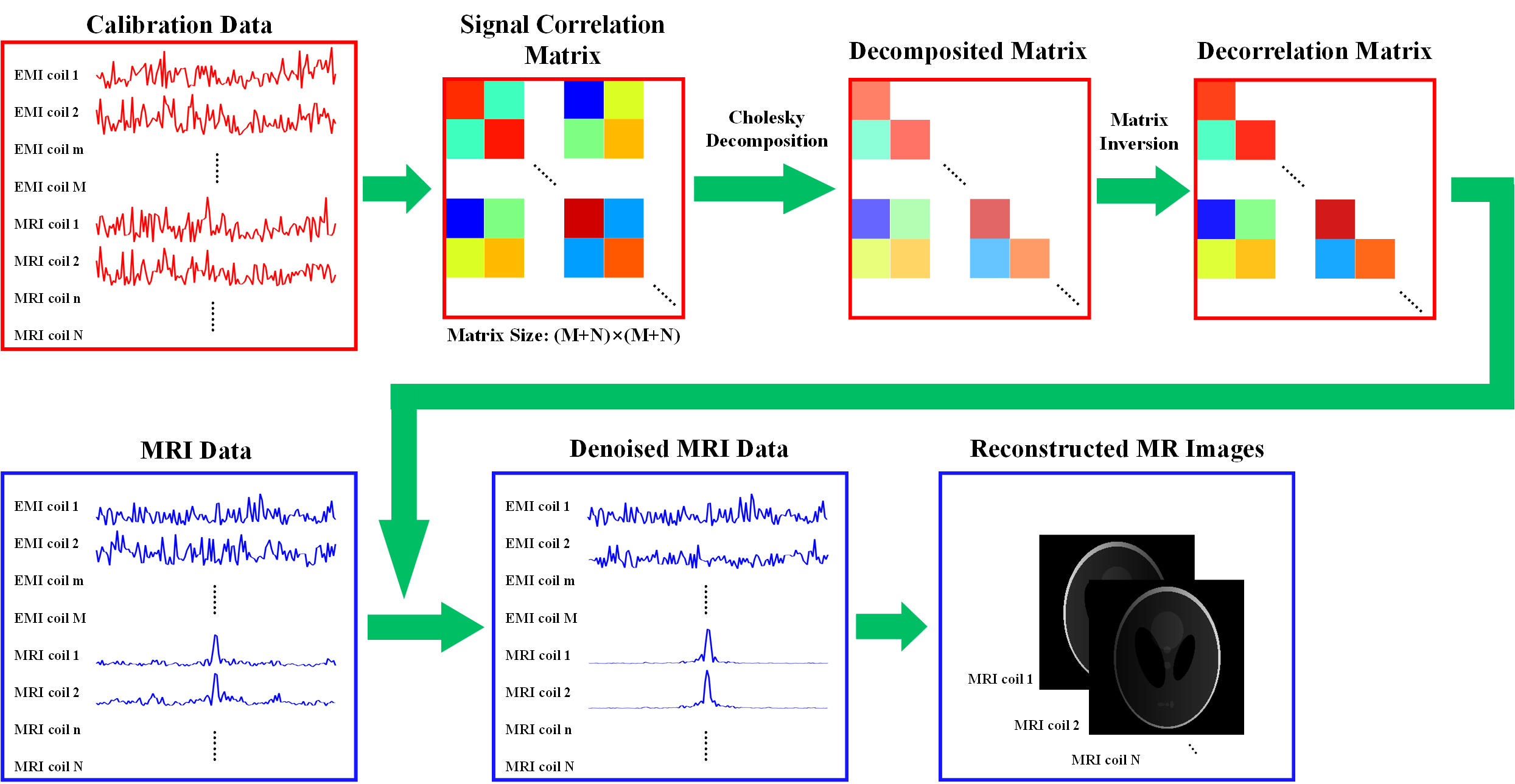
The pipeline of proposed inter-channel correlation-based EMI noise removal method is shown in Figure 1. Pure environmental EMI noise was collected as calibration data from both EMI detectors (EMI coils) and primary RF coils (MRI coils) in the absence of RF excitation before or during MR sampling. These calibration data were then combined as a calibration matrix to calculate signal correlation matrix using matrix inner product:$${\bf{\Psi }} = \left[ {\begin{array}{*{20}{c}}{\left\langle {{{\bf{\eta }}_1},{{\bf{\eta }}_1}} \right\rangle }& \cdots &{\left\langle {{{\bf{\eta }}_1},{{\bf{\eta }}_M}} \right\rangle }&{\left\langle {{{\bf{\eta }}_1},{{\bf{\gamma }}_1}} \right\rangle }& \cdots &{\left\langle {{{\bf{\eta }}_1},{{\bf{\gamma }}_N}} \right\rangle }\\ \vdots & \ddots & \vdots & \vdots & \ddots & \vdots \\{\left\langle {{{\bf{\eta }}_M},{{\bf{\eta }}_1}} \right\rangle }& \cdots &{\left\langle {{{\bf{\eta }}_M},{{\bf{\eta }}_M}} \right\rangle }&{\left\langle {{{\bf{\eta }}_M},{{\bf{\gamma }}_1}} \right\rangle }& \cdots &{\left\langle {{\eta _M},{{\bf{\gamma }}_N}} \right\rangle }\\{\left\langle {{{\bf{\gamma }}_1},{{\bf{\eta }}_1}} \right\rangle }& \cdots &{\left\langle {{{\bf{\gamma }}_1},{{\bf{\eta }}_M}} \right\rangle }&{\left\langle {{{\bf{\gamma }}_1},{{\bf{\gamma }}_1}} \right\rangle }& \cdots &{\left\langle {{{\bf{\gamma }}_1},{{\bf{\gamma }}_N}} \right\rangle }\\ \vdots & \ddots & \vdots & \vdots & \ddots & \vdots \\{\left\langle {{{\bf{\gamma }}_N},{{\bf{\eta }}_1}} \right\rangle }& \cdots &{\left\langle {{{\bf{\gamma }}_N},{{\bf{\eta }}_M}} \right\rangle }&{\left\langle {{{\bf{\gamma }}_N},{{\bf{\gamma }}_1}} \right\rangle }& \cdots &{\left\langle {{{\bf{\gamma }}_N},{{\bf{\gamma }}_N}} \right\rangle }\end{array}} \right]$$Where $$${\bf{\Psi }}$$$ denotes signal correlation matrix, $$${{\bf{\eta }}_i},i = 1,2,...M$$$ is calibration data from EMI coil $$$i$$$ and $$${{\bf{\gamma }}_j},j = 1,2,...,N$$$ is calibration data from MRI coil $$$j$$$, $$$\left\langle { \cdot , \cdot } \right\rangle $$$ is the inner product. Then Cholesky decomposition was conducted on signal correlation matrix followed by matrix inversion to retrieve the decorrelation matrix. The obtained decorrelation matrix was then applied to MRI data contaminated by EMI noise to eliminate accompanied EMI noise in all receive channels. Finally, MR signals were extracted and transformed to reconstruct MR images.Experiments were conducted on a home-built 0.11T portable MR system equipped with four receive channels (two channels were used as MR receivers, and the other two were used as EMI noise sensors) in a regular room (on the ground floor with an area of 80m2) located in an industrial park. T1-weighted images were acquired using a 3D fast field echo sequence with the following parameters: repetition time = 60ms, echo time = 14.7ms, field of view = 256×256×128mm³, voxel size = 2×2×8mm³, average = 2, and total scan time = 4:07min.
Results
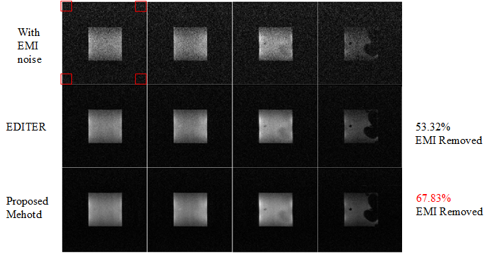
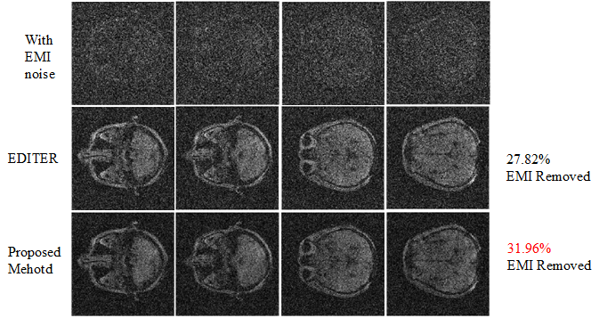
Figure 2 shows a phantom experiment for EMI elimination. Four typical slices with EMI noise, denoised by EDITER [3], and denoised by the proposed method were exhibited the performance of EMI suppression. Figure 3 shows EMI noise removal for four slices of the human brain in vivo, of which the images were contaminated with strong EMI noise and the head structures are nearly invisible. The EDITER and proposed method were able to suppress EMI noise and restore brain structures with improved identification of anatomical structures contours. The proposed inter-channel correlation-base method was able to improve EMI removal efficacy than EDITER.Discussion and Conclusions
The inter-channel correlation-based EMI noise removal technique proves to be effective in eliminating EMI noise on a shielding-free low-field MR scanner. While the residual noise might correspond to uncorrelated relationship among the channels employed, to further reduce the noise level and enhance image SNR, more receive channels can be considered to integrated in RF coil design and manufacturing.Future experiments shall be conducted in a hospital setting to showcase the efficacy of the proposed method. Moreover, our method prefers the design of multi-receive coils such as phased arrays, which can also significantly improve the SNR in MR signal acquisition, enhance the performance of noise removal, and merit accelerated data acquisition such as parallel imaging.
Acknowledgements
This work was supported in part by STI 2030 - Major Projects (2021ZD0200401), National Natural Science Foundation of China (52277232 and 52293424), and Zhejiang Provincial Natural Science Foundation of China (LR23E070001).References
[1] S. Geethanath and J. T. Vaughan Jr, "Accessible magnetic resonance imaging: a review," Journal of Magnetic Resonance Imaging, vol. 49, no. 7, pp. e65-e77, 2019.
[2] Liu, Yilong, et al. A low-cost and shielding-free ultra-low-field brain MRI scanner. Nature Communication. 2021;12(1): 7238.
[3] Srinivas, Sai Abitha, et al. External dynamic interference estimation and removal (EDITER) for low field MRI. Magnetic Resonance Medicine. 2022;87(2): 614-628.
[4] L. Yang, W. He, Y. He, J. Wu, S. Shen, and Z. Xu, "Active EMI suppression system for a 50 mT unshielded portable MRI scanner," IEEE Transactions on Biomedical Engineering, vol. 69, no. 11, pp. 3415-3426, 2022.
[5] M. S. Hansen and P. Kellman, "Image reconstruction: an overview for clinicians," Journal of Magnetic Resonance Imaging, vol. 41, no. 3, pp. 573-585, 2015.
Figures