0157
Finding common ground: Subject grounding to reduce electromagnetic interference at 46 mT1C.J. Gorter MRI Center, Radiology Department, Leids Universitair Medisch Centrum, Leiden, Netherlands
Synopsis
Keywords: Low-Field MRI, Low-Field MRI, EMI reduction
Motivation: Electromagnetic interference (EMI) reduction is essential to utilize low-field point-of-care MRI devices in different environments with different noise conditions.
Goal(s): Improving EMI reduction by subject grounding
Approach: Noise scans and brain images were acquired with and without subject grounding. This is done with normal imaging conditions and when adding broadband noise or single frequency EMI.
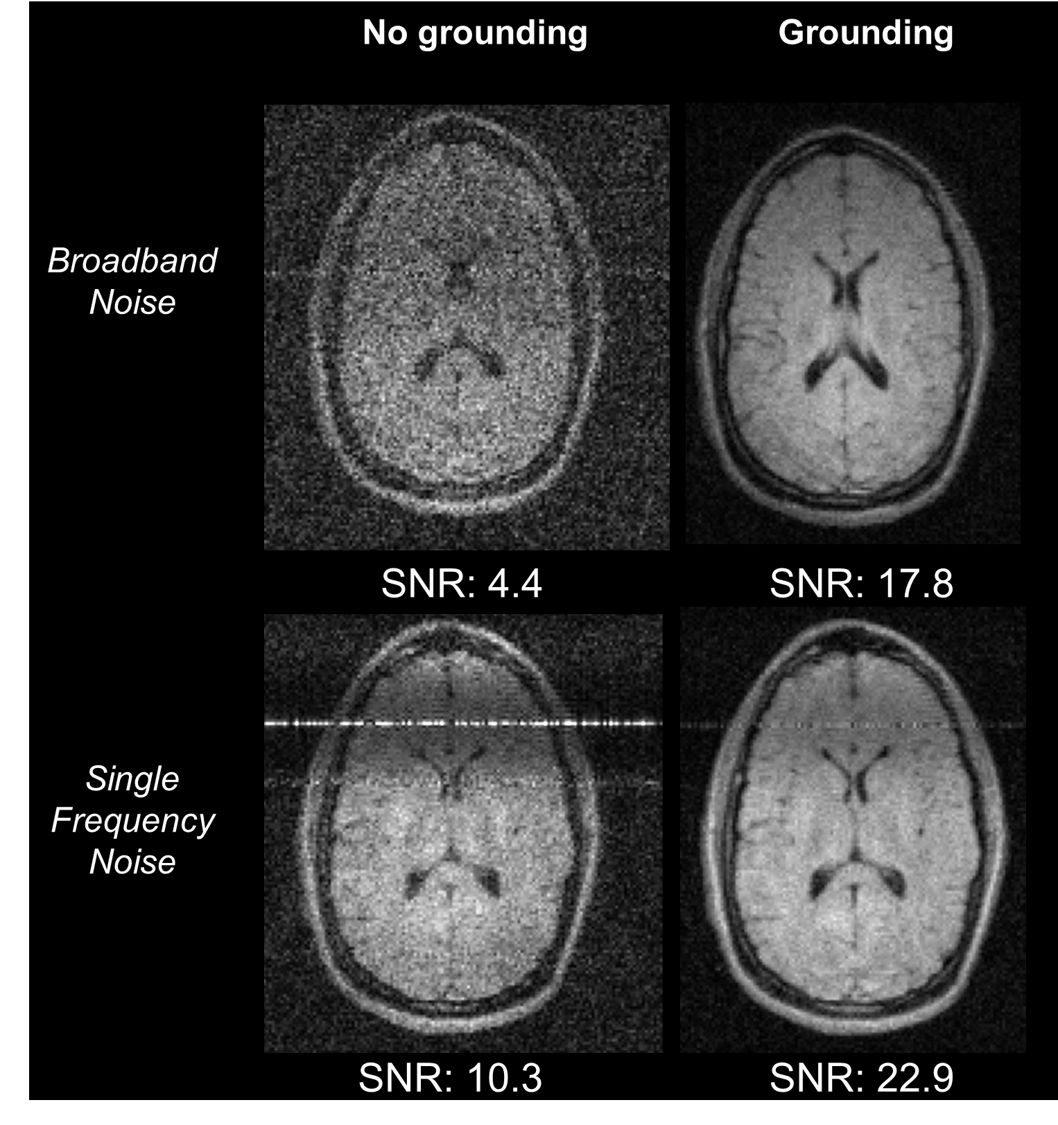
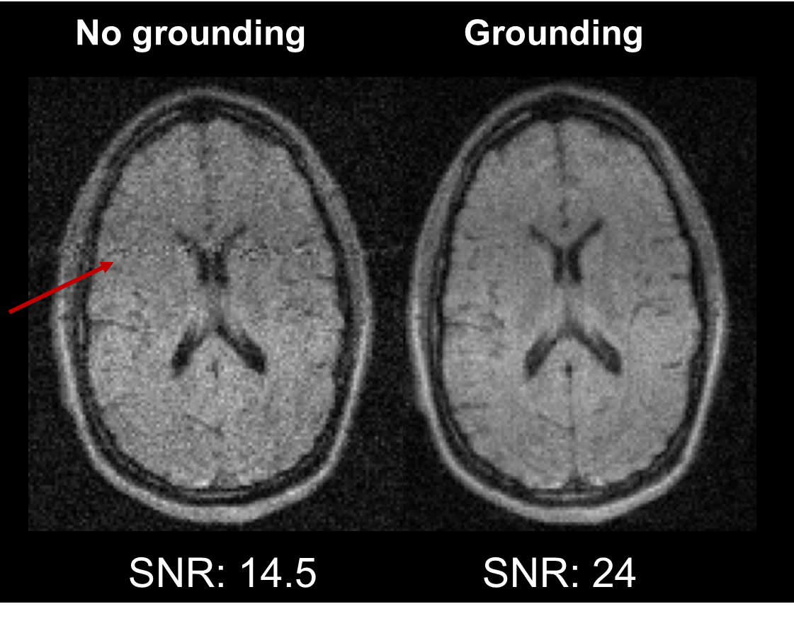
Results: The SNR of the images was improved by a factor of ~4 when grounding the subject and adding broadband EMI to the experiment. A factor ~2 improvement in SNR was observed for the single frequency EMI and a factor of 1.5 improvement for the normal imaging conditions.
Impact: Subject grounding effectively reduced EMI interference. It may be relevant to investigate whether this setup would be able to reduce EMI from medical equipment, or general environmental EM noise in typically challenging POC settings (ICU, emergency room, in remote locations)
Introduction
Advancements in both image processing and hardware have resulted in renewed interest in applying low-field MRI in Point-of-care (POC) settings[1] [2]. One of the challenges low-field POC systems face is that the human body acts as an antenna and couples noise and electromagnetic interference (EMI) into the receiver coil(s). This is especially a problem in the unshielded environments in which these systems may operate. This issue can be partially solved by placing a conductive cloth around the subject [3] or by using AI-based denoisers in post-processing[4–7]. However, cloths tend to lose efficacy over time and denoisers are location dependent require additional hardware. The noise issue can also be addressed by grounding all system components and utilising RF shielding around the subject [8]. This results in a reduction of noise to levels which are almost equivalent to that of an unloaded coil. However, the human body can still couple in a significant amount of EMI and these conditions can vary day to day and with subject size and position. In this work we show that proper grounding of the subject can significantly reduce the noise coupled into the RF coil.Methods
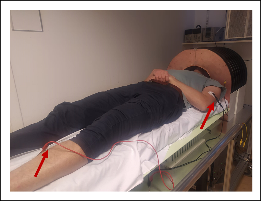
Images were obtained using a 46 mT Halbach based MRI system using a Magritek Kea2 spectrometer [9]. A solenoidal transmit/receive head-coil was used for imaging. The hypothesis of the study was that body acts as an antenna for random noise and EMI arising from specific sources, and that grounding the body to a common ground with the other electronics would decrease the noise coupled into the receiver coil. To confirm this hypothesis, in-vivo data was acquired in which the volunteer was grounded by placing ECG electrodes (3M Red Dot ECG electrodes) on the arm and lower leg (Figure 1). Additionally, two semi-cylindrical aluminium RF shields were added to shield the subject [8]. To verify the efficacy of grounding, images were acquired using a standard turbo spin echo (TSE) sequence with and without grounding the subject, for the following three scenarios:i) Ambient noise propagation via the human antenna effect,
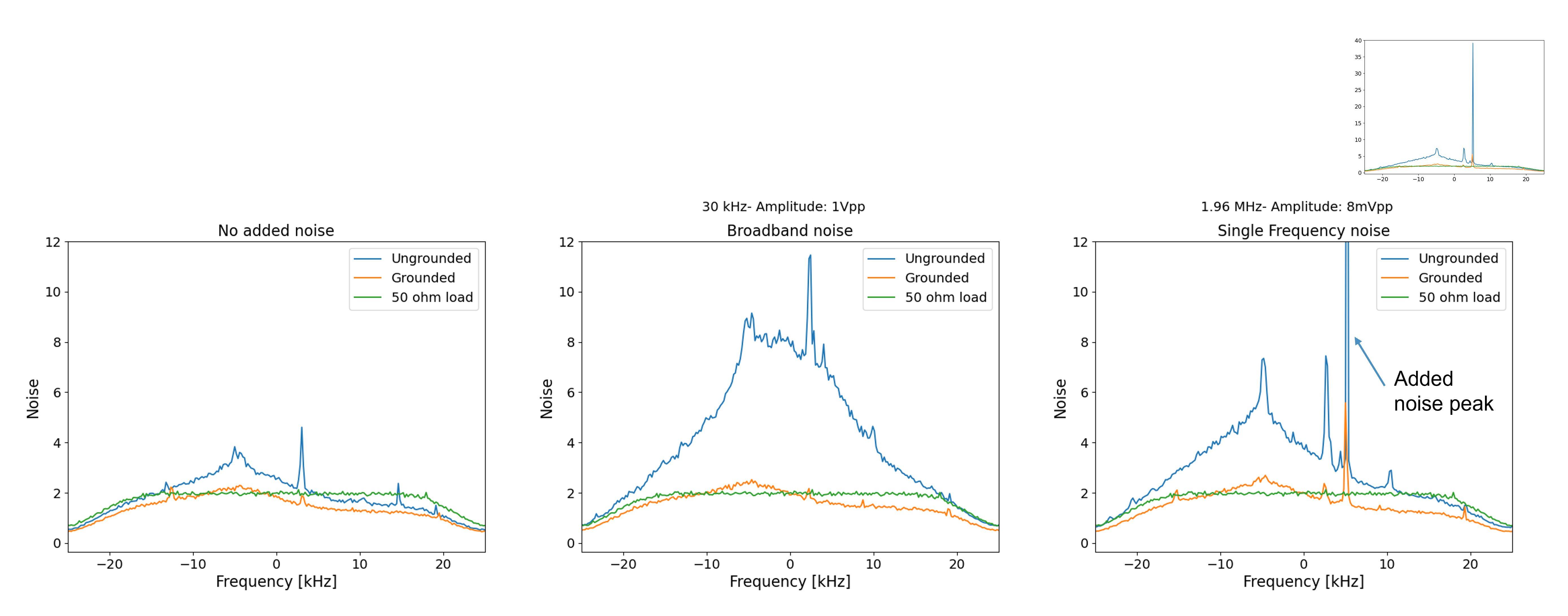
ii) Adding a discrete frequency EMI via a transmitting antenna and function generator. The noise coil was positioned outside the RF cage, sending a 1.96 MHz continuous sine wave with a 4 mV peak to peak voltage,
iii) Adding 30 kHz broadband noise with the same setup as ii) with an amplitude of 1 V peak to peak. Quantification was done by performing a noise scan for each of these scenarios. A polynomial function was fitted to the noise profile to estimate the noise level.
Results
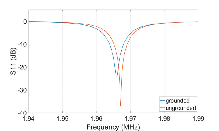
Providing the subject and the coil a common grounding point reduces the coupling between the subject and the coil, representing a decrease in the mutual inductance between the two, which is reflected in a 1 kHz reduction in resonance frequency of the coil (Figure 2). The Q-factor of the coil is unchanged after retuning of the coil. In Figure 3, after subject grounding, the EMI and noise coupled into the coil are very similar to the values of the noise floor (measured with a 50 ohm load). This result is also reflected in the TSE images: when broadband noise is added (Figure 4), grounding the subject increased the SNR by a factor of ~4. With the single frequency EMI is applied, the SNR is improved by a factor of ~2, and the discrete “zipper artefact” is less evident. Figure 5 shows that even without adding additional noise sources, subject grounding improved the SNR by a factor of ~1.5.Discussions and Conclusions
We present a simple but effective method to decrease the noise and EMI coupled into the RF receiver coil by effective grounding of the subject. This leads to a more stable measurement setup which can be used in different environments, in addition to other shielding and denoising methods. Further analysis will explore whether the optimal positioning of electrodes for grounding the subject may eliminate the need for the less practical aluminium RF shield.Acknowledgements
This work was partly funded by the Dutch Science Foundation Open Technology 18981References
1. Arnold TC, Freeman CW, Litt B, Stein JM (2023) Low-field MRI: Clinical promise and challenges. Journal of Magnetic Resonance Imaging 57
2. Sarracanie M, Salameh N (2020) Low-Field MRI: How Low Can We Go? A Fresh View on an Old Debate. Front Phys 8
3. O’Reilly T, Teeuwisse WM, de Gans D, et al (2021) In vivo 3D brain and extremity MRI at 50 mT using a permanent magnet Halbach array. Magn Reson Med 85:. https://doi.org/10.1002/mrm.28396
4. Man C, Lau V, Su S, et al (2023) Deep learning enabled fast 3D brain MRI at 0.055 tesla. Sci Adv 9:. https://doi.org/10.1126/sciadv.adi9327
5. Le DBT, Sadinski M, Nacev A, et al (2021) Deep Learning-based Method for Denoising and Image Enhancement in Low-Field MRI. In: IST 2021 - IEEE International Conference on Imaging Systems and Techniques, Proceedings
6. Zhang Y, He W, Chen F, et al (2022) Denoise ultra-low-field 3D magnetic resonance images using a joint signal-image domain filter. Journal of Magnetic Resonance 344:. https://doi.org/10.1016/j.jmr.2022.107319
7. Parsa J, O’Reilly T, Webb A (2023) A single-coil-based method for electromagnetic interference reduction in point-of-care low field MRI systems. Journal of Magnetic Resonance 346:. https://doi.org/10.1016/j.jmr.2022.107355
8. Webb A, O’Reilly T (2023) Tackling SNR at low-field: a review of hardware approaches for point-of-care systems. Magnetic Resonance Materials in Physics, Biology and Medicine 36
9. O’Reilly T, Teeuwisse WM, Webb AG (2019) Three-dimensional MRI in a homogenous 27 cm diameter bore Halbach array magnet. Journal of Magnetic Resonance 307:. https://doi.org/10.1016/j.jmr.2019.106578
Figures