0153
Motion-Robust Multiparametric MRI of the Liver at 3T: Simultaneous Estimation of Water-Specific T1, PDFF, Motion-Resolved R2*, and QSM1Bernard and Irene Schwartz Center for Biomedical Imaging, Department of Radiology, New York University Grossman School of Medicine, New York, NY, United States, 2Center for Advanced Imaging Innovation and Research (CAI2R), Department of Radiology, New York University Grossman School of Medicine, New York, NY, United States, 3Biomedical Engineering and Imaging Institute, Icahn School of Medicine at Mount Sinai, New York, NY, United States
Synopsis
Keywords: Quantitative Imaging, Quantitative Imaging
Motivation: Quantitative multiparametric MRI has the potential to improve the characterization of liver diseases, but its clinical implementation is limited by challenges such as respiratory motion and slow imaging speed.
Goal(s): To develop a motion-robust multiparametric MRI technique that enables simultaneous estimation of water-specific T1, PDFF, motion-resolved R2*, and QSM of the liver from a single acquisition at 3T.
Approach: Our technique employs inversion recovery-prepared golden-angle multi-echo stack-of-stars sampling in combination with advanced low-rank subspace reconstruction for generating different quantitative parameters.
Results: Free-breathing multiparametric estimation of 3D water-specific T1, PDFF, R2* and QSM with motion compensation has been successfully demonstrated in volunteers and patients.
Impact: This new technique is capable of estimating water-specific T1, PDFF, motion-resolved R2*, and QSM of the liver from a single acquisition at 3T. It holds potential to promote the use of quantitative MRI in moving organs such as the liver.
Introduction
Quantitative MRI techniques for estimating proton density fat fraction (PDFF) and R2* have seen increasing interest in clinical liver exams. Recently, several studies have demonstrated that PDFF, R2* and fat/water-separated T1 can be simultaneously estimated from a single free-breathing inversion recovery (IR)-prepared 3D multi-echo acquisition[1–3]. It has been shown that accurate quantification of R2* in the liver requires effective respiratory motion compensation, particularly at 3T[4]. However, the use of IR preparation introduces significant signal variation in acquired data, posing challenges for accurate motion tracking and motion compensation. In this study, we propose a novel acquisition scheme that combines IR-prepared golden-angle multi-echo stack-of-stars sampling with periodic acquisition of 2D navigators for dependable motion compensation. We introduce GraspT1-Dixon-Nav that enables the simultaneous estimation of water-specific T1, PDFF, motion-resolved R2*, and QSM of the liver from a single acquisition at 3T.Methods
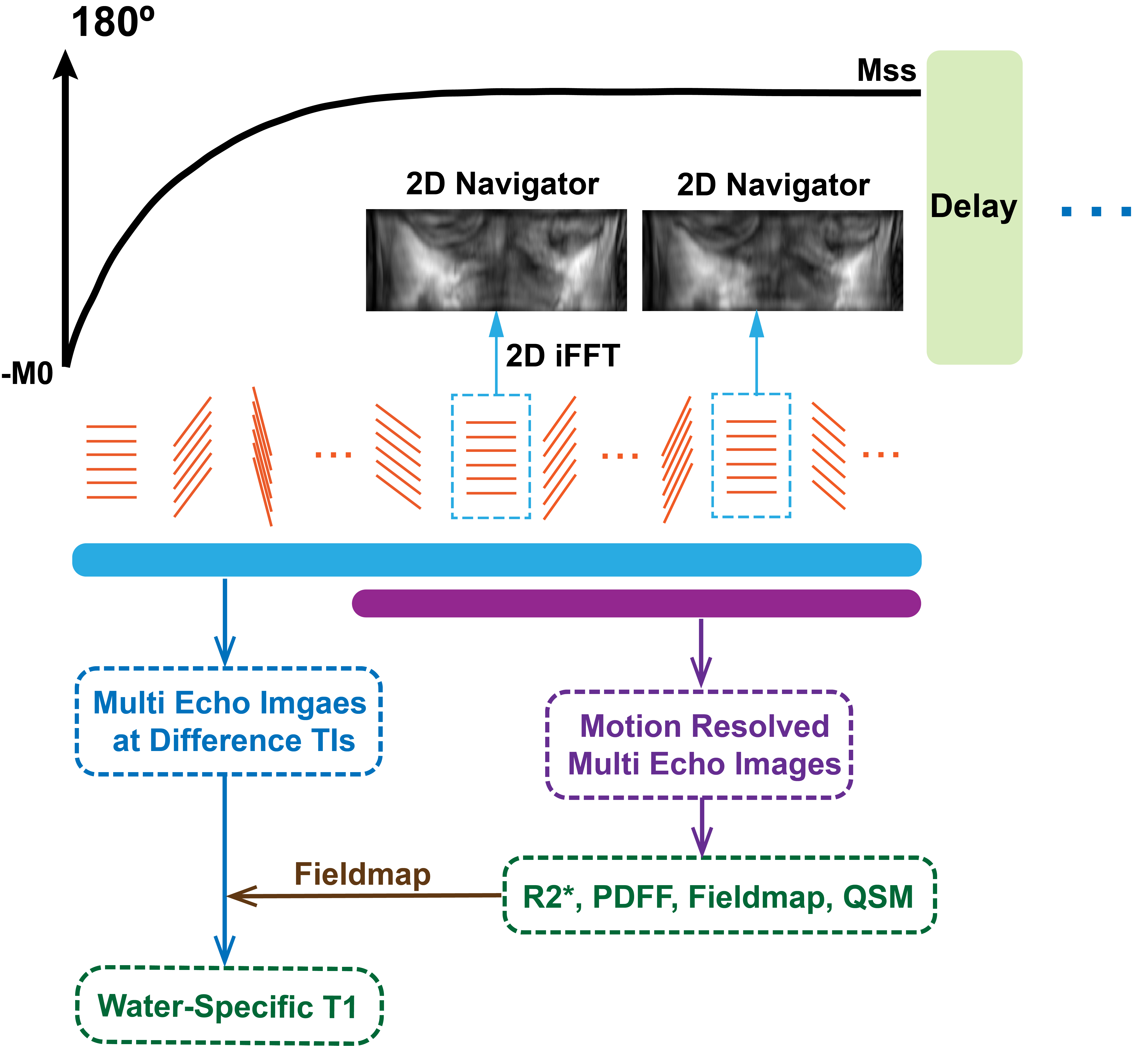
GraspT1-Dixon-NavOur technique, referred to as GraspT1-Dixon-Nav, was developed based on the Golden-angle RAdial Sparse Parallel (GRASP) MRI technique with inversion recovery (IR) preparation and multi-echo acquisition. Within one TR, after 10 inversion times (TIs), 2D navigators are acquired every 6 TIs for motion detection as shown in Figure 1. Free-breathing liver MRI was performed on a healthy volunteer and a patient with confirmed non-alcoholic fatty liver disease (NAFLD). Imaging parameters were as follows: FOV=360x360, matrix size=256x256, slice thickness=6mm, number of slices=16, number of echoes=6, TR/TE1/ΔTE/TE6=10.8/10.8/1.4/1.6/9.4msms, flip angle=5°, number of TIs per TR=53, number of IR preparations=25, slice partial Fourier factor=75%, and total acquisition time (TA)=4min40sec. For comparison, free-breathing multi-echo stack-of-stars imaging without IR preparation was performed to generate reference PDFF, R2* and QSM.
Image reconstruction
T1 mapping is calculated using stack-of-stars at all TIs excluding the 2D navigators. Images at each TI are reconstructed following the pipeline as described in ref[5]. The magnetic field map is estimated using graphcut[6] method and subsequently being used for water-fat separation[7] for images at each TI to generate water-specific T1.
Out of all 53 TIs, the later 42 TIs are used for motion resolved R2* and QSM estimation to minimize the bias due to signal variation during recovery. Specifically, 2D navigators are used to sort 3D multi-echo volumes into four motion states, followed by estimation of PDFF, R2* and field map using graphcut[6] water-fat separation for every motion state. The background field component in the field map is filtered out using the projection-onto-dipole-fields (PDF)[8] method and the remaining local tissue field map is used for QSM calculation with the iLSQR[9] method. QSM values are referenced by setting the magnetic susceptibility of Aortic blood to be -0.085 ppm[10]. The motion averaged images and quantitative maps are calculated using the same method as well for comparison.
Results
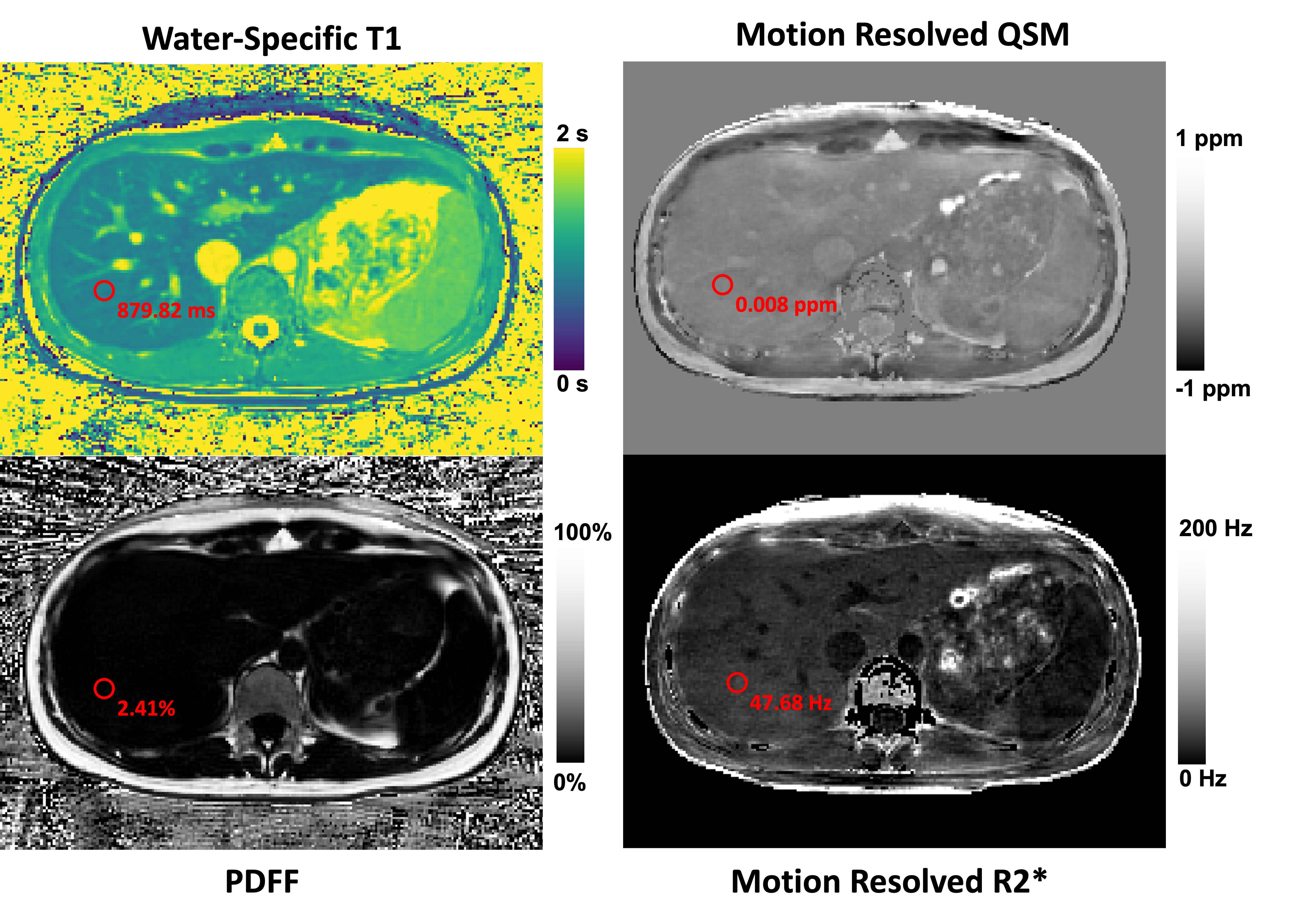
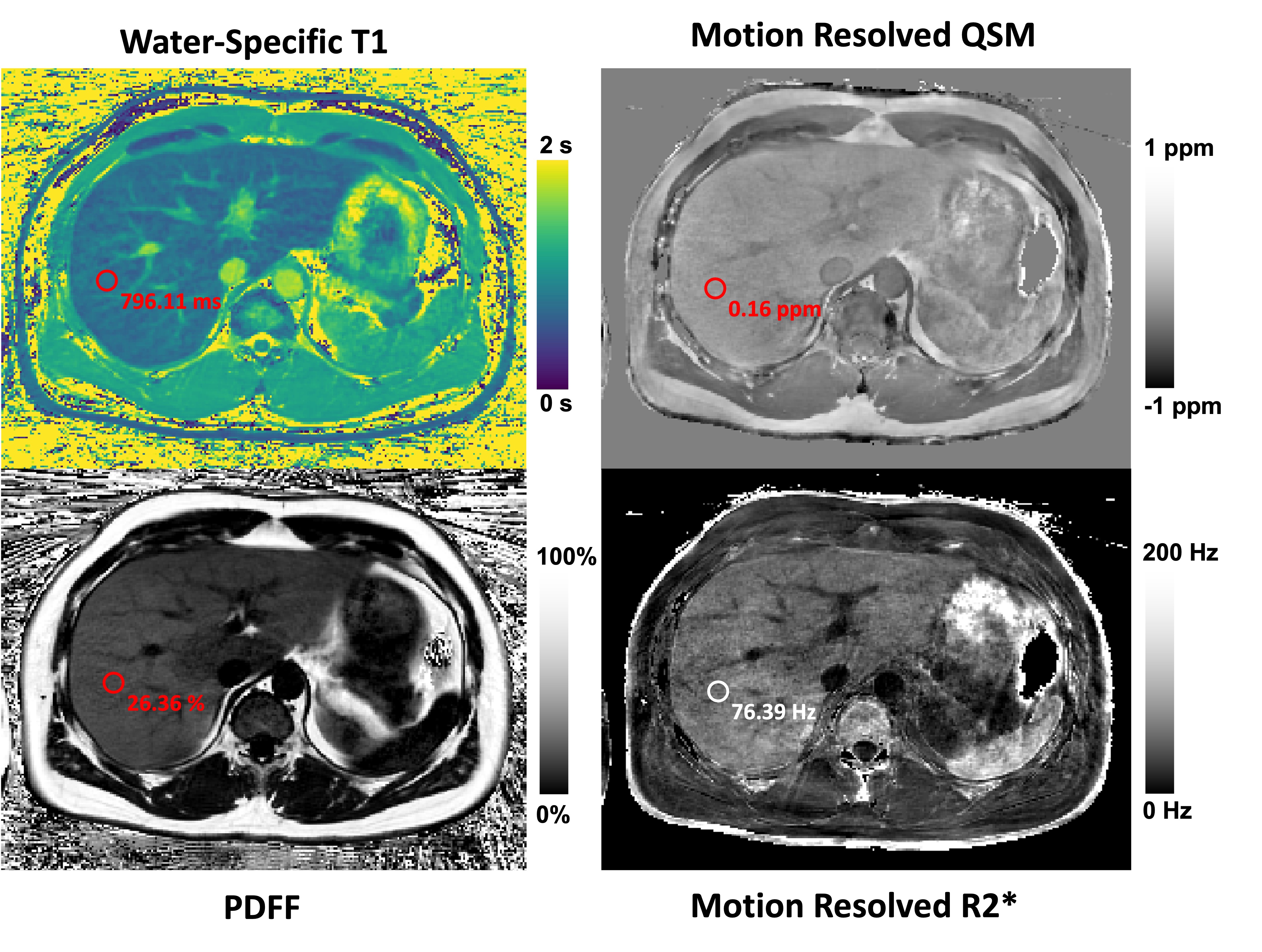
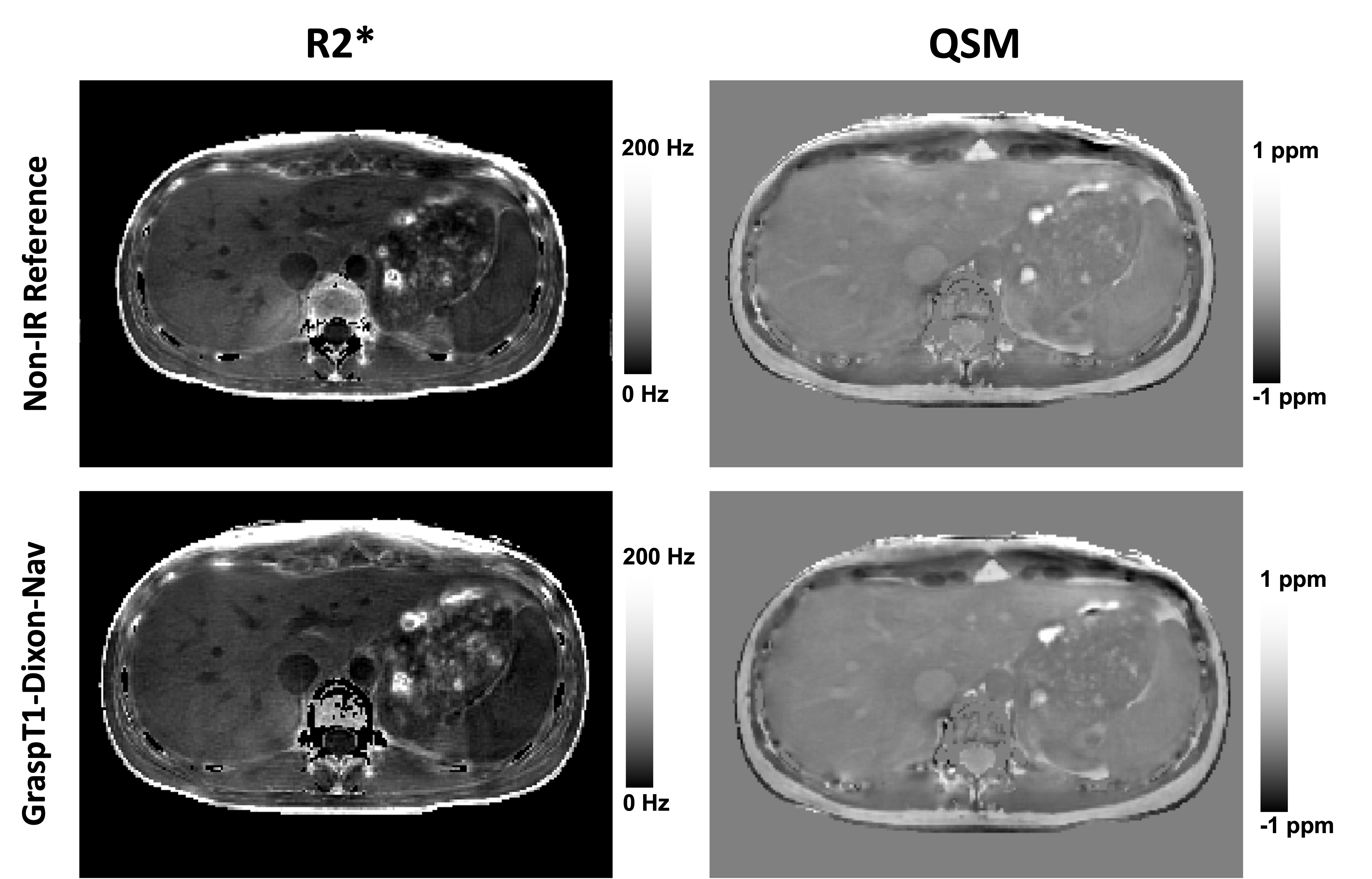
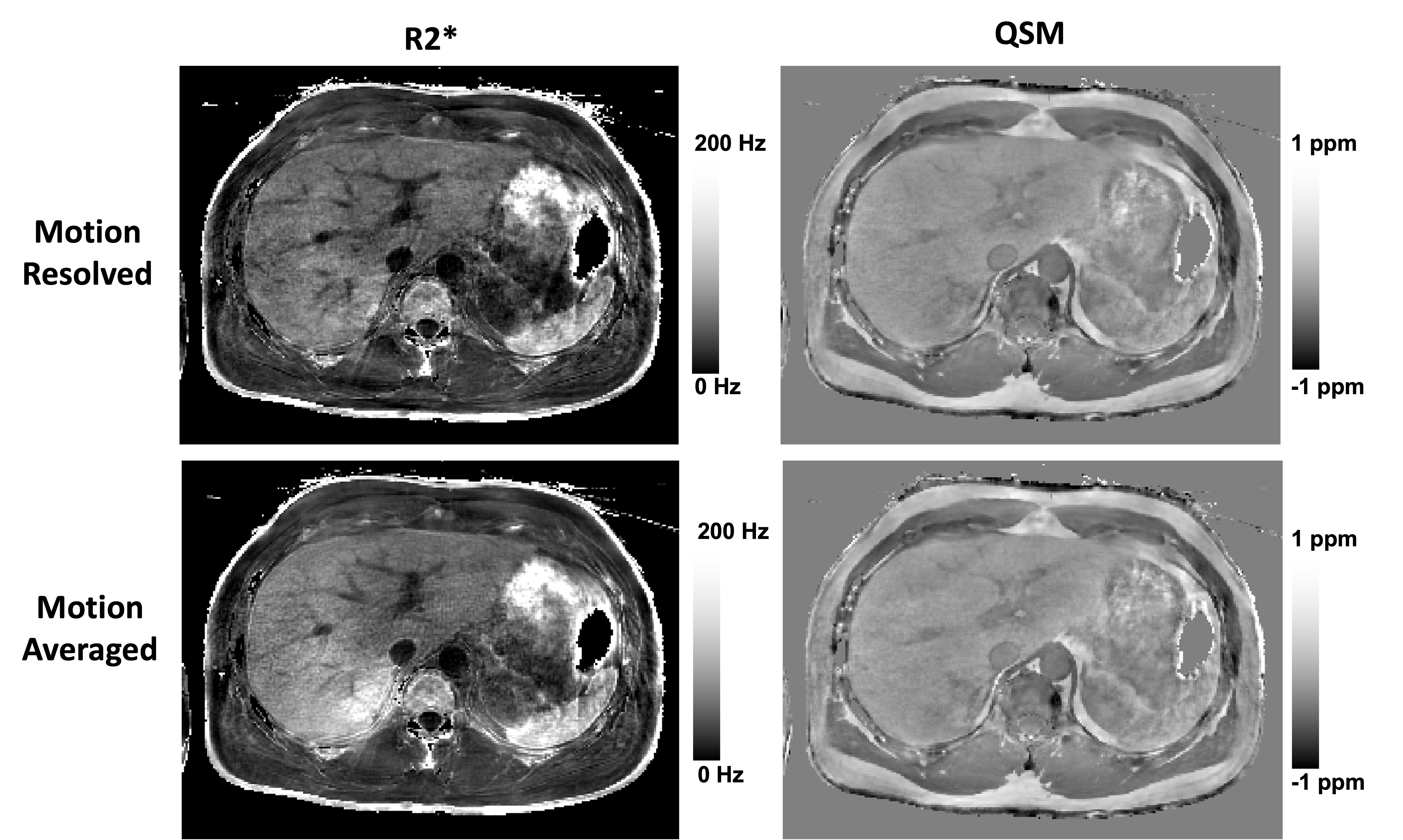
Figure 2 show the simultaneously achieved water-specific T1, PDFF, motion-resolved R2* and motion-resolved QSM for one healthy subject using GraspT1-Dixon-Nav technique. The simultaneously estimated quantitative maps from the patient are shown in Figure 3. The anatomical structure and disease pathology are clearly revealed in all maps without major artifacts. As seen in Figure 4, compared to a regular multi-echo free-breathing liver scan, the additional inversion recovery preparation in the sequence does not compromise the accuracy of the estimated R2* value or the QSM value. In Figure 5, without motion compensation, the motion averaged R2* during a free-breathing exam is artificially elevated due to motion, in agreement with a previous study[4]. The 2D navigators in GraspT1-Dixon-Nav are able to effectively track the respiratory motion and enable motion sorting to allow recovery of accurate and robust liver R2* estimates. Further, it appears that QSM, comparing to R2*, is less sensitive to motion-induced bias.Discussion
Quantitative multiparametric MRI has the potential to improve the characterization of liver diseases, but respiratory motion poses challenges in accurate quantification. This study demonstrated a new acquisition strategy in which water-specific T1, PDFF, motion-resolved R2* and QSM can be simultaneously estimated from a single free-breathing MRI scan in less than 5 min. The 2D navigators provide effective and efficient motion compensation to achieve reliable motion-resolved R2* and QSM maps in the presence of the signal changes due to the IR preparation. Additionally, in our initial demonstration, QSM appears to be more robust to respiratory motion than R2*. It is likely that the motion-induced field inhomogeneity is spatially slowly varying and can therefore be filtered out during the background field removal step. Further work will be performed for to validate the repeatability of our approach and its readiness for clinical translation.Acknowledgements
This work was supported by the NIH (R01EB030549, R01EB031083, R21EB032917 and P41EB017183) and was performed under the rubric of the Center for Advanced Imaging Innovation and Research (CAI2R), an NIBIB National Center for Biomedical Imaging and Bioengineering.References
(1) Jaubert, O.; Arrieta, C.; Cruz, G.; Bustin, A.; Schneider, T.; Georgiopoulos, G.; Masci, P.-G.; Sing-Long, C.; Botnar, R. M.; Prieto, C. Multi-Parametric Liver Tissue Characterization Using MR Fingerprinting: Simultaneous T1 , T2 , T2 *, and Fat Fraction Mapping. Magn Reson Med 2020, 84 (5), 2625–2635. https://doi.org/10.1002/mrm.28311.
(2) Wang, N.; Cao, T.; Han, F.; Xie, Y.; Zhong, X.; Ma, S.; Kwan, A.; Fan, Z.; Han, H.; Bi, X.; Noureddin, M.; Deshpande, V.; Christodoulou, A. G.; Li, D. Free-Breathing Multitasking Multi-Echo (MT-ME) MRI for Whole-Liver Water-Specific T1, Proton Density Fat Fraction, and R2* Quantification. Magn Reson Med 2022, 87 (1), 120–137. https://doi.org/10.1002/mrm.28970.
(3) Thompson, R. B.; Chow, K.; Mager, D.; Pagano, J. J.; Grenier, J. Simultaneous Proton Density Fat-Fraction and Imaging with Water-Specific T1 Mapping (PROFIT1): Application in Liver. Magnetic Resonance in Medicine 2021, 85 (1), 223–238. https://doi.org/10.1002/mrm.28434.
(4) Zhong, X.; Armstrong, T.; Nickel, M. D.; Kannengiesser, S. A. R.; Pan, L.; Dale, B. M.; Deshpande, V.; Kiefer, B.; Wu, H. H. Effect of Respiratory Motion on Free-Breathing 3D Stack-of-Radial Liver Relaxometry and Improved Quantification Accuracy Using Self-Gating. Magnetic Resonance in Medicine 2020, 83 (6), 1964–1978. https://doi.org/10.1002/mrm.28052.
(5) Feng, L.; Liu, F.; Soultanidis, G.; Liu, C.; Benkert, T.; Block, K. T.; Fayad, Z. A.; Yang, Y. Magnetization-Prepared GRASP MRI for Rapid 3D T1 Mapping and Fat/Water-Separated T1 Mapping. Magn Reson Med 2021, 86 (1), 97–114. https://doi.org/10.1002/mrm.28679.
(6) Hernando, D.; Kellman, P.; Haldar, J. P.; Liang, Z.-P. Robust Water/Fat Separation in the Presence of Large Field Inhomogeneities Using a Graph Cut Algorithm. Magn Reson Med 2010, 63 (1), 79–90. https://doi.org/10.1002/mrm.22177.
(7) Benkert, T.; Feng, L.; Sodickson, D. K.; Chandarana, H.; Block, K. T. Free-Breathing Volumetric Fat/Water Separation by Combining Radial Sampling, Compressed Sensing, and Parallel Imaging. Magnetic Resonance in Medicine 2017, 78 (2), 565–576. https://doi.org/10.1002/mrm.26392.
(8) Liu, T.; Khalidov, I.; de Rochefort, L.; Spincemaille, P.; Liu, J.; Tsiouris, A. J.; Wang, Y. A Novel Background Field Removal Method for MRI Using Projection onto Dipole Fields (PDF). NMR Biomed 2011, 24(9), 1129–1136. https://doi.org/10.1002/nbm.1670.
(9) Li, W.; Wang, N.; Yu, F.; Han, H.; Cao, W.; Romero, R.; Tantiwongkosi, B.; Duong, T. Q.; Liu, C. A Method for Estimating and Removing Streaking Artifacts in Quantitative Susceptibility Mapping. Neuroimage 2015, 108, 111–122. https://doi.org/10.1016/j.neuroimage.2014.12.043.
(10) Jafari, R.; Sheth, S.; Spincemaille, P.; Nguyen, T. D.; Prince, M. R.; Wen, Y.; Guo, Y.; Deh, K.; Liu, Z.; Margolis, D.; Brittenham, G. M.; Kierans, A. S.; Wang, Y. Rapid automated liver quantitative susceptibility mapping. J Magn Reson Imaging 2019, 50 (3), 725–732. https://doi.org/10.1002/jmri.26632.
Figures