0151
Water/fat separated Echo Planar Time-resolved Imaging (EPTI) for efficient distortion-free multi-contrast imaging1Athinoula A. Martinos Center for Biomedical Imaging, Massachusetts General Hospital, Boston, MA, United States, 2Department of Radiology, Harvard Medical School, Boston, MA, United States, 3Harvard-MIT Division of Health Sciences and Technology, Massachusetts Institute of Technology, Cambridge, MA, United States
Synopsis
Keywords: Fat & Fat/Water Separation, Fat
Motivation: Echo Planar Time-resolved Imaging (EPTI) can produce distortion- and blurring-free multi-echo images with high efficiency. For its broader application such as in body imaging, the challenge of fat suppression/separation needs to be addressed.
Goal(s): Achieving efficient water/fat separation using EPTI for high-quality fast multi-contrast/quantitative imaging in the presence of fat tissues.
Approach: In this study, water/fat separated EPTI (WFS-EPTI) was proposed to achieve this by: (1) designing a novel in-phase and out-of-phase EPTI acquisition and encoding scheme; and (2) adopting a k-space-based water/fat separation method.
Results: Experimental results demonstrated the efficacy of WFS-EPTI for water/fat separation and fat-robust distortion-free multi-contrast/quantitative imaging.
Impact: The proposed WFS-EPTI effectively separates water and fat signals, while providing efficient acquisition of high-resolution, distortion-free multi-contrast images and quantitative maps. It can extend EPTI to a broader range of applications.
INTRODUCTION
Conventional EPI has been widely used due to its fast speed. However, it suffers from T2*-blurring and geometric-distortions, which hinder its use in high-resolution imaging. In addition, it is prone to chemical-shift artifacts when there is insufficient fat suppression, especially in body imaging. EPTI1 addresses EPI’s distortion and blurring by exploiting the spatial-temporal correlation in the encoding process. It produces distortion- and blurring-free multi-contrast images with high efficiency, making it a practical, ultra-fast acquisition in many brain MRI applications, including multi-parametric quantitative imaging2-4. However, the challenge of fat suppression/separation needs to be addressed for its broader application in body imaging.Here, we proposed a Water/Fat Separated EPTI (WFS-EPTI) technique that enables effective water/fat separation and provides efficient distortion-free multi-contrast/quantitative imaging even in the presence of fat. Specifically, the multi-echo data acquired by EPTI readout intrinsically encodes the phase of water and fat signals, therefore making it suitable for water/fat separation5-8. Nevertheless, there are several challenges: (1) the phase due to the chemical-shift of fat evolves rapidly across the EPTI readout, which might compromise the temporal correlation and violate the low-rank assumption underpinning subspace reconstruction9, leading to poor reconstruction performance; (2) the bipolar echo-planar readout in EPTI could result in chemical-shift induced misregistration, necessitating rectification during water/fat separation. We have addressed these issues by: (1) designing a novel in-phase and out-of-phase acquisition and encoding scheme for EPTI to ensure accurate subspace reconstruction while preserving acquisition efficiency; (2) adopting a k-space-based water/fat separation method to address the issue caused by the bipolar readout10, 11. We demonstrated the efficacy of the WFS-EPTI for water/fat separation and ultra-fast multi-contrast/quantitative imaging in both phantom and in-vivo human brain without any fat-saturation as proof-of-concept experiments.
METHODS
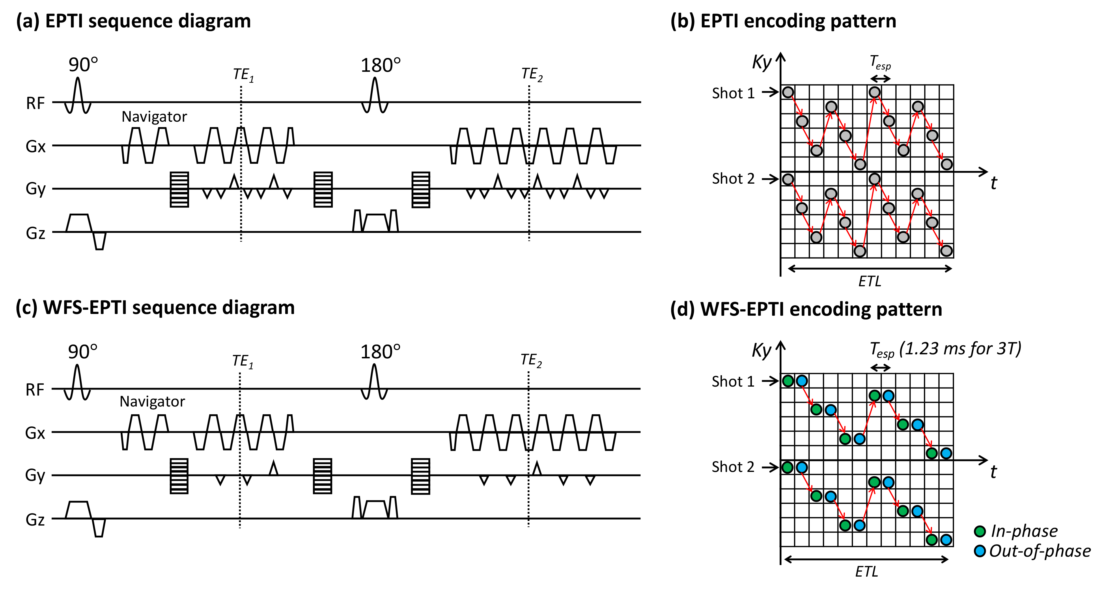
Fig. 1 shows the GESE sequence and encoding used by conventional EPTI and the proposed WFS-EPTI. As an example, when considering the chemical-shift of fat, the gradient-echo image $$$I_{m}$$$ at the (m+1)-th echo can be expressed as:$$I_{m}=(\rho_{W}+\rho_{F}e^{-i2\pi f_{F}(t_{0}+mT_{esp})})e^{-(t_{0}+mT_{esp})/T_2^*}e^{-i\gamma \triangle B(t_{0}+mT_{esp})}$$Here, $$$\rho_{W}$$$ is the intensity of the water component; $$$\rho_{F}$$$ is the intensity of the fat component with a frequency shift $$$f_{F}$$$ (in Hz); $$$t_{0}$$$ is the echo time of the first echo; $$$T_{esp}$$$ is the echo spacing (ESP); $$$e^{-i2\pi f_{F}(t_{0}+mT_{esp})}$$$ is the chemical-shift encoding; $$$e^{-(t_{0}+mT_{esp})/T_2^*}$$$ is the $$$T_2^*$$$ signal decay; $$$e^{-i\gamma \triangle B(t_{0}+mT_{esp})}$$$ is the B0-inhomogeneity-induced phase.The fast-evolving fat phase $$$ e^{-i2\pi f_{F}(t_{0}+mT_{esp})}$$$ may violate the low-rank assumption in subspace reconstruction. To resolve this problem, Dixon method12 was incorporated into the EPTI acquisition, where TEs and $$$T_{esp}$$$ (~1.23ms@3T) were chosen such that the odd and even echoes acquired in-phase and out-of-phase images, respectively. Images acquired in WFS-EPTI can then be defined as:$$I_{m}=\begin{cases}(\rho_{W}+\rho_{F})e^{-(t_{0}+mT_{esp})/T_2^*}e^{-i\gamma \triangle B(t_{0}+mT_{esp})} & m \in odd\space\ echoes\\(\rho_{W}-\rho_{F})e^{-(t_{0}+mT_{esp})/T_2^*}e^{-i\gamma \triangle B(t_{0}+mT_{esp})} & m \in even\space\ echoes\end{cases}$$By eliminating the fast-evolving chemical-shift term, in-phase and out-of-phase images can be reconstructed separately with significantly improved reconstruction conditioning. To improve image quality, encoding patterns for WFS-EPTI were further optimized by implementing spatiotemporal CAIPI trajectory separately for odd and even echoes (Fig. 1d).
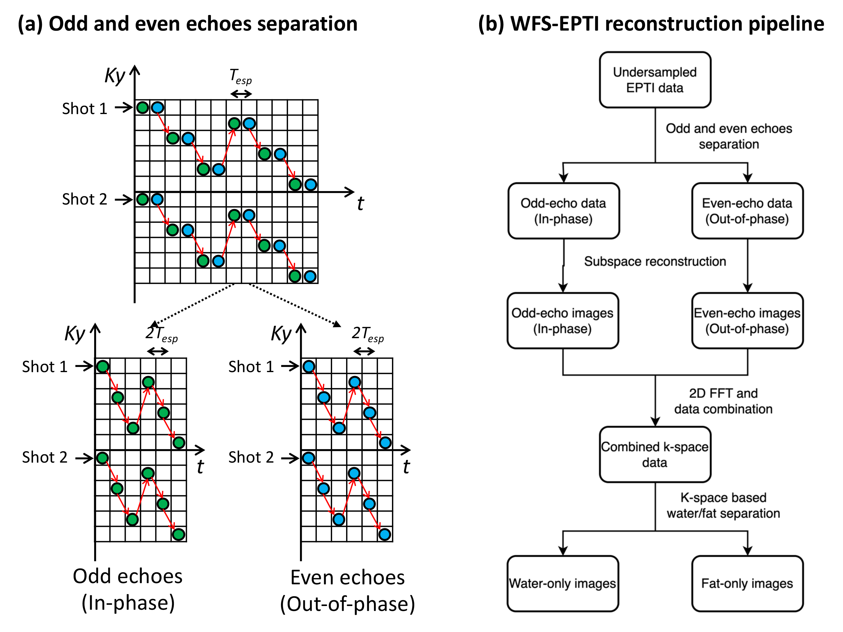
Fig. 2 shows the flowchart of WFS-EPTI reconstruction. Firstly, the odd and even echoes (in- and out-of-phase data) were separated, and each underwent independent subspace reconstruction. The reconstructed images were then transformed into the k-space domain. Using the multi-echo k-space data, a k-space-based water/fat separation method10 was employed to address the chemical-shift misregistration caused by the bipolar readout. Quantitative maps (T2,T2*,PD,B0 maps) can be derived from multi-echo images. Phantom and in-vivo experiments were implemented on a Siemens Prisma 3T scanner with a 32-channel head coil.
RESULTS and DISCUSSION
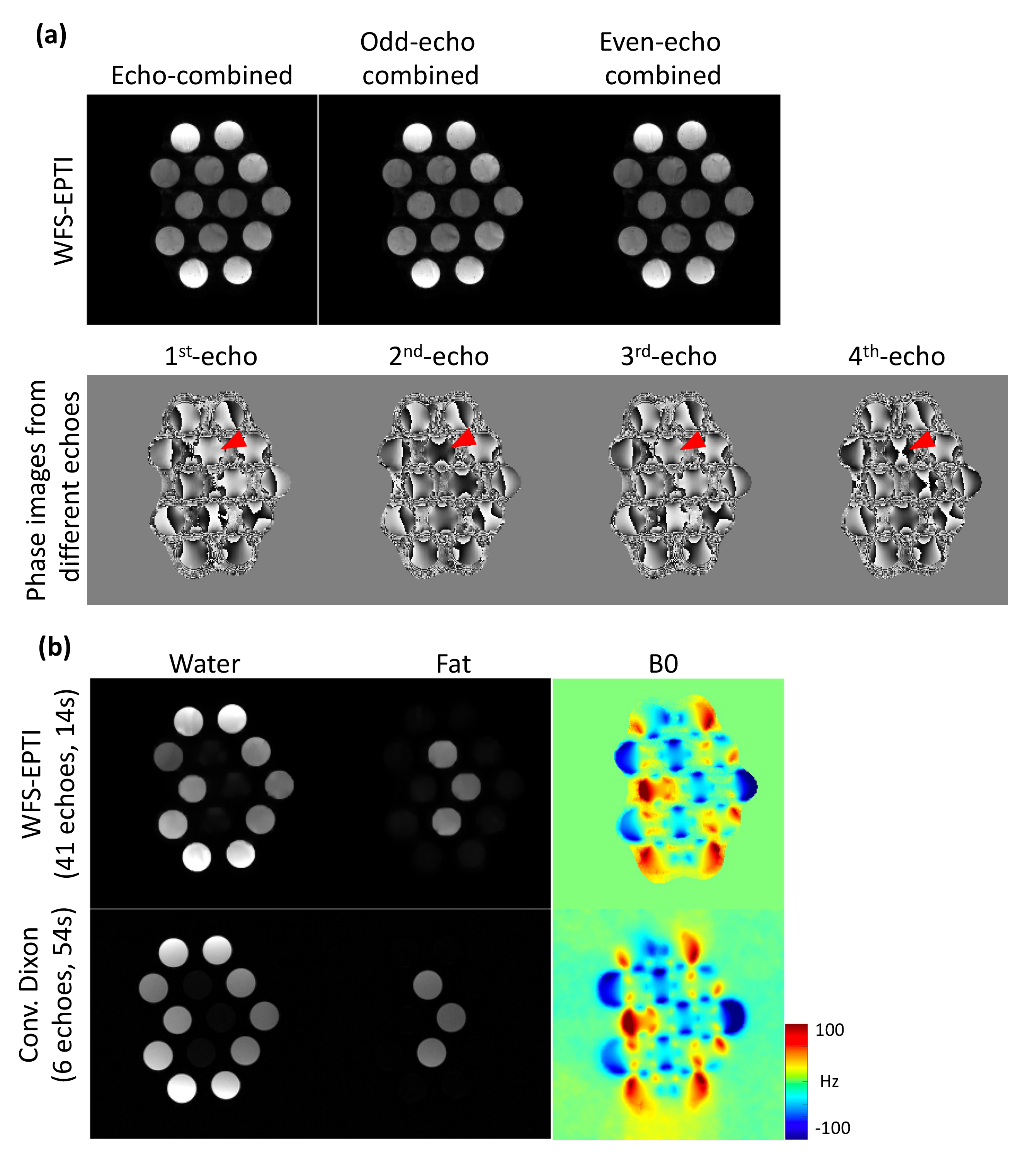
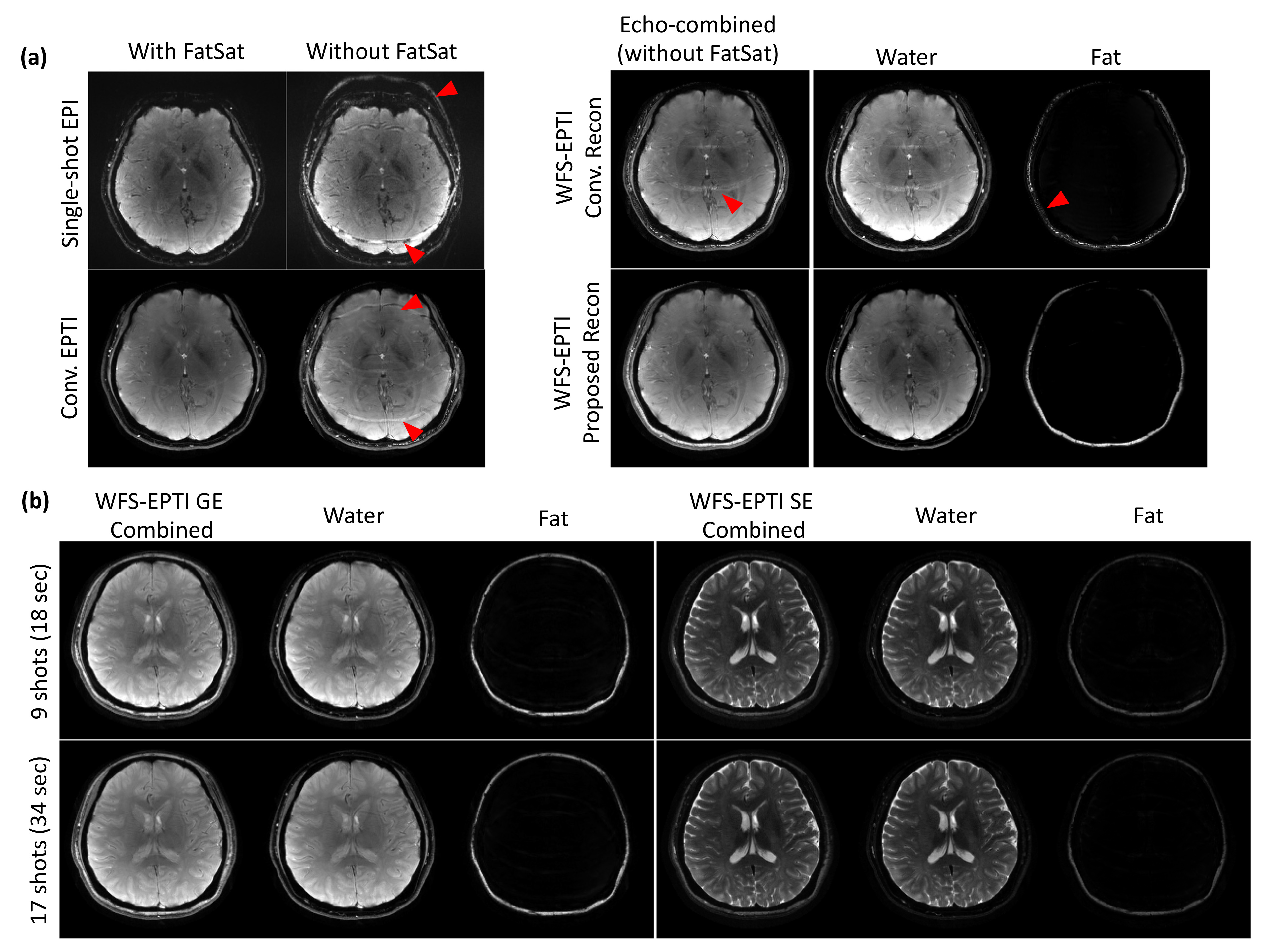
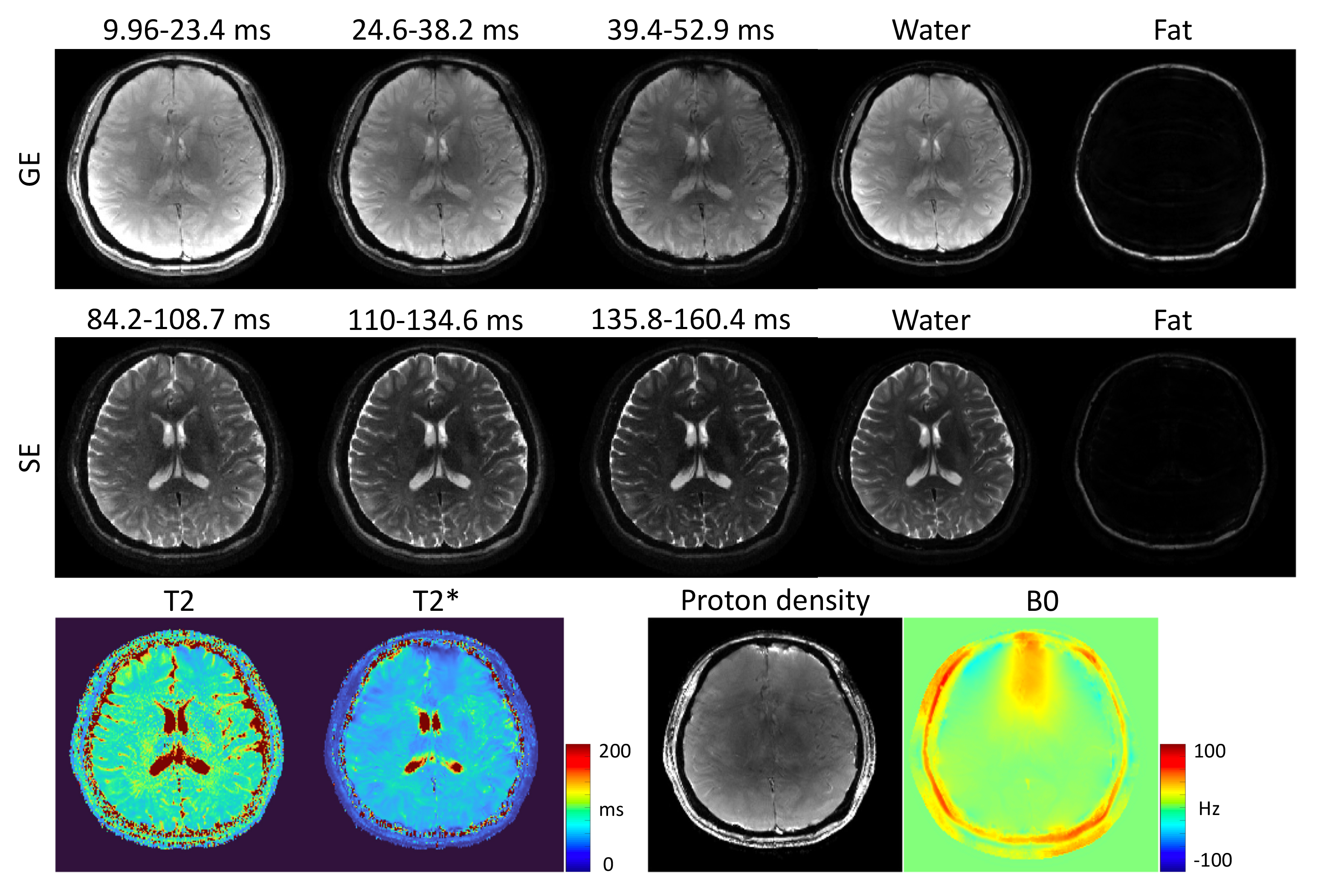
In a water/fat phantom, WFS-EPTI obtained images with minimal fat artifacts (Fig.3a), and reconstructed water/fat images and B0 maps were comparable to those acquired by the standard lengthier GRE acquisition (Fig.3b), demonstrating the efficacy of the proposed method in water/fat separation and the acquisition efficiency (14s vs. 54s). In the in-vivo experiment, without fat-saturation, conventional EPI and EPTI resulted in severe fat artifacts (Fig.4a-left). WFS-EPTI encoding with conventional reconstruction was able to mitigate the fat artifacts but not completely. The proposed WFS-EPTI integrating both the new encoding and reconstruction effectively removes the fat artifacts and results in good water/fat separation (Fig.4a-right-bottom). Fig.4b shows the results from a 9-shot and a 17-shot GESE WFS-EPTI with high image quality. Fig.5 shows the multi-contrast images and quantitative maps (T2, T2*, PD, B0) obtained from the 9-shot WFS-EPTI, all acquired in 18s.CONCLUSION
The proposed WFS-EPTI can separate water/fat effectively, while inheriting the high-resolution, distortion-free, and fast-acquisition features from EPTI. It can extend EPTI to a broader range of applications such as providing fast body imaging while addressing challenges including fat and distortion artifacts.Acknowledgements
This work was supported by the NIH (U24-NS129893, K99-AG083056, R01-EB019437, P41-EB030006), and made possible by resources provided by Shared Instrumentation Grants (S10-OD023637).References
1. Wang F, Dong Z, Reese TG, et al. Echo planar time-resolved imaging (EPTI). Magn Reson Med 2019;81:3599-3615.2. Dong Z, Wang F, Reese TG, Bilgic B, Setsompop K. Echo planar time-resolved imaging with subspace reconstruction and optimized spatiotemporal encoding. Magn Reson Med 2020;84:2442-2455.
3. Wang F, Dong Z, Wald LL, Polimeni JR, Setsompop K. Simultaneous pure T(2) and varying T(2)'-weighted BOLD fMRI using Echo Planar Time-resolved Imaging for mapping cortical-depth dependent responses. NeuroImage 2021;245:118641.
4. Wang F, Dong Z, Reese TG, Rosen B, Wald LL, Setsompop K. 3D Echo Planar Time-resolved Imaging (3D-EPTI) for ultrafast multi-parametric quantitative MRI. NeuroImage 2022;250:118963.
5. Reeder SB, Pineda AR, Wen Z, et al. Iterative decomposition of water and fat with echo asymmetry and least-squares estimation (IDEAL): application with fast spin-echo imaging. Magn Reson Med 2005;54:636-644.
6. Hernando D, Kellman P, Haldar J, Liang ZP. Robust water/fat separation in the presence of large field inhomogeneities using a graph cut algorithm. Magnetic resonance in medicine 2010;63:79-90.
7. Hu Z, Wang Y, Dong Z, Guo H. Water/fat separation for distortion-free EPI with point spread function encoding. Magn Reson Med 2019;82:251-262.
8. Glover G, Schneider E. Three‐point dixon technique for true water/fat decomposition with B0 inhomogeneity correction. Magnetic resonance in medicine 1991;18:371-383.
9. Wang X, Tan Z, Scholand N, Roeloffs V, Uecker M. Physics-based reconstruction methods for magnetic resonance imaging. Philos Trans A Math Phys Eng Sci 2021;379:20200196.
10. Lu W, Yu H, Shimakawa A, Alley M, Reeder SB, Hargreaves BA. Water-fat separation with bipolar multiecho sequences. Magn Reson Med 2008;60:198-209.
11. Soliman AS, Wiens CN, Wade TP, McKenzie CA. Fat quantification using an interleaved bipolar acquisition. Magn Reson Med 2016;75:2000-2008.
12. Dixon WT. Simple proton spectroscopic imaging. Radiology 1984;153:189-194.
Figures