0149
Black blood cine vessel wall imaging based dynamic quantitative mapping of carotid artery vessel wall1Center for Biomedical Imaging Research, Department of Biomedical Engineering, School of Medicine Tsinghua Univeristy, Beijing, China
Synopsis
Keywords: Quantitative Imaging, Quantitative Imaging, vessel wall imaging
Motivation: Dynamic quantitative carotid artery vessel wall imaging can effectively reduce the blurring effect caused by vascular pulsation.
Goal(s): This study aims to develop a black blood cine sequence based dynamic quantitative carotid artery vessel wall imaging method.
Approach: A VFA and ViMSDE duration-based BB cine quantitative sequence was proposed to acquire dynamic multi-contrast images. Dictionary matching method was introduced to estimate quantitative parameters from complicate signal equation.
Results: The proposed protocol was in excellent agreement with standard mapping sequence in both phantom and volunteer experiment. Dynamic T1 and T2 maps has shown its potential in eliminating pulsation resulting blurring
Impact: Dynamic T1 and T2 maps can be acquired with less pulsation related blurring. Dynamic and accurate quantitative information is expected to better assist clinical decision-making.
Introduction
Multi-contrast black blood (BB) vessel wall imaging (VWI) can characterize carotid vulnerable atherosclerotic plaques1. Recently, quantitative imaging has shown its potential in plaque components identification and segmentation2-3. However, vascular pulsation and flow artifacts leads to blurring of the vessel wall which may further affect the accuracy of quantitative mapping. It has been proven that the BB cine VWI can provide images with effective blood suppression4 with potential in generating more accurate dynamic T1 and T2 maps. However, due to the limited acquisition time within a cardiac cycle, the evolution of T1 and T2 resulted signal of k space central line becomes complicate and reaches a new steady state in cine mode. In this work, we proposed a BB cine based dynamic quantitative mapping sequence of carotid vessel wall and evaluated its accuracy and feasibility in generating dynamic T1 and T2 maps.Methods
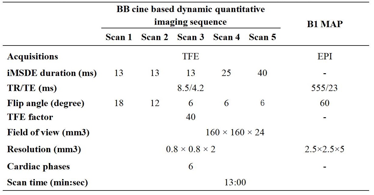
Sequence:The improved motion sensitized driven equilibrium (iMSDE) prepulse based BB VWI sequence was optimized to acquire cine carotid vessel wall images (Figure 1,gif). SPGR acquisition with low-high trajectory was performed after spir and iMSDE prepulse. Five scans with variable flip angles (FA) and iMSDE duration were combined to generate multi-contrast images with different T1- and T2-weighted images (Table 1). B1 map was included for FA correction. Retrospective gating was used. High intensity TFE shots were performed to acquire multi- cardiac phases images with largely shortened echo trains which may affect the steady central line k space signal in low-high trajectory according to Eq.1.
$$M_z^n = M_z^0(1-e^{-\frac{TR}{T1}})\frac{1-(cos\alpha e^{-\frac{TR}{T1}})^{TFE_factor}}{1-cos\alpha e^{-\frac{TR}{T1}}}+M_z^{(n-1)}{({cos\alpha e^{-\frac{TR}{T1}}})}^{TFE_factor} n\geq1 (1)$$
In this study, the cardiac phase was optimized to 6 with a maximum TFE factors of 40. The steady k space central line signal with T1 and T2 mixed with each other is shown in Eq. 2(shot interval not included).
$$ M_{SS} = M_z^0(1-e^{-\frac{TR}{T1}})\frac{1-(cos\alpha e^{-\frac{TR}{T1}})^{TFE_factor}}{1-cos\alpha e^{-\frac{TR}{T1}}}\frac{e^{-\frac{TE_{prep}}{T2}}}{1-{(cos\alpha e^{-\frac{TR}{T1}})}^{TFE_factor}e^{{-\frac{TE_{prep}}{T2}}}} (2)$$
T1&T2 estimation :
Dictionary matching method is preferred with high efficiency compared with numerical solving method. The iMSDE resulting T2 decay and shot parameters (TFE factor and shot interval) determined longitudinal recovery was included in the simulated signal dictionary. Dynamic signal curves generated by varying T1 and T2 value (5ms step for T1 and 1 ms step for T2) were matched with the acquired dynamic curves using orthogonal algorithm after normalization.
Experiments:
Phantom experiments were performed to validate the accuracy of the proposed method on static subject. Volunteer experiments were carried out to evaluate the accuracy and feasibility of the proposed method in generating dynamic T1 and T2 maps.
Phantom study:
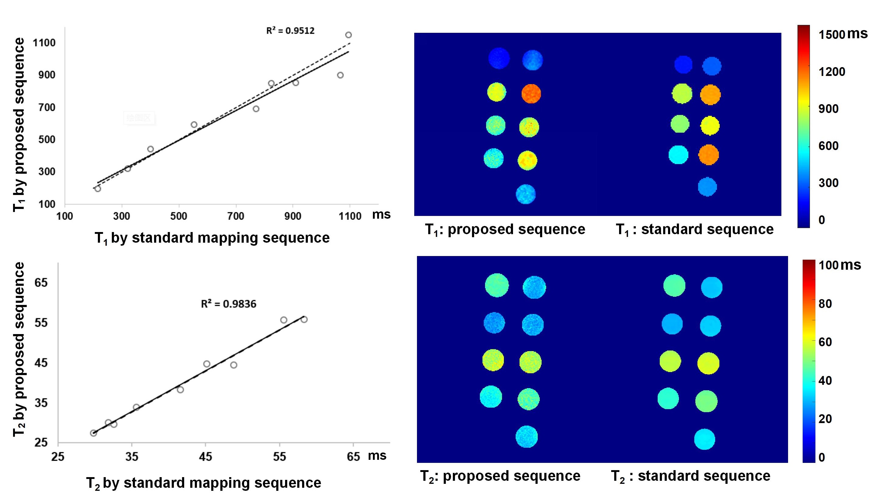
A phantom with different T1 and T2 values was scanned using the proposed BB cine quantitative imaging sequence and standard mapping sequences (IR-SE for T1 mapping and SE for T2 mapping) on a 3.0 T MR scanner (Ingenia CX, Philips Healthcare, Best, The Netherlands) with the 20-channel head coil.
Volunteer study:
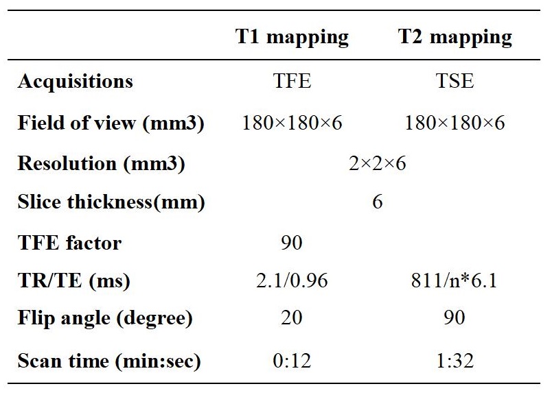
Seven healthy volunteers (26.4±2.9 yrs, 4 males) were recruited in this study. The proposed sequence and clinical referenced mapping (Table 2) sequences were acquired for all subjects on a 3.0 T MR scanner with the 16-channel carotid artery coil.
Statistical analysis:
The correlation of T1 and T2 values measured by the two mehods in phantom experiment was assessed by linear regression. The differences in T1 and T2 value of cervical muscle measured on the two mehods was assessed by paired t-test.
Results:
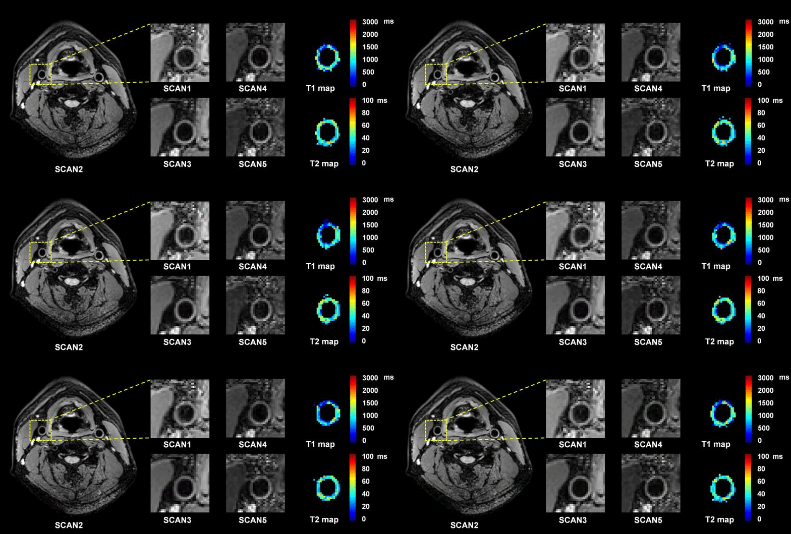
Phantom: Figure 2 showed that the proposed method had an excellent agreement with the standard maps in measuring T1 (R2 =0.95) and T2(R2 =0.98) values of phantoms.Volunteer : No significant difference was found in the T1 (1082.72± 58.24 v.s. 1104.40±29.75 ms, P = 0.23) and T2 (29.88 ± 0.94 v.s. 30.45±1.53 ms, P = 0.24) value of the cervical muscle between the proposed and clinical referenced sequence. The T1 and T2 values of carotid vessel wall of healthy volunteers were 913.02±10.56 ms and 40.53±1.66 ms, respectively. The dynamic T1 and T2 maps of carotid vessel wall are shown in Figure 3(gif), which clearly depict the pulsation of carotid vessel wall without blurring.
Discussion and conclusion:
In this study, a VFA and VMSDE duration-based BB cine quantitative sequence was proposed and validated. The echo train length was optimized to meet the trade off between the SNR and cardiac phase numbers within one RR interval. Dictionary matching based mapping method was proved to be accurate in both phantom and volunteer study. Dynamic T1 and T2 maps has shown its potential in eliminating pulsation resulting blurring. Further study to compare the BB cine based quantitative imaging with static imaging mode and investigate the clinical application for dynamic mapping in carotid artery is warranted.Acknowledgements
No acknowledgement found.References
1 Saba L, Yuan C, Hatsukami TS, Balu N, Qiao Y, DeMarco JK, Saam T, Moody AR, Li D, Matouk CC, Johnson MH, Jäger HR, Mossa-Basha M, Kooi ME, Fan Z, Saloner D, Wintermark M, Mikulis DJ, Wasserman BA; Vessel Wall Imaging Study Group of the American Society of Neuroradiology. Carotid Artery Wall Imaging: Perspective and Guidelines from the ASNR Vessel Wall Imaging Study Group and Expert Consensus Recommendations of the American Society of Neuroradiology. AJNR Am J Neuroradiol. 2018 Feb;39(2):E9-E31.
2 Qiao H, Li D, Cao J, Qi H, Han Y, Han H, Xu H, Wang T, Chen S, Chen H, Wang Y, Zhao X. Quantitative evaluation of carotid atherosclerotic vulnerable plaques using in vivo T1 mapping cardiovascular magnetic resonaonce: validation by histology. J Cardiovasc Magn Reson. 2020 May 21;22(1):38.
3 Jiang Y, Zhu C, Peng W, Degnan AJ, Chen L, Wang X, Liu Q, Wang Y, Xiang Z, Teng Z, Saloner D, Lu J. Ex-vivo imaging and plaque type classification of intracranial atherosclerotic plaque using high resolution MRI. Atherosclerosis. 2016 Jun;249:10-6.
4 Dai E, Dong L, Zhang Z, Li L, Zhang H, Zhao X, Wang J, Yuan C, Guo H. Technical Note: Measurement of common carotid artery lumen dynamics using black-blood MR cine imaging. Med Phys. 2017 Mar;44(3):1105-1112.
Figures
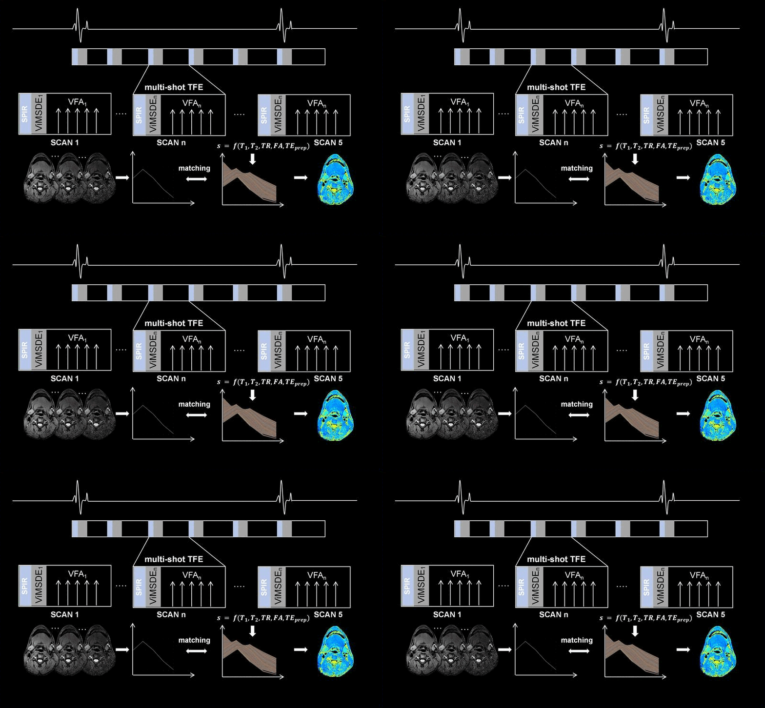
Figure 1 Diagram of the BB cine based dynamic quantitative mapping method: sequence and dictionary matching (gif)