0146
Hybrid Multi-Echo Radial Look-Locker (hME-rLL) Acquisition for Joint Estimation of water-T1, PDFF, and R2*1Electrical and Computer Engineering, University of Arizona, Tucson, AZ, United States, 2Siemens Healthineers, Tucson, AZ, United States, 3Applied Mathematics, University of Arizona, Tucson, AZ, United States, 4Department of Medical Imaging, University of Arizona, Tucson, AZ, United States, 5Siemens Healthineers, Austin, TX, United States, 6Department of Radiological Sciences, University of California Los Angeles, Los Angeles, CA, United States, 7Biomedical Engineering, University of Arizona, Tucson, AZ, United States, 8Medical Imaging, University of Arizona, Tucson, AZ, United States
Synopsis
Keywords: Quantitative Imaging, Liver, T1 mapping, R2* mapping, PDFF mapping
Motivation: Reducing the time required to achieve comprehensive liver evaluation will improve scanning efficiency and increase access to non-invasive diagnostic tools
Goal(s): To develop an acquisition protocol which allows accurate estimation of water-only T1 ($$$T1w$$$), PDFF, and R2*
Approach: Combing dual-echo and extended-echo (>2) readout into a single acquisition, using the extended-echo acquisition to estimate the field-map, R2*, and PDFF. Then using the R2* and field maps to perform fat/water decomposition on the dual-echo acquisition for $$$T1w$$$ estimation.
Results: Both phantom and in vivo results demonstrated that $$$T1w$$$, R2*, and PDFF can be accurately estimated using the proposed approach.
Impact: Increasing the efficiency of MRI sequence and protocols allows for reduced scan time and improved scanner efficiency. These contributions make the diagnostic process easier for patients and physicians, which should result in improved healthcare.
Introduction
Quantitative MRI techniques allow for non-invasive liver assessment. T1-mapping enables diagnosis of conditions like focal liver lesions [1][2], liver fibrosis [3], and cirrhosis [4]. Proton density fat-fraction (PDFF) allows evaluation of patients with fatty liver disease [5][6], while R2* has utility in monitoring patients with iron overload [7][8]. Obtaining these metrics typically requires multiple acquisition protocols, which adds complexity and reduces scanning efficiency. Single-shot inversion recovery radial Look-Locker (rLL) sequences have demonstrated the ability to achieve motion-robust T1 mapping of the abdomen [9][10]. Recent extensions to multi-echo rLL (ME-rLL) in the form of dual-echo acquisition [11][12] combined with Dixon based fat/water decomposition [13] have enabled mapping of the T1 of the water component, referred here as $$$T1w$$$. For accurate estimation of PDFF and R2*, an extended echo (>2) acquisition is required. This work presents a hybrid ME-rLL (hME-rLL) sequence, in which short sampling of the first portion of the T1 recovery curve (T1RC) is performed using a dual-echo acquisition, followed by sampling of the later T1RC using a P-echo (P>2) acquisition. The extended echo at the end of dual-echo readout is used to enable estimation of $$$T1w$$$, PDFF, and R2* from a single pulse sequence.Methods
Technique: A 2D hME-rLL protocol was implemented (Figure 1) to perform slice selective inversion (ssIR) followed by dual-echo gradient echo (GRE) sampling of the early T1RC and ending with P-echo (P>2) GRE sampling of the later T1RC.Radial views acquired for each echo during P-echo sampling are grouped ($$$K_C[p], p=1,...,P$$$) and used with scanner sensitivity maps to reconstruct a set of composite echo images ($$$C[p]$$$). The GRAPHCUT [14] algorithm is then applied to produce estimates of PDFF, R2*, and field-map.
Radial views acquired during the dual-echo acquisition are divided into 16-view groups to produce N TI groups ($$$K[e]_n, e=1,2; n=1,...,N$$$). The TI groups per echo are used in a locally low rank [15] reconstruction to obtain a set of TI images ($$$I[e]_n$$$). The R2* and field-map computed from P-echo data are taken as fixed inputs to the GRAPHCUT fat/water decomposition method, and the echo images for each TI ($$$I[1]_n$$$, $$$I[2]_n$$$) are used to compute a water-only TI image ($$$I_n$$$). The water-only TI images are then taken as input into a deep-learning (DL) based T1-mapping algorithm designed for accurate T1 estimation given a short T1RC [10].
Imaging experiments: Experiments were conducted using Calimetrix R2* and PDFF phantoms, along with vials containing 2% agarose and NiCl2 solutions to produce a range of T1 values. In vivo data was acquired in normal volunteers after informed consent.
ME-rLL and hME-rLL data were acquired at 3T (Skyra, Siemens) with $$$\alpha$$$=8°, TE=1.68ms, and TR=4.16ms (2-echo), 9.58ms (6-echo), 12.22ms (8-echo), 14.86ms (10-echo), 17.5ms (12-echo). Reference 2-echo ME-rLL data was acquired with 1184 views to produce a well sampled T1RC of 4925ms. For the hME-rLL protocol the 2-echo portion of the T1RC was acquired using 256 views (T1RC=1065ms), followed by a 112 (6-echo), 96 (8-echo), 80 (10-echo), or 64 (12-echo) view P-echo acquisition corresponding to a ~1s sampling. The total per-slice acquisition was ~2s.
Results and Discussion
Figures 2-3 demonstrate the impact of $$$P$$$ on $$$T1w$$$, PDFF, and R2* estimation in phantoms. For all metrics there is little observable difference when varying $$$P$$$. All vials show close agreement to the reference as evidenced by the correlation analysis (Figure 3) which shows $$$r^2$$$≥0.96 (0.92≤slope≤1.04) for all $$$P$$$ values.Figures 4-5 demonstrates the proposed method in vivo. Again for all metrics there is little qualitative difference between results produces at different $$$P$$$ values (Figure 4). For quantitative comparison, representative ROIs (Figure 5a) were selected from tissue regions of each acquired slice. The $$$T1w$$$ estimates across $$$P$$$ values was within 1.5 standard deviations of the reference for all tissue types. For PDFF, the absolute error with respect to the reference was within 3.1% for all tissue (excluding fat) for all $$$P$$$ values. For fat, the error was within 6.1% for all $$$P$$$ values. For R2*, the hME-rLL estimated values were within 1.5 standard deviations of the reference for liver, muscle, and kidney. For spleen, increase variability within both the hME-rLL and reference datasets make quantification of agreement challenging.
Conclusions
This work proposed a hybrid multi-echo radial Look-Locker (hME-rLL) sequence which enables accurate estimation of registered $$$T1w$$$, PDFF, and R2* maps within a single acquisition. This will assist in interpretation and improve scanning efficiency for the evaluation of conditions such as fatty liver disease.Acknowledgements
We would like to acknowledge grant support from the National Institutes of Health (CA245920 and EB031894), Arizona Biomedical Research Centre (CTR056039), and the Technology and Research Initiative Fund (TRIF) Improving Health Initiative.References
[1] N. Yoshimura, K. Saito, T. Saguchi, T. Funatsu, Y. Araki, S. Akata and K. Tokuuye, "Distinguishing hepatic hemangiomas from metastatic tumors using T1 mapping on gadoxetic-acid-enhanced MRI," Magn Reson Imaging., vol. 31, no. 1, pp. 23-27, 2013.
[2] M. Mio, Y. Fujiwara, K. Tani, T. Toyofuku, T. Maeda and T. Inoue, "Quantitative evaluation of focal liver lesions with T1 mapping using a phase-sensitive inversion recovery sequence on gadoxetic acid-enhanced MRI," European Journal of Radiology, vol. 8, p. 100312, 2021.
[3] J. A. Luetkens, S. Klein, F. Traeber, F. C. Schmeel, A. M. Sprinkart, D. L. R. Kuetting, W. Block, K. Hittatiya, F. E. Uschner, R. Schierwagen, J. Gieseke, H. H. Schild, J. Trebicka and G. M. Kukuk, "Quantitative liver MRI including extracellular volume fraction for non-invasive quantification of liver fibrosis: a prospective proof-of-concept study.," Gut, vol. 67, no. 3, pp. 593-594, 2018.
[4] K. A. Kim, M.-S. Park, I.-S. Kim, B. Kiefer, W.-S. Chung, M.-J. Kim and K. W. Kim, "Quantitative evaluation of liver cirrhosis using T1 relaxation time with 3 tesla MRI before and after oxygen inhalation.," Journal of magnetic resonance imaging, vol. 36, no. 2, pp. 405-410, 2012.
[5] J. Starekova, D. Hernando, P. J. Pickhardt, S. B. Reeder, "Quantification of Liver Fat Content with CT and MRI.," Radiology, vol. 301, no. 2, 2021.
[6] T. Armstrong, X. Zhong, S. Shih, E. Felker, D. Lu, B. Dale, H. Wu, "Free-breathing 3D stack-of-radial MRI quanitification of liver fat and R2* in adults withfatty liver disease.," Magnetic Resonance Imaging, vol. 85, pp. 141-152, 2022.
[7] J. C. Wood, C. Enriquez, N. Ghugre, J. M. Tyzka, S. Carson, M. D. Nelson, T. D. Coates, "MRI R2 and R2* mapping accurately estimates hepatic iron concentration in transfusion-dependent thalassemia and sickle cell disease patients,." Blood, vol. 106, no. 4, 2005.
[8] M. S. Sussman, R. Ward, K. H. Kuo, G. Tomlinson, K. S. Jhaveri, "Impact of MRI technique on clinical decision-making in patients with liver iron overload: comparison of FerriScan- versus R2*-derived liver iron concentration,." European Radiology, vol. 30, pp 1959-1968, 2020.
[9] X. Wang, S. Rosenzweig, N. Scholand, H. C. M. Holme and M. Uecker, "Model-based reconstruction for simultaneous multi-slice T1 mapping using single-shot inversion-recovery radial FLASH," Magnetic Resonance in Medicine, vol. 85, no. 3, pp. 1258-1271, 2021.
[10] E. Ahanonu, K. Johnson, U. Goerke, B. Toner, V. Deshpande, A. Bilgin and M. Altbach, "T1 mapping of the entire abdomen using a time efficient free breathing neural network radial Look Locker approach," in Proceedings of the International Society for Magnetic Resonance in Medicine, Toronto, 2023.
[11] Z. Li, M. Mathew, A. B. Syed, L. Feng, R. Brunsing, J. M. Pauly, S. S. Vasanawala, "Rapid fat-water separated T1 mapping using a sigle-shot radial inversion-recovery spoiled gradient recalled pulse sequence,." NMR in Biomedicine, 2022
[12] E. H. Moquilaza, K. Weiss, Y. J. Lee, J. Stelter, T. Amthor, P. Koken, M. R. Makowski, R. Braren, M. Doneva, D. C. Karampinous, "Fast multi-slice liver water T1 mapping using single-shot continuous inversion recovery spiral imaging,." ISMRM Sedona Workshop, Janurary 2023.
[13] Berglund J, Ahlström H, Johansson L, Kullberg J. Two-point Dixon method with flexible echo times. Magn Reson Med. 2011
[14] D. Hernando, P. Kellman, J. P. Halder, Z-P. Liang, "Robust water/fat separation in the presence of large field inhomogeneities using a graph cut algorithm,." Magn Reson Med., 63(10, pp 79-90, 2010.
[15] https://mrirecon.github.io/bart
Figures
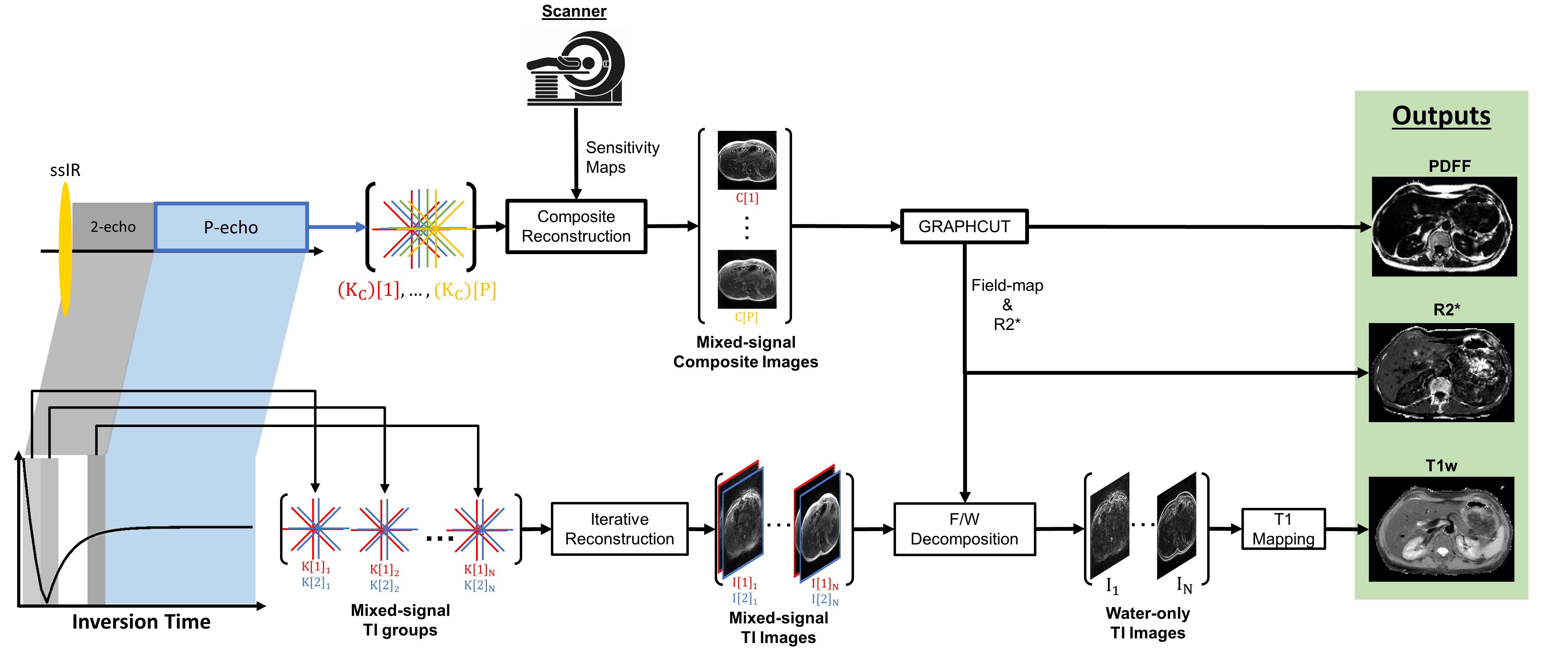
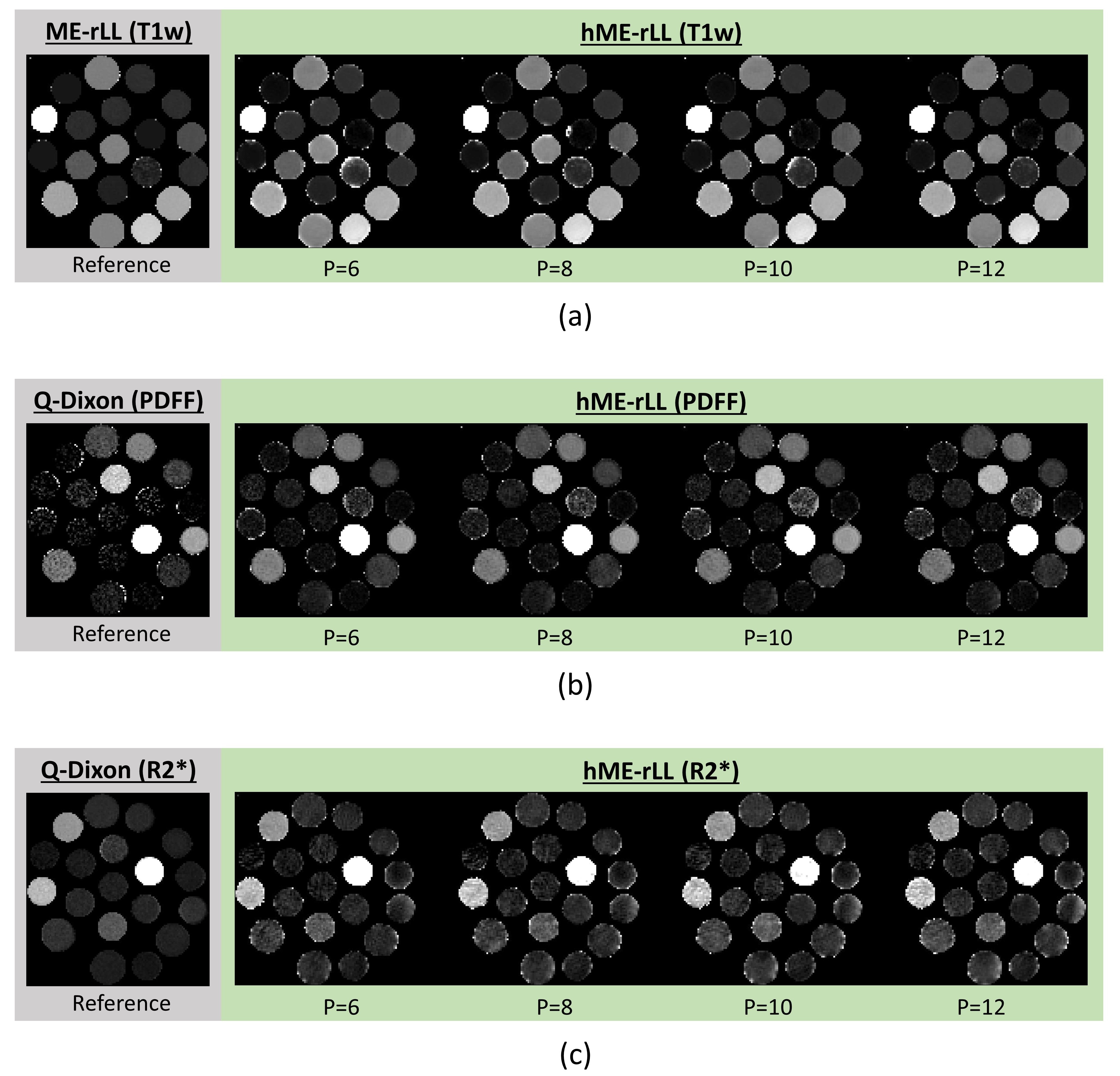
Figure 2. Comparing $$$T1w$$$ (a), PDFF (b), and R2* (c) generated values across choice of $$$P$$$ for phantom data. Reference data for $$$T1w$$$ comes from a dual-echo ME-rLL acquisition with a 5s T1RC and Dixon based fat/water separation, followed by Bloch based T1 fitting. The refence values for PDFF and R2* are computed using the Q-Dixon method utilizing a 6-echo acquisition.
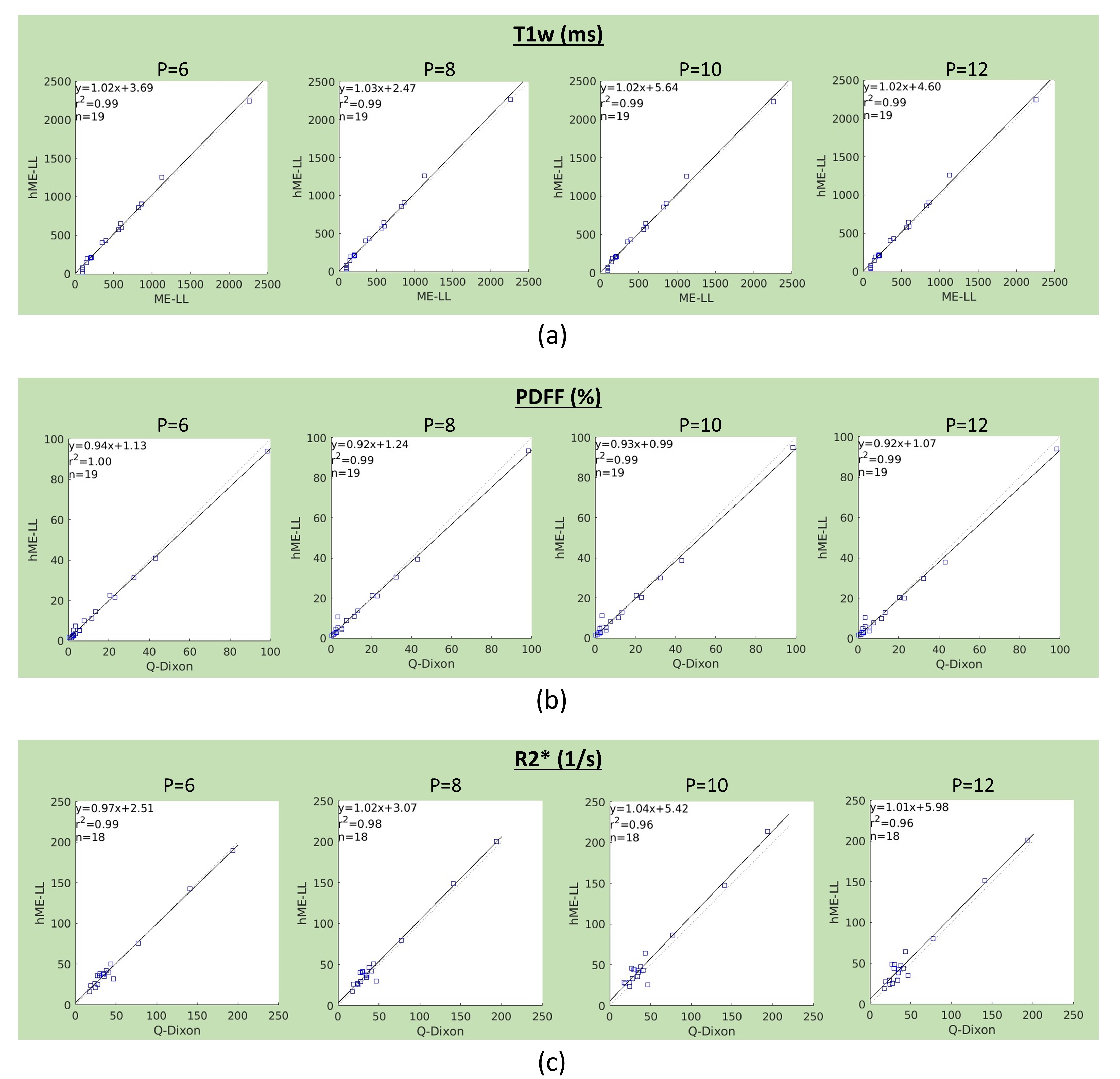
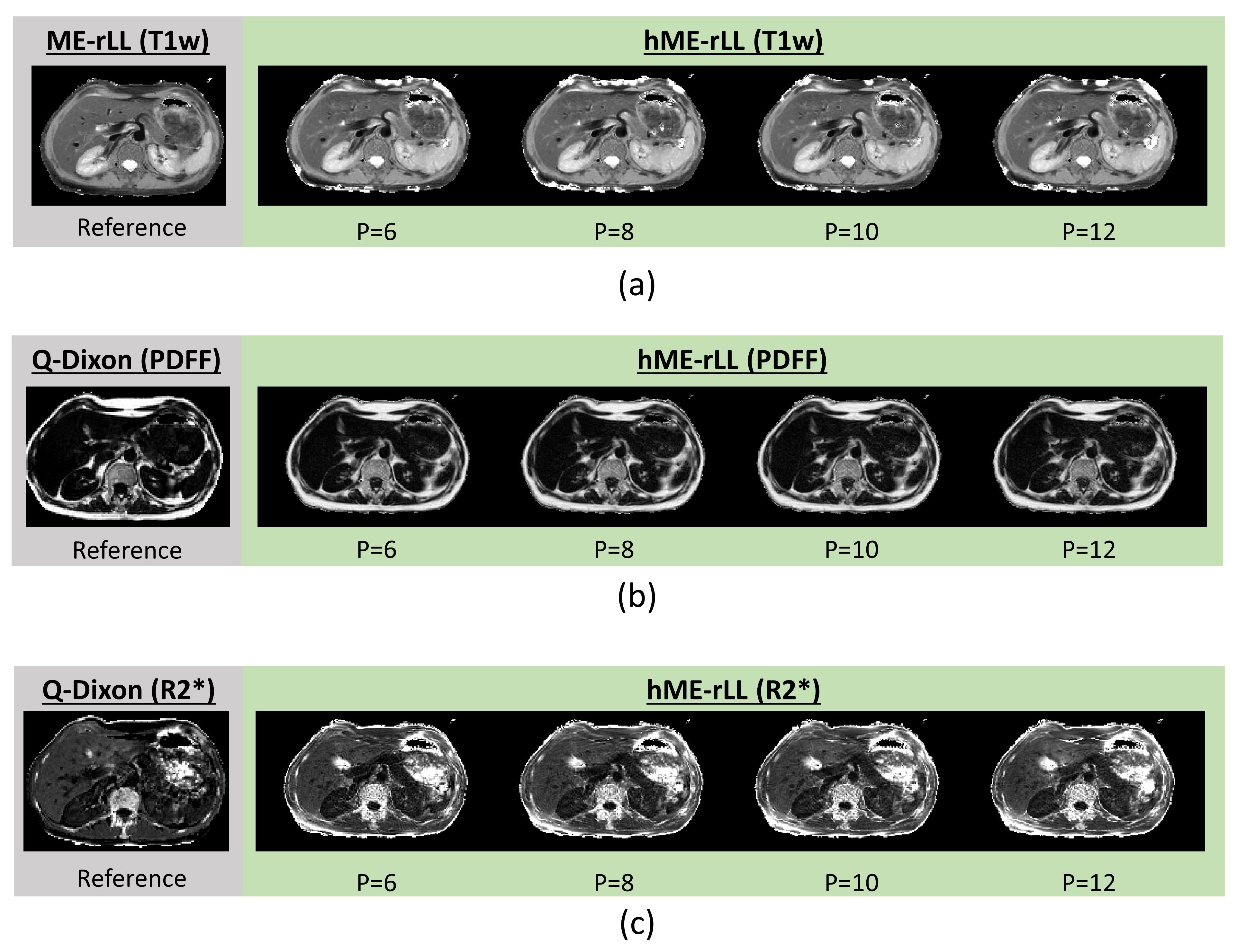
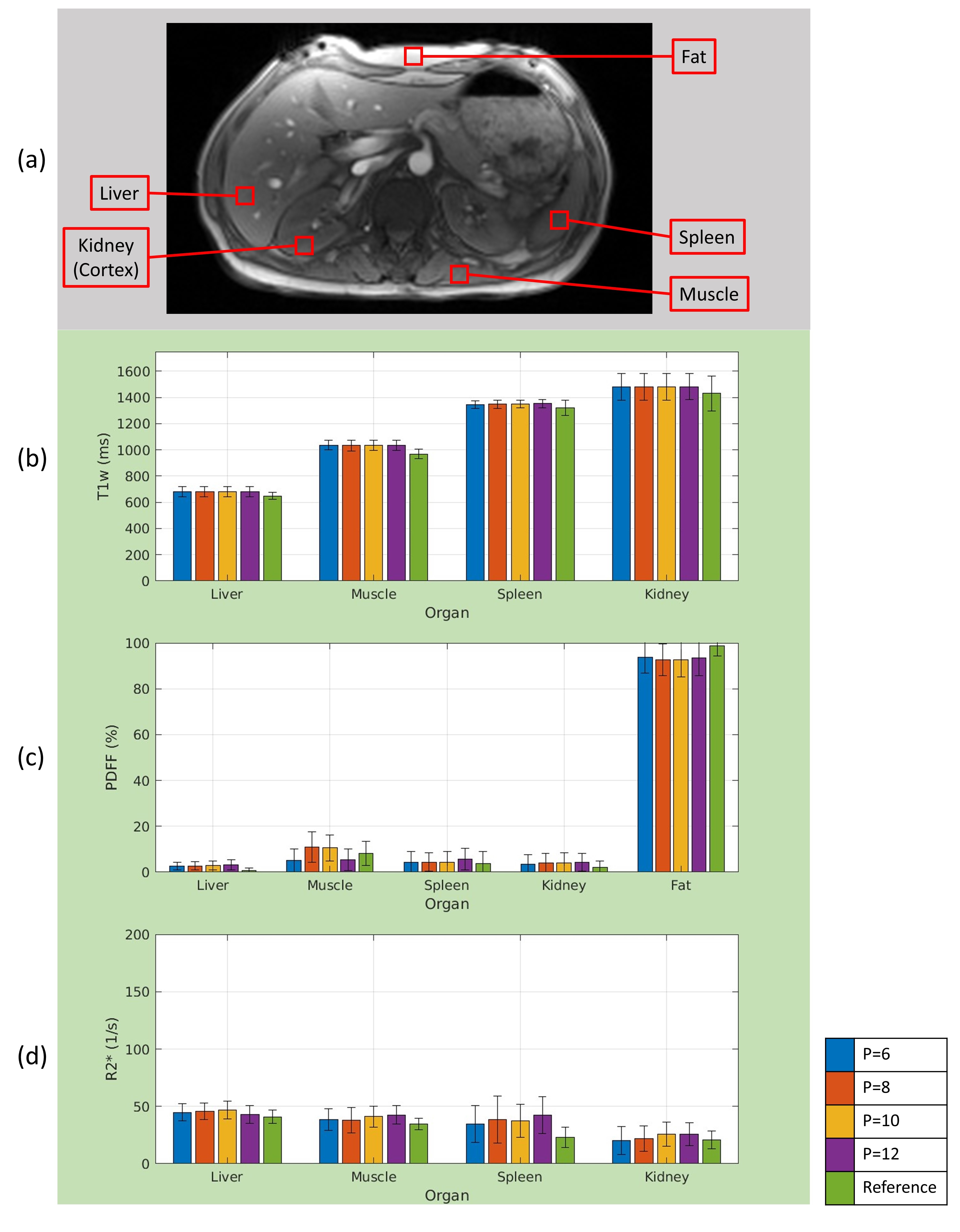
Figure 5. (a) Abdominal image showing the approximate locations used to generate the tissue specific bar-plots in (b)-(d). The average value within each tissue ROI for each metric and $$$P$$$ value define the bar graphs.