0144
The Influence of Accelerated Brain Aging on Coactivation Pattern Dynamics in Parkinson's Disease1Tongji Hospital, Tongji Medical College, Huazhong University of Science and Technology, Wuhan, China
Synopsis
Keywords: Parkinson's Disease, Parkinson's Disease
Motivation: Aging has been widely recognized as the primary risk factor for brain degeneration, and Parkinson’s disease (PD) tends to follow accelerated aging trajectories.
Goal(s): The aim of this study was to investigate the influence of structural brain aging on large-scale functional network temporal dynamics in PD.
Approach: The level of brain aging was assessed by calculating global and local brain age gap estimates from T1-weighted images. Coactivation patterns of the whole brain were identified from fMRI to capture neural network activity.
Results: Accelerated structural brain aging in PD affected brain function, which manifested as aberrant brain network dynamics.
Impact: These findings relate whole-brain coactivation patterns to spatial variation in accelerated brain aging, providing insights into the neuropathological mechanisms in neurodegenerative diseases and implying the possibility of intervention for PD progression by slowing the brain aging process.
Introduction
Parkinson’s disease (PD) is primarily driven by the degeneration of dopaminergic neurons within substantia nigra. Although the etiology and pathogenesis of PD is unknown, it has been acknowledged that aging is the predominant risk factor for brain degeneration1, 2. Several major contributors to PD development, including mitochondrial dysfunction, oxidative stress, neuroinflammation, and protein homeostasis disruption, overlap with molecular hallmarks in the normal brain aging process3. The trajectories of brain aging follow specific patterns of brain morphological alterations in the lifespan4. Neurodegenerative diseases tend to accelerate regional brain atrophy beyond the normal aging effect5, 6. On the other hand, age-related functional brain changes also reflect on static and dynamic functional connectivity7-10. Considering the convergence of structural and functional alterations in PD and normal aging, our study aims to investigate the impact of structural brain aging, as evidenced by global and local brain-age-gap-estimates (G-brainAGE and L-brainAGE) on network dynamics in PD.Methods
With approval of the Institutional Review Board and signed informed consent of all subjects, a total of 62 PD patients and 32 healthy controls (HCs) were recruited in our study. We obtained High resolution 3D T1-weighted brain volume images and resting-state functional images using a 3.0 T scanner (Discovery MR 750, GE Medical Systems, Waukesha, WI) with the 32-channel head coil. The severity of PD and motor disability was assessed using the Hoehn & Yahr (H&Y) stage and the third part of the Unified Parkinson’s Disease Rating Scale (UPDRS-III). With T1-weighted images as inputs, we yielded voxel‐wise L-brainAGE and G-brainAGE using a convolutional neural network (U‐Net) with the parameters pre-trained in an independent sample of 3463 healthy individuals aged 18–90 years (https://github.com/SebastianPopescu/U-NET-for-LocalBrainAge-prediction)11. A whole-brain seed-free voxel-wise coactivation pattern (CAP) analysis was conducted using the TbCAPs Toolbox12. To evaluate the dynamic properties within and between CAPs, we calculated the following CAP metrics including fraction of time, persistence, and transition probability. The significance of the spatial correspondence between L-brainAGE t-value map and CAPs maps was estimated using a spatial autocorrelation-preserving permutation test (spin tests), which generates randomly rotated brain maps preserving spatial covariance13.Results
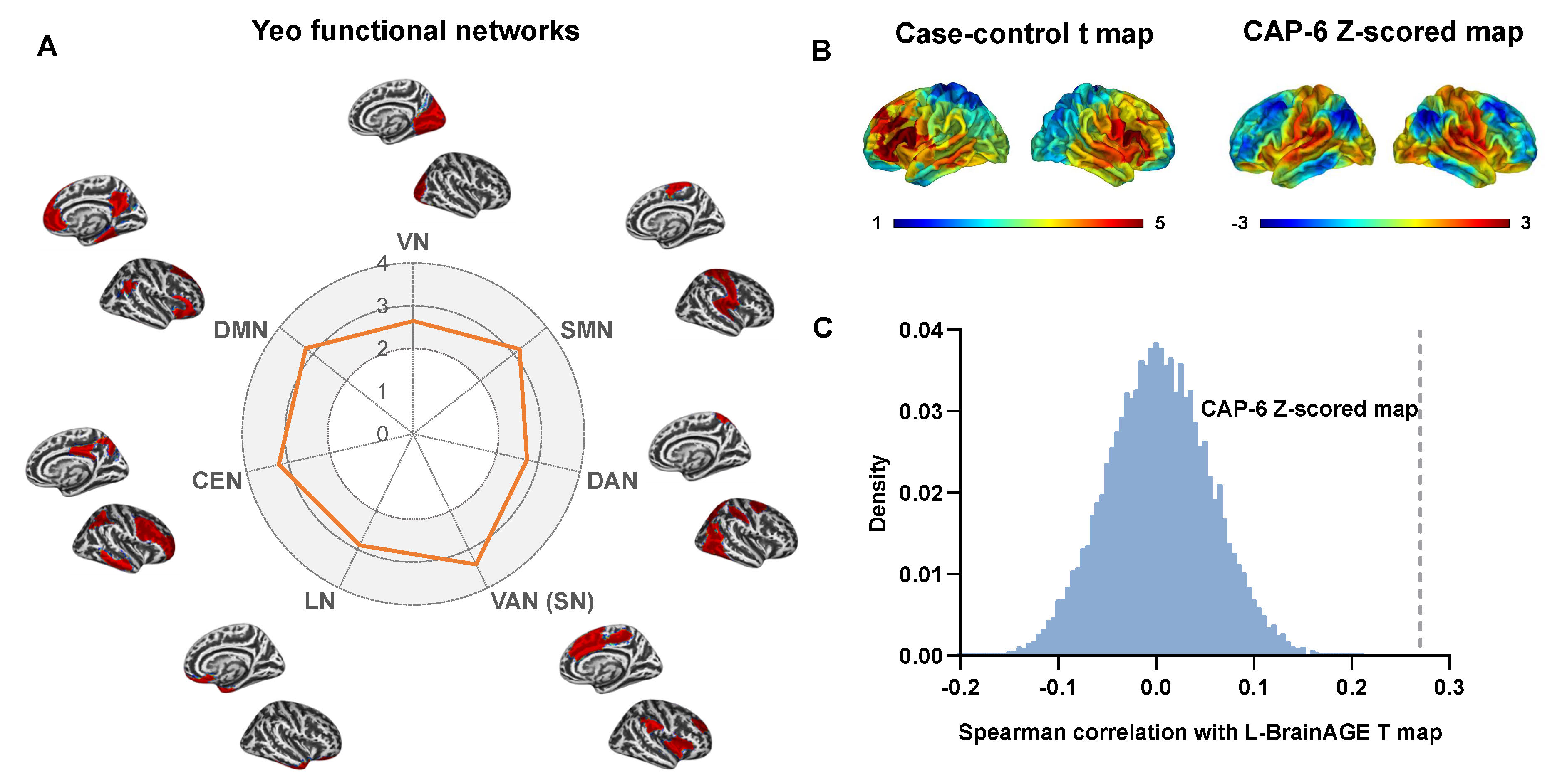
Significant increased brainAGE was indicated in patients with PD at the whole brain level and in the specific areas covering the middle and inferior frontal lobe, superior and middle temporal lobe, insular gyrus, precentral gyrus, and putamen. And the G-brainAGE was correlated with disease severity and duration. The six coactivation patterns were generated from all HCs using k-means clustering. PD patients remained less fraction of time in CAP 2 with the default mode network (DMN) and frontoparietal network (FPN) activated and CAP 6 with the sensorimotor network (SMN) and salience network (SN) activated, and had the lower transition probability to these CAPs. Notably, the pattern of localized brain aging shared spatial similarities with the CAP 6, which may offer structural basis for functional network dynamics alterations.Discussion
Neurodegenerative diseases generally deviate from the normal aging trajectory14. Consistent with our findings, prior studies demonstrated that the advanced brain age in PD was mild to moderate compared with HCs15-17. Since numerous neuropsychiatric disorders related with increased brain age, L-brainAGE could characterize more disease-specific regional information than individual metrics. The findings of L-brainAGE showed a pattern of widespread increased brainAGE that was most pronounced in the frontal, temporal, and putamen regions. The spatial variations of accelerated brain aging were similar to atrophy patterns of caudate, putamen, temporal/hippocampal, frontal and parietal areas in de novo PD18.Most CAP states demonstrated aberrant dynamic characteristics, reflecting susceptible functional networks in PD. These results extend the current understanding of PD-related dynamic abnormalities that brain network reconfiguration in PD involves not only primary (VN, SMN) but also higher-order processing networks (DMN, FPN, SN). Regional brain atrophy of the frontotemporal lobes, insular gyrus, and precentral gyrus indicated that the process of accelerated brain aging is characterized by selective susceptibility in brain networks19. Gray matter losses tend to result in inefficient recruitment of brain functional networks in PD20. Therefore, the spatial similarity between the L-brainAGE t map and CAP 6 could explain, to some extent, the decreased fraction of time and persistence in CAPs for PD patients.
Conclusions
This study revealed the influence of accelerated brain aging pattern on abnormal brain network dynamics in PD. Abnormal network dynamics in the DMN-FPN and SMN-SN states of PD, which are linked to reduced structural brain integrity as indicated by BrainAGE. These findings could emphasize the importance of interventions to prevent or slow age-related deterioration and enhance our understanding of the associations between normal brain aging and neurodegenerative diseases.Acknowledgements
This study was supported by the Regional Innovation and Development Joint Fund of National Natural Science Foundation of China (U22A20354), National Key Research and Development Program of China (2022YFC2406903) and Key Research and Development Project of Hubei Province (2021BCA123).
References
1 Collier TJ, Kanaan NM, Kordower JH (2011) Ageing as a primary risk factor for Parkinson's disease: evidence from studies of non-human primates. NATURE REVIEWS NEUROSCIENCE 12:359-366
2 Phillipson OT (2014) Management of the aging risk factor for Parkinson's disease. NEUROBIOLOGY OF AGING 35:847-857
3 Coleman C, Martin I (2022) Unraveling Parkinson's Disease Neurodegeneration: Does Aging Hold the Clues? J Parkinsons Dis 12:2321-2338
4 Lemaitre H, Goldman AL, Sambataro F et al (2012) Normal age-related brain morphometric changes: nonuniformity across cortical thickness, surface area and gray matter volume? NEUROBIOLOGY OF AGING 33:617.e611-619
5 Lewis MM, Du G, Lee EY et al (2016) The pattern of gray matter atrophy in Parkinson's disease differs in cortical and subcortical regions. JOURNAL OF NEUROLOGY 263:68-75
6 McDonald CR, McEvoy LK, Gharapetian L et al (2009) Regional rates of neocortical atrophy from normal aging to early Alzheimer disease. NEUROLOGY 73:457-465
7 Ferreira LK, Busatto GF (2013) Resting-state functional connectivity in normal brain aging. NEUROSCIENCE AND BIOBEHAVIORAL REVIEWS 37:384-400
8 Damoiseaux JS (2017) Effects of aging on functional and structural brain connectivity. NEUROIMAGE 160:32-40
9 Zhang H, Lee A, Qiu A (2017) A posterior-to-anterior shift of brain functional dynamics in aging. Brain Struct Funct 222:3665-3676
10 Tian L, Li Q, Wang C, Yu J (2018) Changes in dynamic functional connections with aging. NEUROIMAGE 172:31-39
11 Popescu SG, Glocker B, Sharp DJ, Cole JH (2021) Local Brain-Age: A U-Net Model. Frontiers in Aging Neuroscience 13:761954
12 Bolton TAW, Tuleasca C, Wotruba D et al (2020) TbCAPs: A toolbox for co-activation pattern analysis. NEUROIMAGE 211:116621
13 Alexander-Bloch AF, Shou H, Liu S et al (2018) On testing for spatial correspondence between maps of human brain structure and function. NEUROIMAGE 178:540-551
14 Anderson VM, Schott JM, Bartlett JW, Leung KK, Miller DH, Fox NC (2012) Gray matter atrophy rate as a marker of disease progression in AD. NEUROBIOLOGY OF AGING 33:1194-1202
15 Beheshti I, Mishra S, Sone D, Khanna P, Matsuda H (2020) T1-weighted MRI-driven Brain Age Estimation in Alzheimer's Disease and Parkinson's Disease. Aging and Disease 11:618-628
16 Eickhoff CR, Hoffstaedter F, Caspers J et al (2021) Advanced brain ageing in Parkinson's disease is related to disease duration and individual impairment. Brain Commun 3:fcab191
17 Charissé D, Erus G, Pomponio R et al (2022) Brain age and Alzheimer's-like atrophy are domain-specific predictors of cognitive impairment in Parkinson's disease. NEUROBIOLOGY OF AGING 109:31-42
18 Sarasso E, Agosta F, Piramide N, Filippi M (2021) Progression of grey and white matter brain damage in Parkinson's disease: a critical review of structural MRI literature. JOURNAL OF NEUROLOGY 268:3144-3179
19 Zhang HY, Chen WX, Jiao Y, Xu Y, Zhang XR, Wu JT (2014) Selective vulnerability related to aging in large-scale resting brain networks. PLoS One 9:e108807
20 Lucas-Jiménez O, Ojeda N, Peña J et al (2016) Altered functional connectivity in the default mode network is associated with cognitive impairment and brain anatomical changes in Parkinson's disease. PARKINSONISM & RELATED DISORDERS 33:58-64
Figures
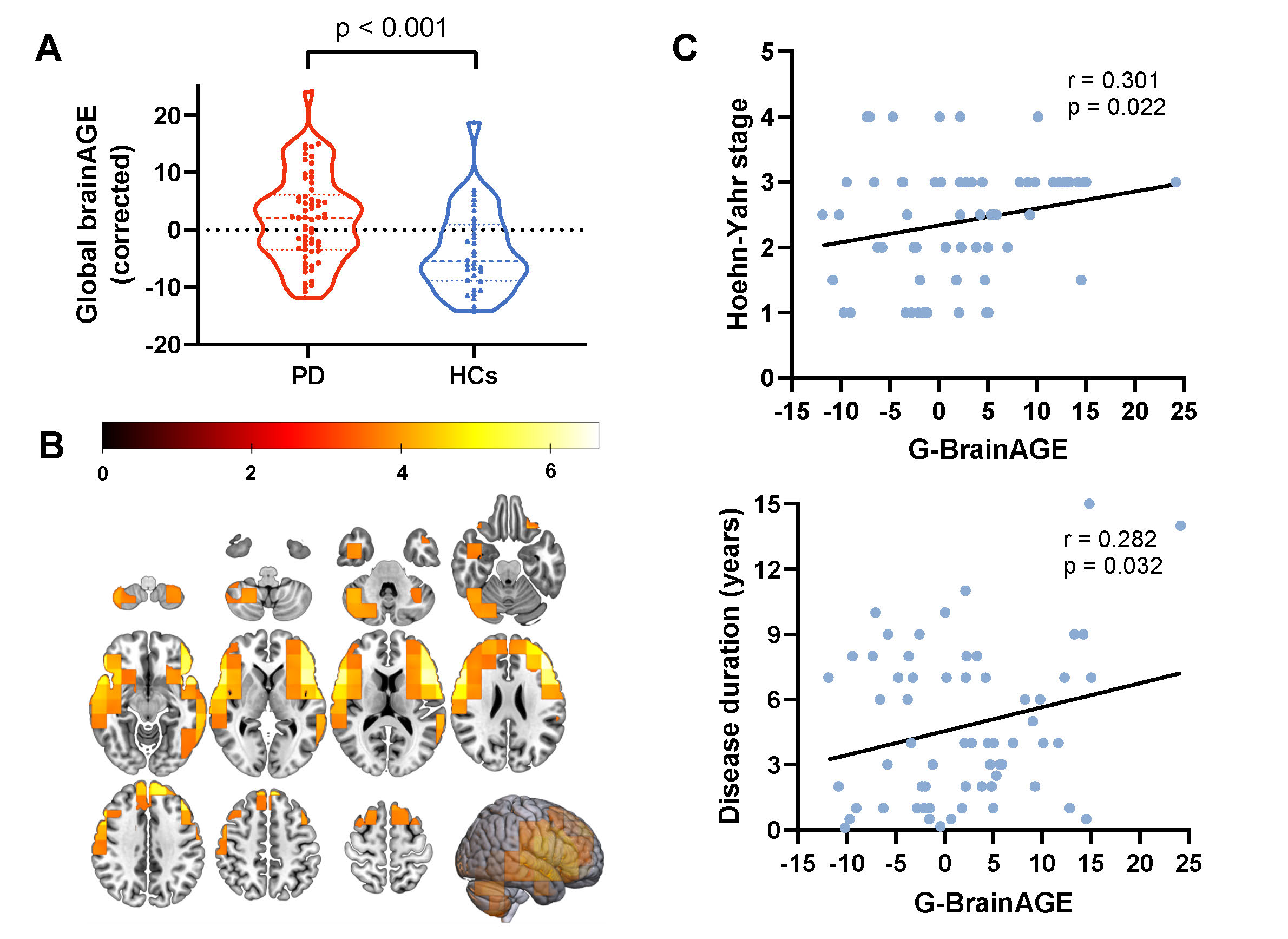
Figure 1 Abnormal brain aging patterns in PD. (A) G-brainAGE were significantly higher in PD group than in HCs group (T = 3.658, p < 0.001). (B) Differences in L-brainAGE between HCs and PD patients. The T-value map was overlayed to show brain regions with statistically significant group differences in L-brainAGE (p < 0.05, voxel-level FWE corrected). (C) The associations of G-brainAGE with Hoehn-Yahr stage and disease duration time (p < 0.05). PD, Parkinson’s disease; HCs, health controls; G-brainAGE, global brain-age-gap-estimates; L-brainAGE, local brain-age-gap-estimates.
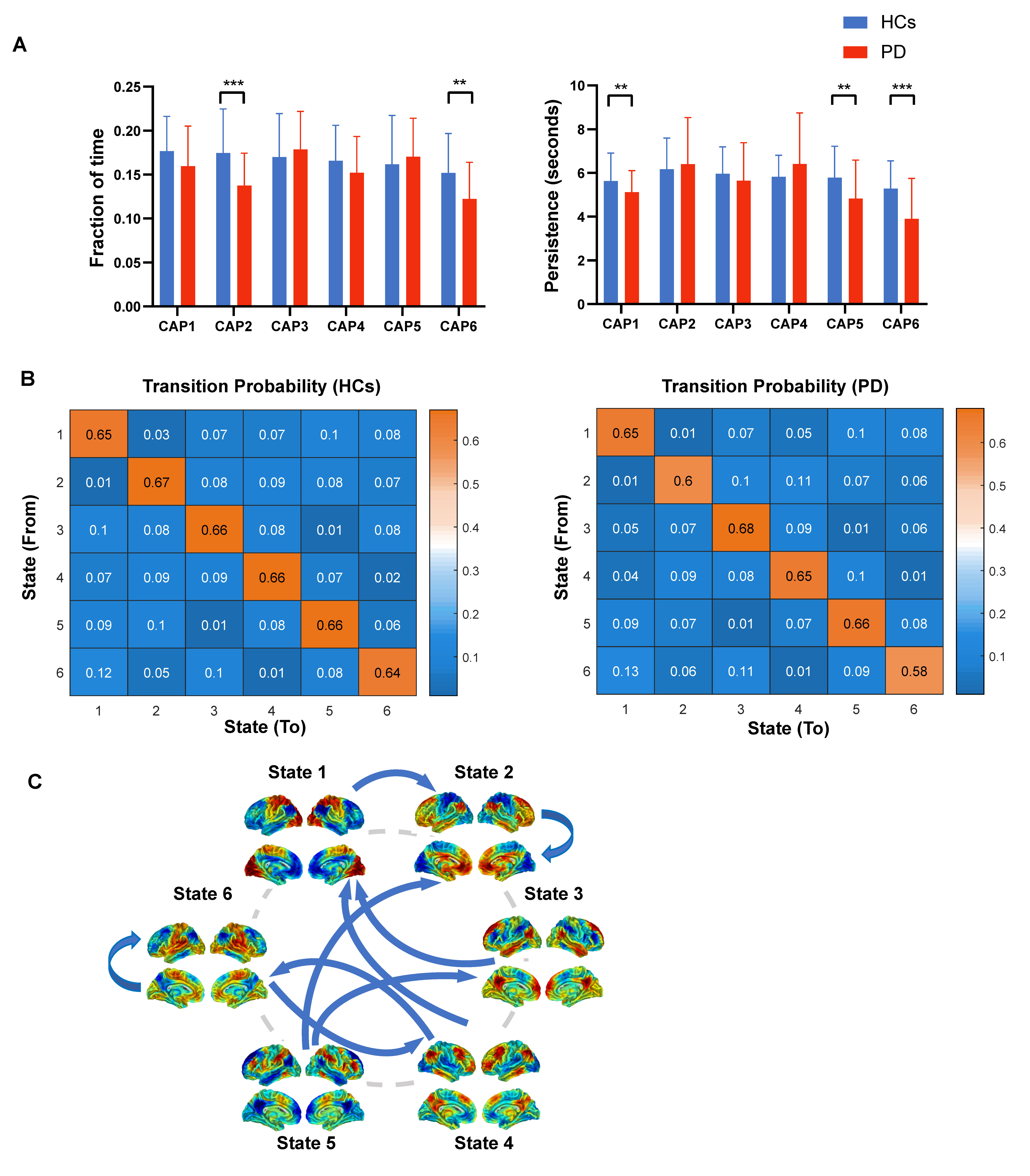
Figure 2 CAP dynamic differences between PD patients and HCs. (A) The group differences in fraction of time and persistence. (B) The group average transition probability matrix for HCs and PD patients. C) Decreased transition probability between certain CAPs in PD patients compared with HCs. Two-sample t-tests were performed with age, sex, and education years as covariates. * p < 0.05, ** p < 0.01, *** p < 0.001 with FDR correction. CAP, co-activation pattern; PD, Parkinson’s disease; HCs, health controls.