0143
Brain arteriolosclerosis in community-based older adults is associated with lower gray matter volume1Department of Biomedical Engineering, Illinois Institute of Technology, Chicago, IL, United States, 2Rush Alzheimer’s Disease Center, Rush University Medical Center, Chicago, IL, United States
Synopsis
Keywords: Dementia, Blood vessels, Arteriolosclerosis, Brain, Pathology, Ex-vivo applications, Gray matter, Neurodegeneration, Vascular
Motivation: Despite brain arteriolosclerosis being one of the most prevalent small vessel diseases in older adults, its association with regional brain volumes has not been investigated.
Goal(s): To investigate the association of brain arteriolosclerosis with regional gray matter volumes.
Approach: Regional brain volumetry on ex-vivo MRI and detailed neuropathological examination were combined in a large number of community-based older adults that came to autopsy.
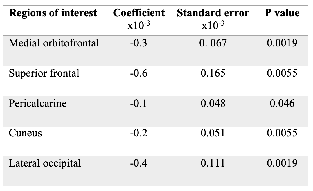
Results: More severe brain arteriolosclerosis was associated with lower volume in a number of gray matter regions, including medial orbitofrontal, superior frontal, pericalcarine, cuneus, and lateral occipital areas, independently of the effects of other neuropathologies.
Impact: The finding that brain arteriolosclerosis is associated with lower regional gray matter volumes independently of the effects of other neuropathologies enhances our understanding of the brain anomalies associated with this common small vessel disease pathology.
Introduction
Brain arteriolosclerosis is characterized by thickening of vessel walls and arteriolar stenosis and is one of the primary pathologies of cerebral small vessel disease1. It has a higher occurrence among older adults and is more severe in women2 and black individuals3. Arteriolosclerosis is linked to lower cognitive and motor function4, as well as an elevated risk of dementia5. Despite its prevalence and deleterious effects, the impact of arteriolosclerosis on brain macrostructure has not been investigated. Therefore, the aim of this study was to investigate the association of brain arteriolosclerosis with regional gray matter volumes in a large number of community-based older adults.Methods
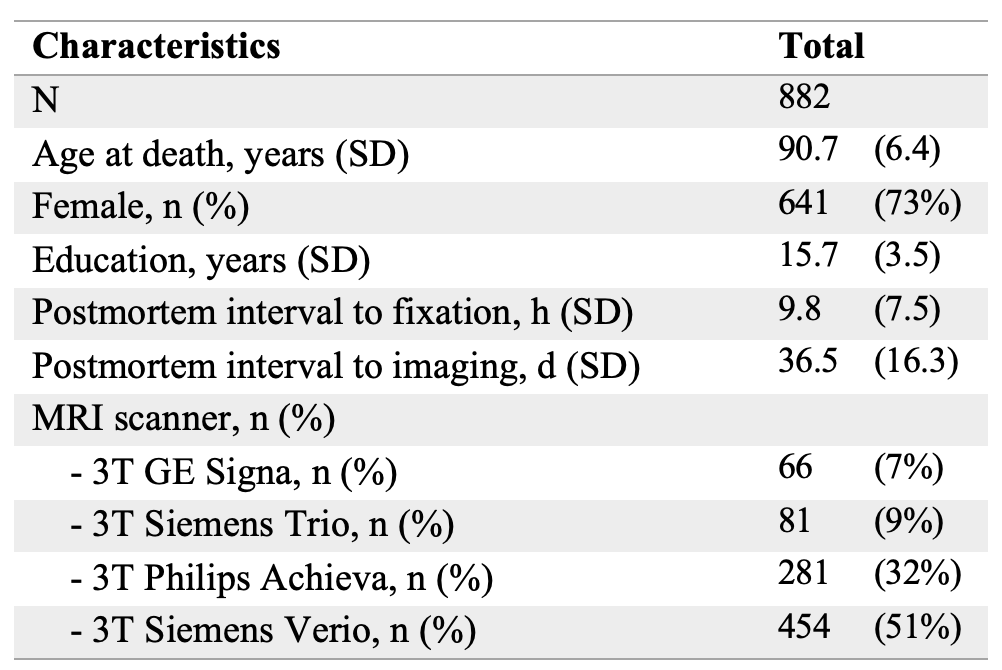
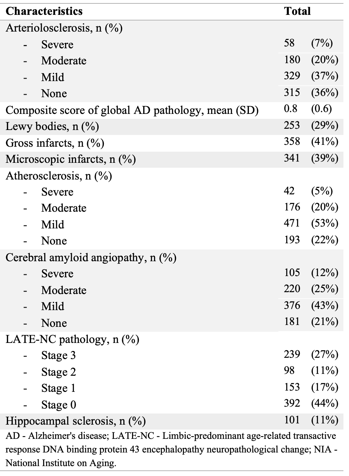
Participants, MRI, neuropathologyEx-vivo MRI and detailed neuropathological evaluation were combined in 882 older adults participating in four longitudinal, clinical-pathologic cohort studies of aging6,7: Rush Memory and Aging Project (MAP), Religious Orders Study (ROS), Minority Aging Research Study (MARS), and Clinical Core (CC) of the Rush Alzheimer’s Disease Research Center (Fig.1). Cerebral hemispheres from all participants were obtained at autopsy and imaged ex-vivo with a multi-echo spin-echo (ME-SE) sequence on 3T clinical MRI scanners approximately one month postmortem8. The acquired voxel size was 0.6mm × 0.6mm × 1.5mm, and the scan time was approximately 30 minutes8. Gray and white matter were segmented in the ex-vivo MRI data, and gray matter was further divided into 42 cortical and subcortical regions using multi-atlas segmentation9. The volume of each region was measured and normalized by the participant’s cerebral hemisphere volume10. Following ex-vivo MRI, all hemispheres underwent detailed neuropathologic examination. The assessed pathologies included arteriolosclerosis11, atherosclerosis, cerebral amyloid angiopathy, gross and microscopic infarcts, Alzheimer's pathology, Lewy bodies, limbic-predominant age-related TDP-43 encephalopathy neuropathological change (LATE-NC), and hippocampal sclerosis (Fig. 2).
Statistical analysis
Linear regression was used to investigate the association of brain arteriolosclerosis with regional gray matter volumes (normalized by cerebral hemisphere volume) controlling for all other neuropathologies (Fig. 2), demographics (age at death, sex, years of education), postmortem intervals, and scanner (Fig. 1). Statistical analysis was conducted using FSL’s PALM tool, with 10,000 permutations12. After correcting for multiple testing using the false discovery rate (FDR), significance was set at p<0.05.
Results & Discussion
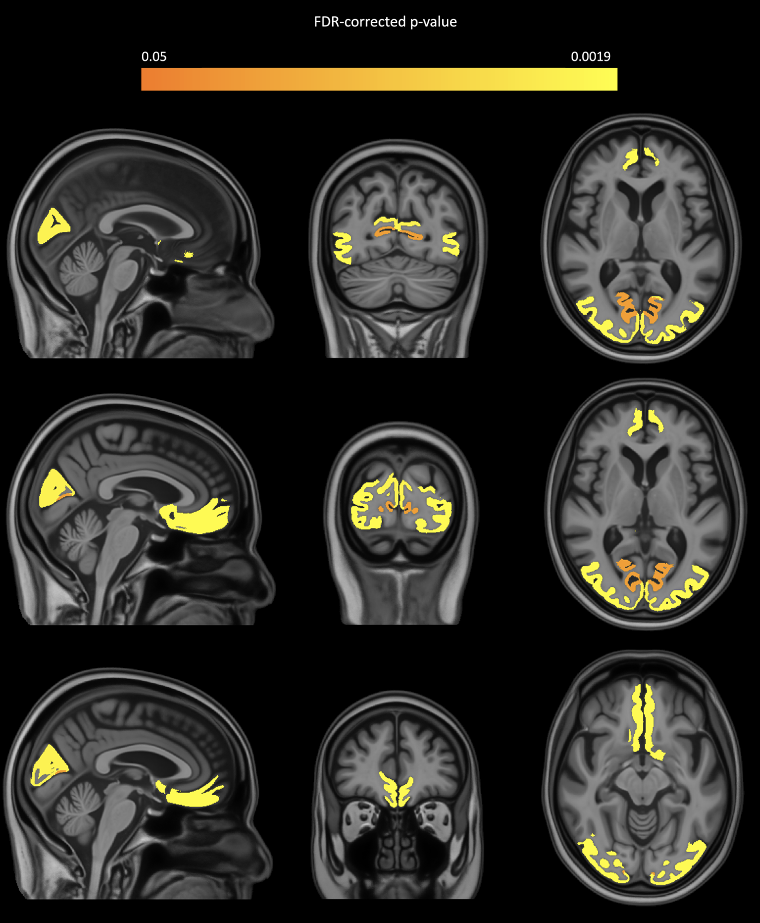
More severe brain arteriolosclerosis was associated with lower volume in a number of gray matter regions, including medial orbitofrontal, superior frontal, pericalcarine, cuneus, and lateral occipital areas, independently of the effects of other neuropathologies (Figs. 3,4). No regions showed higher volume with more severe arteriolosclerosis. These findings substantially expand our understanding of arteriolosclerosis-related brain anomalies since, so far, arteriolosclerosis was known to be associated mainly with white matter hyperintensities8. The present work in a large number of community-based older adults provides strong evidence that arteriolosclerosis is also related to neurodegenerative changes in gray matter. Furthermore, the brain regions involved are distributed in both anterior and posterior parts of the brain in line with the more widespread distribution of arteriolosclerosis pathology in the brain.Conclusion
The present work combined ex-vivo MRI and detailed neuropathological examination in a large number of community-based older adults and demonstrated that brain arteriolosclerosis is associated with lower volume in multiple gray matter regions independently of the effects of other vascular or neurodegenerative pathologies. This link between one of the main small vessel disease pathologies with neurodegeneration in gray matter is a novel finding that enhances our understanding of the impact of arteriolosclerosis on the brain. Finally, the regional volumes that are linked to arteriolosclerosis may be used as additional features in ARTS13, a recently-developed in-vivo marker of arteriolosclerosis, which may enhance its performance in predicting the pathology in-vivo.Acknowledgements
This study was supported by the following grants:
National Institute of Neurological Disorders and Stroke (NINDS): UH2-UH3NS100599, UF1NS100599
National Institute on Aging (NIA): R01AG064233, R01AG067482, R01AG017917, R01AG015819, RF1AG022018, R01AG056405, R01AG052200, P30AG010161, P30AG072975
References
1. Blevins BL, Vinters HV, Love S, et al. Brain arteriolosclerosis. Acta Neuropathol (Berl). 2021;141(1):1-24.2. Oveisgharan S, Arvanitakis Z, Yu L, Farfel J, Schneider JA, Bennett DA. Sex differences in Alzheimer’s disease and common neuropathologies of aging. Acta Neuropathol (Berl). 2018;136(6):887-900.
3. Barnes LL, Leurgans S, Aggarwal NT, et al. Mixed pathology is more likely in black than white decedents with Alzheimer dementia. Neurology. 2015;85(6):528-534.
4. Buchman AS, Yu L, Boyle PA, et al. Microvascular brain pathology and late-life motor impairment. Neurology. 2013;80(8):712-718.
5. Arvanitakis Z, Capuano AW, Leurgans SE, Bennett DA, Schneider JA. Relation of cerebral vessel disease to Alzheimer’s disease dementia and cognitive function in elderly people: a cross-sectional study. Lancet Neurol. 2016;15(9):934-943.
6. Bennett DA, Buchman AS, Boyle PA, Barnes LL, Wilson RS, Schneider JA. Religious Orders Study and Rush Memory and Aging Project. Perry G, Avila J, Moreira PI, Sorensen AA, Tabaton M, eds. J Alzheimers Dis. 2018;64(s1):S161-S189.
7. L. Barnes L, C. Shah R, T. Aggarwal N, A. Bennett D, A. Schneider J. The Minority Aging Research Study: Ongoing Efforts to Obtain Brain Donation in African Americans without Dementia. Curr Alzheimer Res. 2012;9(6):734-745.
8. Arfanakis K, Evia AM, Leurgans SE, et al. Neuropathologic Correlates of White Matter Hyperintensities in a Community-Based Cohort of Older Adults. Flanagan M, ed. J Alzheimers Dis. 2020;73(1):333-345.
9. Kotrotsou A, Bennett DA, Schneider JA, et al. Ex vivo MR volumetry of human brain hemispheres. Magn Reson Med. 2014;71(1):364-374.
10. Dawe RJ, Bennett DA, Schneider JA, Arfanakis K. Neuropathologic Correlates of Hippocampal Atrophy in the Elderly: A Clinical, Pathologic, Postmortem MRI Study. Pant H, ed. PLoS ONE. 2011;6(10):e26286.
11. Buchman AS, Leurgans SE, Nag S, Bennett DA, Schneider JA. Cerebrovascular Disease Pathology and Parkinsonian Signs in Old Age. Stroke. 2011;42(11):3183-3189.
12. Winkler AM, Ridgway GR, Webster MA, Smith SM, Nichols TE. Permutation inference for the general linear model. NeuroImage. 2014;92:381-397.
13. Makkinejad N, Evia AM, Tamhane AA, et al. ARTS: A novel In-vivo classifier of arteriolosclerosis for the older adult brain. NeuroImage Clin. 2021;31:102768.
14. Wu Y, Ridwan AR, Niaz MR, Bennett DA, Arfanakis K. High resolution 0.5mm isotropic T1-weighted and diffusion tensor templates of the brain of non-demented older adults in a common space for the MIITRA atlas. NeuroImage. 2023;282:120387.
Figures