0141
Submillimeter Isotropic in vivo Quantitative Susceptibility Mapping: Application to Women with Suspected Coronary Microvascular Dysfunction1Department of Neurology, Cedars-Sinai Medical Center, Los Angeles, CA, United States, 2Department of Biomedical Sciences and Imaging, Biomedical Imaging Research Institute (BIRI), Cedars-Sinai Medical Center, Los Angeles, CA, United States, 3Barbra Streisand Women's Heart Center, Smidt Heart Institute, Cedars-Sinai Medical Center, Los Angeles, CA, United States, 4Department of Neurology, Mayo Clinic College of Medicine and Science, Scottsdale, AZ, United States, 5Department of Kinesiology, The University of Texas at Arlington, Arlington, TX, United States, 6Departments of Neurology and Medicine, Cedars-Sinai Medical Center, Los Angeles, CA, United States, 7Department of Physical Medicine and Rehabilitation, Cedars-Sinai Medical Center, Los Angeles, CA, United States
Synopsis
Keywords: Aging, Quantitative Susceptibility mapping, Heart, Brain, women, INOCA, Aging, Dementia
Motivation: The study's motivation lies in overcoming the low-resolution limitations of Quantitative susceptibility mapping (QSM) in 3T-MRI, which is critical for investigating brain aging and neurodegeneration.
Goal(s): We aimed to construct high-resolution QSM using submillimeter T2*-3D-EPI sequence to measure iron deposition in the cortical and deep gray matter of women with suspected coronary microvascular dysfunction.
Approach: The approach involved a novel imaging protocol, TGV-based QSM reconstruction, and statistical analysis correlating iron deposition with cardiovascular health markers.
Results: Results indicate a significant association between brain iron accumulation and microvascular heart conditions, pointing to a potential interconnected pathology in women with suspected coronary microvascular dysfunction.
Impact: The study's high-resolution QSM technique could revolutionize neuroimaging, allowing clinicians to detect microvascular changes early and personalize treatments. It opens avenues for exploring the systemic nature of microvascular diseases, potentially altering approaches to managing neurodegenerative and cardiovascular conditions.
Background and Aim
Quantitative susceptibility mapping (QSM) is an advanced post-processing MRI technique that can measure the local susceptibility properties of the brain tissues and provide unique insights into the role of iron deposition in normal aging and dementia-related neurodegeneration1-3.However, the acquisition of the images necessary for QSM currently relies on a conventional gradient-echo sequence which is inherently slow and limited to the millimeter scale resolution preventing the accurate quantification of small brain structures such as the cortical grey matter and deep gray matter (DGM)4. We recently developed a novel acquisition method to collect rapidly gradient-echo images at submillimeter isotropic (650 µm) levels using a multi-shot 3-dimensional echo-planar-imaging (3D-EPI) sequence5. In this study, we introduce for the first time the use of the 3D-EPI sequence to generate submillimeter isotropic QSM of the whole brain and measure in vivo the magnetic susceptibility of the cortical and DGM tissues in women with suspected coronary microvascular dysfunction in the Women’s Ischemia Syndrome Evaluation (WISE) Pre- Heart Failure With Preserved Ejection Fraction (Pre-HFpEF) (NCT03876223) study.
Material and Methods
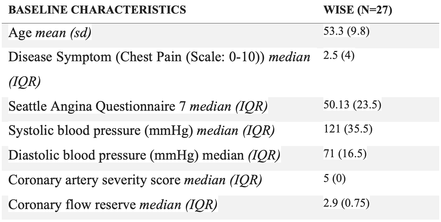
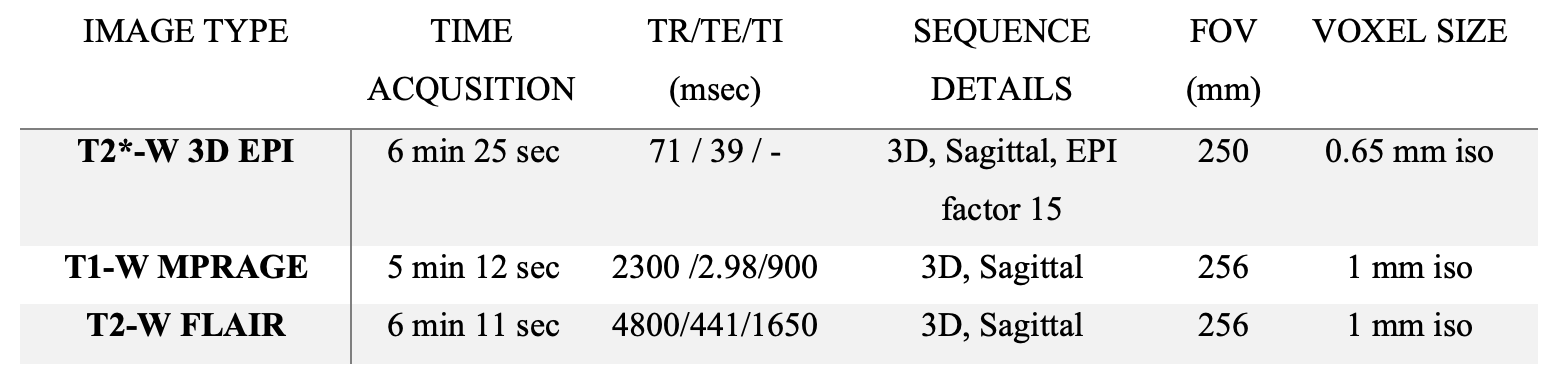
Twenty-seven women with suspected ischemia and no obstructive coronary arteries (INOCA) underwent Seattle Angina Questionnaire 7 chest pain rating and coronary flow testing6 (Table-1).Brain MRI protocol in 3T-MRI (Siemens, Vida) included submillimeter (650 µm) isotropic sagittal 3D-EPI, millimeter (1mm) isotropic T2-FLAIR, and T1-MPRAGE (see parameters in Table-2).
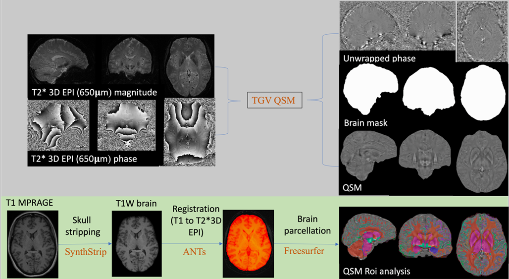
To reconstruct QSM, a total generalized variation (TGV)-based7 method was utilized (Figure-1). TGV-based QSM reconstruction mainly combines phase unwrapping, background field removal, and dipole inversion into a single integrated step. To calculate QSM values, T1-MPRAGE was initially aligned with the high-resolution 3D-EPI via advanced normalization tools (ANTs)8, after skull stripping using SynthStrip9 (Figure-1). Subsequently, an automated procedure in Freesurfer was used to parcellate the brain based on the Destrieux atlas10 in the 3D-EPI space (Figure-1). To enable inter-subject analysis of the QSM data, the unwrapped QSM values were normalized by subtracting the values corresponding to cerebrospinal fluid for each participant. Then, tissue iron deposition was assessed on QSM in both the cortex and in six DGM regions using a custom MATLAB script. Among the parcellated cortex and the six DGM regions, fifteen regions with the highest mean QSM values were identified across the cohort. The associations between whole-brain iron deposition and clinical characteristics in Table-1 were determined through robust linear regression analysis in Python, adjusting for age.
Results
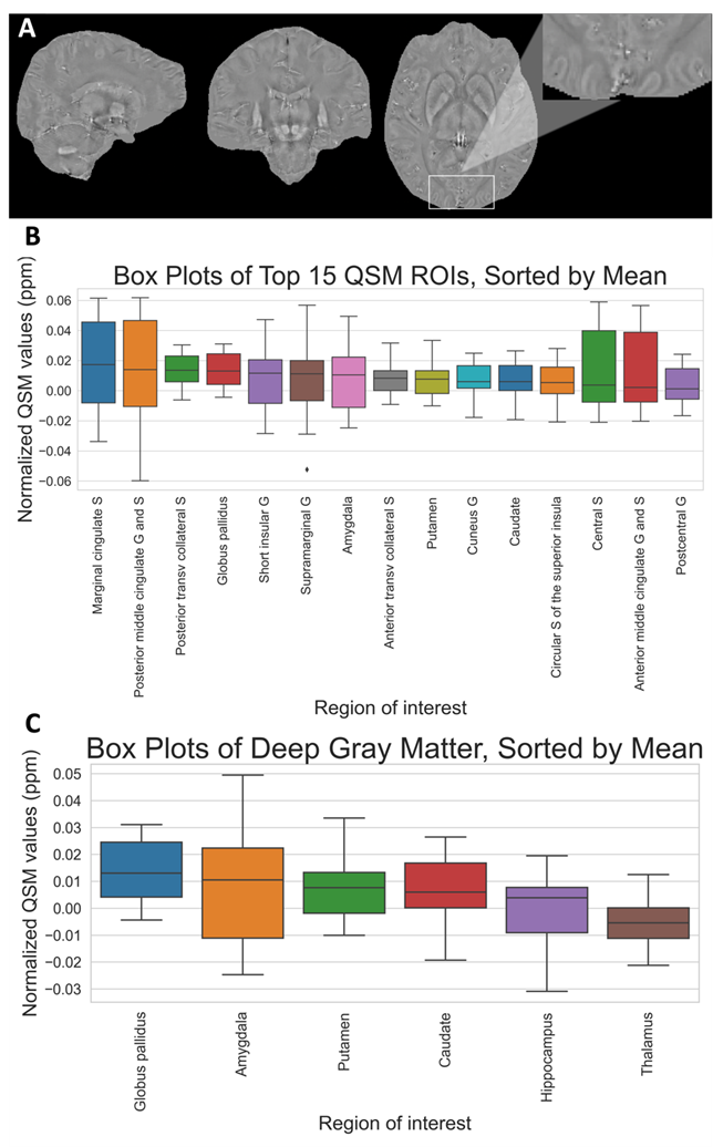
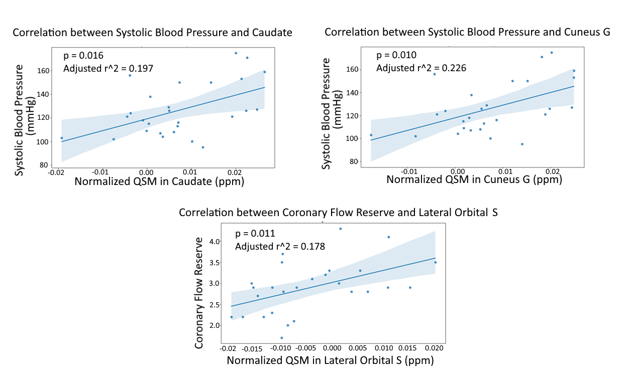
For each participant, QSM was constructed using submillimeter isotropic 3D-EPI. A representative example is shown for one of the participants (Figure-2A). The cortical and DGM regions that exhibited the highest iron accumulation were: cingulate marginal sulcus, anterior and posterior middle cingulate gyrus and sulcus, anterior and posterior transverse collateral sulcus, central sulcus, short insular, supramarginal, cuneus and postcentral gyrus of the cortex, and globus pallidus (GP), amygdala, putamen and caudate parts of the DGM (Figure-2B). Thalamus, hippocampus, caudate, putamen, amygdala and GP displayed a consistent pattern of QSM values with a gradual increase in iron deposition (Figure-2C). Systolic blood pressure showed significant associations with the iron content in the cuneus gyrus (p=0.016, adjusted-r2=0.197) and caudate (p=0.010, adjusted-r2=0.226) in women with suspected INOCA (Figure-3). Moreover, there was a positive relationship between coronary flow reserve (CFR) and lateral orbital sulcus (p=0.011, adjusted-r2=0.178) (Figure-3).Discussion
The submillimeter scale used in this study allowed the measurement of QSM values in the cortical and DGM tissues with high confidence. Amongst the different DGM regions, GP displayed the highest QSM values as reported previously11. In the cortex, QSM regions with the highest mean values included the cingulate, postcentral gyrus, insula, amygdala and caudate, which is aligned with previous studies indicating elevated QSM values in these brain areas of subjects with Alzheimer's disease12. Moreover, our findings are consistent with the cortical regions playing a mediating role in the age‐related cognitive decline in healthy subjects13. This may imply an accelerated aging process in these women with suspected INOCA.Overall, our initial findings suggest a potential link between brain iron deposition and systolic blood pressure, which bear similarities with prior research on hypertensive patients14. Moreover, a unique association between CFR - an indicator of coronary microvascular dysfunction15- and brain iron content was observed in this study. These relationships may indicate a connection between heart and brain conditions marked by microvascular disease.
Conclusions
In summary, our study provides first insights into the potential link between brain iron deposition in the cortex and DGM measured by submillimeter 3D-EPI-derived QSM, and cardiovascular regulations determined by systolic blood pressure and CFR, in women with suspected INOCA. Future investigations on the full cohort are further warranted to confirm our preliminary findings.Acknowledgements
The women’s ischemia syndrome evaluation (WISE) Pre- Heart Failure With Preserved Ejection Fraction (Pre-HFpEF) (NCT03876223) study funded under grant number R01-HL-146148-01.
References
1. Wang Y, Liu T. Quantitative susceptibility mapping (QSM): Decoding MRI data for a tissue magnetic biomarker. Magn Reson Med. 2015;73(1):82-101. Epub 20140717. doi: 10.1002/mrm.25358. PubMed PMID: 25044035; PMCID: PMC4297605.2. Acosta-Cabronero J, Betts MJ, Cardenas-Blanco A, Yang S, Nestor PJ. In Vivo MRI Mapping of Brain Iron Deposition across the Adult Lifespan. J Neurosci. 2016;36(2):364-74. doi: 10.1523/JNEUROSCI.1907-15.2016. PubMed PMID: 26758829; PMCID: PMC4710766.
3. Ayton S, Fazlollahi A, Bourgeat P, Raniga P, Ng A, Lim YY, Diouf I, Farquharson S, Fripp J, Ames D, Doecke J, Desmond P, Ordidge R, Masters CL, Rowe CC, Maruff P, Villemagne VL, Australian Imaging B, Lifestyle Research G, Salvado O, Bush AI. Cerebral quantitative susceptibility mapping predicts amyloid-beta-related cognitive decline. Brain. 2017;140(8):2112-9. doi: 10.1093/brain/awx137. PubMed PMID: 28899019.
4. Haller S, Haacke EM, Thurnher MM, Barkhof F. Susceptibility-weighted Imaging: Technical Essentials and Clinical Neurologic Applications. Radiology. 2021;299(1):3-26. doi: 10.1148/radiol.2021203071. PubMed PMID: WOS:000631703800014.
5. Sati P, Patil S, Krueger G, Derbyshire JA, Reich DS. Fast Sub-Millimeter Whole-Brain Magnetic Resonance Imaging of Novel Biomarkers for Multiple Sclerosis. Multiple Sclerosis Journal. 2019;25:12-3. PubMed PMID: WOS:000468918500024.
6. Wei J, Mehta PK, Johnson BD, Samuels B, Kar S, Anderson RD, Azarbal B, Petersen J, Sharaf B, Handberg E, Shufelt C, Kothawade K, Sopko G, Lerman A, Shaw L, Kelsey SF, Pepine CJ, Merz CN. Safety of coronary reactivity testing in women with no obstructive coronary artery disease: results from the NHLBI-sponsored WISE (Women's Ischemia Syndrome Evaluation) study. JACC Cardiovasc Interv. 2012;5(6):646-53. doi: 10.1016/j.jcin.2012.01.023. PubMed PMID: 22721660; PMCID: PMC3417766.
7. Langkammer C, Bredies K, Poser BA, Barth M, Reishofer G, Fan AP, Bilgic B, Fazekas F, Mainero C, Ropele S. Fast quantitative susceptibility mapping using 3D EPI and total generalized variation. Neuroimage. 2015;111:622-30. doi: 10.1016/j.neuroimage.2015.02.041. PubMed PMID: WOS:000352224100054.
8. Avants BB, Tustison NJ, Song G, Cook PA, Klein A, Gee JC. A reproducible evaluation of ANTs similarity metric performance in brain image registration. Neuroimage. 2011;54(3):2033-44. doi: 10.1016/j.neuroimage.2010.09.025. PubMed PMID: WOS:000286302000027.
9. Hoopes A, Mora JS, Dalca AV, Fischl B, Hoffmann M. SynthStrip: skull-stripping for any brain image. Neuroimage. 2022;260. doi: 10.1016/j.neuroimage.2022.119474. PubMed PMID: WOS:000843819500004.
10. Destrieux C, Fischl B, Dale A, Halgren E. Automatic parcellation of human cortical gyri and sulci using standard anatomical nomenclature. Neuroimage. 2010;53(1):1-15. doi: 10.1016/j.neuroimage.2010.06.010. PubMed PMID: WOS:000280818900001.
11. Treit S, Naji N, Seres P, Rickard J, Stolz E, Wilman AH, Beaulieu C. R2* and quantitative susceptibility mapping in deep gray matter of 498 healthy controls from 5 to 90 years. Hum Brain Mapp. 2021;42(14):4597-610. Epub 20210629. doi: 10.1002/hbm.25569. PubMed PMID: 34184808; PMCID: PMC8410539.
12. Kim HG, Park S, Rhee HY, Lee KM, Ryu CW, Rhee SJ, Lee SY, Wang Y, Jahng GH. Quantitative susceptibility mapping to evaluate the early stage of Alzheimer's disease. Neuroimage Clin. 2017;16:429-38. Epub 20170824. doi: 10.1016/j.nicl.2017.08.019. PubMed PMID: 28879084; PMCID: PMC5577408.
13. Howard CM, Jain S, Cook AD, Packard LE, Mullin HA, Chen NK, Liu C, Song AW, Madden DJ. Cortical iron mediates age-related decline in fluid cognition. Hum Brain Mapp. 2022;43(3):1047-60. Epub 20211202. doi: 10.1002/hbm.25706. PubMed PMID: 34854172; PMCID: PMC8764476.
14. Li X, Jin D, Zhu Y, Liu L, Qiao Y, Qian Y, Tian J, Jiang B, Hou C, Geng J, Li X, Gao X, Ma Y, Wang S, Zong J, Qin Y. Quantitative susceptibility mapping to evaluate brain iron deposition and its correlation with physiological parameters in hypertensive patients. Ann Transl Med. 2021;9(20):1582. doi: 10.21037/atm-21-5170. PubMed PMID: 34790788; PMCID: PMC8576670.
15. Suppogu N, Wei J, Nelson MD, Cook-Wiens G, Cheng S, Shufelt CL, Thomson LEJ, Tamarappoo B, Berman DS, Samuels B, Azarbal B, Anderson RD, Petersen JW, Handberg EM, Pepine CJ, Merz CNB. Resting coronary velocity and myocardial performance in women with impaired coronary flow reserve: Results from the Women's Ischemia Syndrome Evaluation-Coronary Vascular Dysfunction (WISE-CVD) study. Int J Cardiol. 2020;309:19-22. Epub 20200123. doi: 10.1016/j.ijcard.2020.01.053. PubMed PMID: 32037132; PMCID: PMC7195998.
Figures