0137
Choroid Plexus Structural and vascular Changes Associated with Aging in the HCP Dataset1Bernard and Irene Schwartz Center for Biomedical Imaging, Department of Radiology, NYU Grossman School of Medicine, New York, NY, United States, 2Center for Advanced Imaging Innovation and Research (CAI2R), Department of Radiology, NYU Grossman School of Medicine, New York, NY, United States, 3Vilcek Institute of Graduate Biomedical Sciences, NYU Grossman School of Medicine, New York, NY, United States, 4Center for Cognitive Neurology, NYU Grossman School of Medicine, New York, NY, United States, 5Departments of Pathology and Psychiatry, NYU Grossman School of Medicine, New York, NY, United States
Synopsis
Keywords: Aging, Aging, Human Connectome Project, choroid plexus
Motivation: Analyzing ChP changes in normal aging is essential for grasping its role in neurological disorders.
Goal(s): To evaluate ChP changes with age using diffusion and perfusion MRI in a lifespan HCP-aging dataset.
Approach: MR images of 641 healthy participants aged from 36 to 90 years old were analyzed to extract diffusion and perfusion measurements of ChP to investigate their age-related changes.
Results: With age, the ChP undergoes significant changes, including increased volume, reduced blood flow, elevated MD values, and a more rapid decline in blood flow compared to gray and white matter.
Impact: This study offers a comprehensive evaluation of age-related changes in the ChP, enhancing our comprehension of its potential involvement in age-related cognitive decline. Furthermore, age-related ChP alterations exhibit distinct patterns compared to changes in gray and white matter.
INTRODUCTION
The choroid plexus (ChP) has gained increasing attention due to its vital roles in cerebrospinal fluid (CSF) production and waste removal. Unlike the blood-brain barrier (BBB), ChP capillaries are fenestrated, allowing for free water exchange between the blood vessels and surrounding stroma. Recent studies have investigated ChP's involvement in neurological disorders such as Alzheimer's disease, epilepsy, and multiple sclerosis[1-3], primarily about its volumetric and proteomic changes. However, a comprehensive exploration of age-related microstructure and microvasculature changes remains lacking. In this study encompassing a large sample size and wide age range from HCP-aging dataset, we assessed age-related changes of mean diffusivity (MD) derived from diffusion MRI as well as cerebral blood flow (CBF) and arterial transit time (ATT) derived from arterial spin labeling (AS) MRI. Our hypothesis is that age-related changes in ChP microstructure and blood perfusion could better explain the previously documented volumetric alterations, which potentially affects cerebrospinal fluid (CSF) dynamics in the aging population.METHODS
Participants and imaging protocolWe exploited 641 healthy participants aged from 36 to 90 years old (60±16 years; 282 males) from HCP-Aging dataset [4]. The following imaging protocols were performed on a Connectome Skyra 3T MRI: 1) T1-weighted MPRAGE: 0.8mm isotropic, TR/TE=2500/2.22ms; 2) 2D multiband spin-echo EPI for diffusion-weighted MRI: 1.5mm isotropic, TR/TE=3230/89.2ms, multiband factor=3, b=0, 1500, 3000 s/mm2, 98 directions; 3) Pseudo-continuous arterial spin labeling (pcASL) MRI: 2.5mm isotropic, TR/TE=8000/40ms, simultaneous multi-slice acquisition with multi-band factor=6, post labelling delays=0.2, 0.7, 1.2, 1.7, 2.2s [5].
Data processing
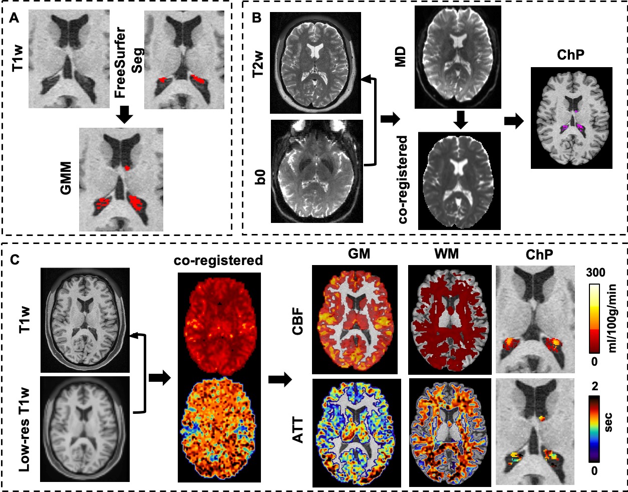
We applied Gaussian Mixture Models to T1w-based FreeSurfer result to improve ChP segmentation [6]. MRtrix3 was applied to preprocess the dMRI data with eddy current and Gibbs ringing correction. The b0 DW image was registered to T2w image and the calculated transform matrix was applied to MD maps. Multiple pCASL data preprocessing steps such as motion and distortion correction were involved to generate calibrated CBF and ATT maps (https://github.com/physimals/hcp-asl). The pipeline-derived low resolution ASL grid T1w image was registered to original T1 and the calculated transform matrix was applied to the corresponding CBF and ATT maps. The ChP mask was then applied to co-registered MD, CBF, and ATT maps to acquire corresponding values (Fig. 1).
Statistical analysis
Pearson's correlation was applied to investigate the age effect on LV and ChP volume, MD, CBF, and ATT after adjusting for sex. One-way ANOVA was applied to reveal the different perfusion properties of GM, WM, and ChP. The effect of CBF on MD was also evaluated using multiple linear regression with age as covariate.
RESULTS
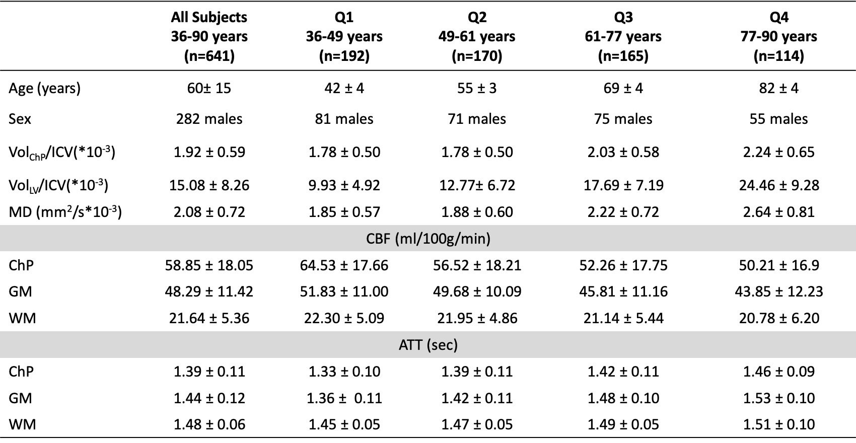
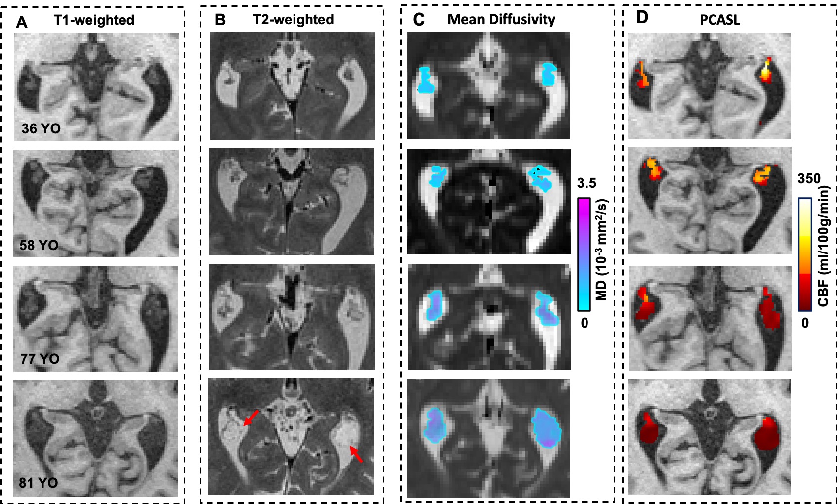
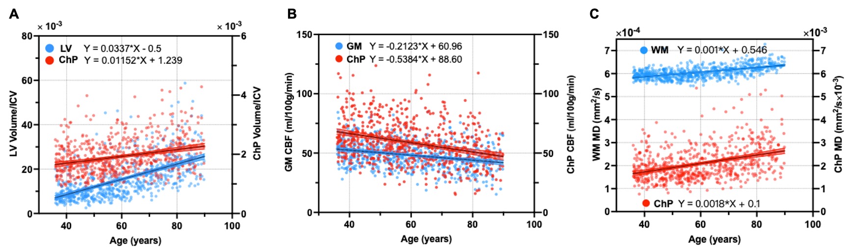
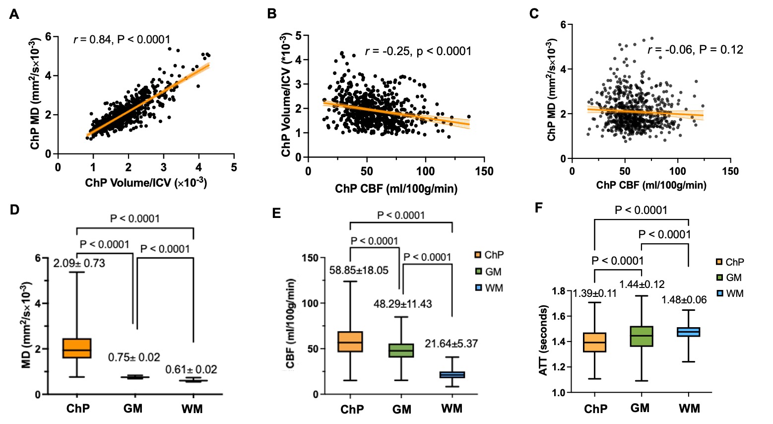
Table 1 showed the demographic data and MRI measurements from different age quartiles. Figure 2 demonstrated representative cases of ChP imaging from different age group showing age-related increased volume and MD but decreased blood flow. As shown in Figure 3, after correcting for ICV, both ChP and LV volumes were positively correlated with age (r = 0.31, p<0.0001; r = 0.61, p<0.0001, respectively), with LV showing a faster enlargement (=0.034 and =0.012, respectively) (Fig. 3A). CBF exhibited a linearly decline with age in ChP and GM (r = -0.4, p<0.0001; r=-0.28, p< 0.0001, respectively), with ChP displaying a faster decline (=-0.54 vs =-0.21) (Fig. 3B). The MD was positively correlated with aging in ChP and WM (r = 0.38, p<0.0001; r = 0.56, p<0.0001, respectively), indicating increased water diffusivity (Fig. 3C). After adjusting for age and sex, MD was significantly correlated with ChP volume (r = 0.84, p<0.0001), indicating a compromised microstructure accompanied with a structural change (Fig. 4A). A negative correlation was observed between ChP CBF and volume (r = -0.25, p < 0.0001) (Fig. 4B). However, there was no correlation between MD and CBF (Fig. 4C).One-way ANOVA analyses followed by Tukey’s multiple comparison correction revealed significant differences in MD, CBF and ATT among ChP, GM, and WM (p<0.0001 for all comparisons) (Fig. 4D-4F).
DISCUSSION AND CONCLUSIONS
Similar to previous research, we observed ChP volume increase with age, likely due to factors such as stromal fibrosis, calcification, and lipofuscin deposition [7]. Despite the increase of ChP volume, there was a reduction in blood flow, which inversely correlates with ChP volume, suggesting degenerative hyperplasia as a compensatory response to the reduction of perfusion and CSF secretion. We also observed higher MD with age, indicating structural loosening with increased cyst-like fluid in ChP [8, 9]. In comparison to GM and WM, the ChP has highest CBF and shortest ATT, indicating its highly vascularized structure. The results obtained from an extensive and well-regarded dataset focused on normal aging offer a crucial benchmark for forthcoming studies on age-related neurodegenerative disorders.Acknowledgements
This work was supported in part by the NIH U01AG052564, P30AG066512, RF1 NS110041, R01 NS108491, U24 NS135568, and was performed under the rubric of the Center for Advanced Imaging Innovation and Research (CAI2R, www.cai2r.net), an NIBIB National Center for Biomedical Imaging and Bioengineering (NIH P41 EB017183). The HCP-Aging 2.0 Release data used in this report came from DOI: 10.15154/1520707 and funded by the McDonnell Center for Systems Neuroscience at Washington University in St. Louis.References
1. Choi, J.D., et al., Choroid Plexus Volume and Permeability at Brain MRI within the Alzheimer Disease Clinical Spectrum. Radiology, 2022. 304(3): p. 635-645.
2. Bergsland, N., et al., Association of Choroid Plexus Inflammation on MRI With Clinical Disability Progression Over 5 Years in Patients With Multiple Sclerosis. Neurology, 2023. 100(9): p. e911-e920.
3. Leitner, D.F., et al., Localized proteomic differences in the choroid plexus of Alzheimer's disease and epilepsy patients. Front Neurol, 2023. 14: p. 1221775.
4. Bookheimer, S.Y., et al., The Lifespan Human Connectome Project in Aging: An overview. Neuroimage, 2019. 185: p. 335-348.
5. Li, X., et al., Evaluation of 3D GRASE and 2D MB-EPI for Multi-Delay PCASL Imaging. Proc. Intl. Soc. Mag. Reson. Med. 25, 2017.
6. Tadayon, E., et al., Improving Choroid Plexus Segmentation in the Healthy and Diseased Brain: Relevance for Tau-PET Imaging in Dementia. J Alzheimers Dis, 2020. 74(4): p. 1057-1068.
7. Alisch, J.S.R., et al., Characterization of Age-Related Differences in the Human Choroid Plexus Volume, Microstructural Integrity, and Blood Perfusion Using Multiparameter Magnetic Resonance Imaging. Front Aging Neurosci, 2021. 13: p. 734992.
8. Kant, S., et al., Choroid plexus genes for CSF production and brain homeostasis are altered in Alzheimer’s disease. Fluids and Barriers of the CNS, 2018. 15(1): p. 34.
9. Zhao, L., et al., Non-invasive measurement of choroid plexus apparent blood flow with arterial spin labeling. Fluids and Barriers of the CNS, 2020. 17(1): p. 58.
Figures