0134
Quantum-Sensing MRI: Neuronal Firings in Human Brains under Finger-Tapping in a Wide Range of Ages1Radiology, New York University Grossman School of Medicine, New York, NY, United States, 2Vilcek Institute of Graduate Biomedical Sciences, New York University Grossman School of Medicine, New York, NY, United States, 3Neurology, New York University Grossman School of Medicine, New York, NY, United States
Synopsis
Keywords: Aging, Nerves, quantum sensing
Motivation: The qsMRI has the potential for non-invasive detection of neuronal electrical activities (action potentials or firings) in the human brain. This emerging technique, however, is still in infant stage and needs more studies to show its potentials.
Goal(s): This study explores whether qsMRI detects the change in neuronal firings during a finger-tapping task in a wide range of ages.
Approach: A group of healthy subjects (27–84 years old) were studied on a 3T MRI scanner, using three tasks: finger-tapping, no tapping, and resting state.
Results: Firing rate varied with age, and older people showed higher firing rate during tapping than resting.
Impact: These positive results further demonstrated the potential of qsMRI to detect neuronal firings in humans, and will encourage researchers to use the technique in a wide range of studies on brain functions and neurological disorders including aging and Alzheimer’s disease.
INTRODUCTION
Neuronal electrical activities (action potentials or firings) are the physical basis of neuronal communication in the brain. Measuring these activities is a key step toward the understanding of mechanisms behind neuronal functions or dysfunctions. Current functional MRI (fMRI) employs the blood oxygenation level dependent (BOLD) signals1,2 to indirectly detect neuronal activities through an unclear neuro-hemodynamic mechanism. fMRI is also limited to the detection of slow neuronal activities, such as postsynaptic potentials, due to relatively low temporal resolutions (40–100ms).3 Recent research efforts have improved temporal resolution to milliseconds4 or sub-milliseconds,5 under the assumption that neuronal activities are exactly repeatable in time, though unlikely to be true. To tackle these challenges, a new concept was recently proposed in which quantum sensing is used to detect tiny magnetic fields induced by the fast (action) and slow (postsynaptic) neuronal currents at high temporal resolution.6,7 The feasibility of quantum-sensing (qs) MRI was demonstrated last year.8 Here we further explore the qsMRI and see whether it detects the changes in neuronal firings in a wide range of ages.METHODS
The study design is shown in Fig.1. A group of 25 healthy subjects (age 58.8±18.2 years in 27–84 years) were studied (after excluding three subjects for incomplete data or epilepsy history), with an IRB-approved consent. Finger-tapping was performed by subject’s index finger in the dominant hand (Fig. 1a). Tapping rate was instructed at 1.0Hz, lasting for 1.5min. FID signals were acquired during tapping, no tapping, and resting, using a product sequence fid on a 3T MRI scanner (Prisma, Siemens), with a standard Head/Neck 20-channel coil (Figs. 1b). FID readout time was 819.2ms at a 0.2-ms sampling, with TE/TR=0.2/1500ms, repeats=64, and manual B0 shimming. The tapping and no-tapping were immediately followed each other (~15s), and shared the same B0 shim and field of view (FOV). The resting state was ~27min later from the no-tapping, with the same FOV but a re-shimming. A custom-developed software in MATLAB R2021a (MathWorks, Natick, MA) calculated neuronal magnetic field Bn,z (Fig. 1c).7,8 The firing rate was measured in peaks per second (Hz) in a time window of the first 500ms for each FID, i.e., Rf = peaks/500ms, through peak search.9 Mean and standard deviation (SD) was then calculated over the 64 FIDs at individual channels.RESULTS
The sensing volumes of the qsMRI (Fig. 2) were defined in the brain by the MRI images for each of the FID signals at individual channels. Fig. 3 summarizes firing rate and its change during the three task states for the whole brain (averaged over all the 16 head channels), and indicates a clear division of the subjects at age 58 years into two age groups, with a large increase (up to 200%) in the old group for the tap and non-tap tasks, compared to the resting state, while they had a similar firing rate in the resting state. Fig. 4 presents a big picture about firing rate in all the head channels across the subjects (age increases with the subject’s index). Compared with the resting state, the tapping task activated more firings, and the channels covering the motor cortex (index #6, 10, 11, and 13) shows an increased firing rate (in pink) during the tapping. However, firing rate is comparable between the tapping and immediate no-tapping. At the motor cortex channels (Fig. 5), firing rate has a pattern similar to that in the whole brain, but having larger values.DISCUSSION
The firing rate was varying with the tapping task and subject age, both in whole brain and in motor cortex region, suggesting a high sensitivity of the qsMRI to neuronal firings. The firing rates in the tapping and no-tapping states are similar due to the immediately following of the no-tapping task. The firing rate during tapping higher in the old subjects than in the young might reflect the fact that experienced people use their brains more efficiently.10 At the motor channels (Fig. 5), larger firing rate confirms the source of firings from motor cortex regions. However, quantitative comparison among these firing rates has not yet performed, and more efforts are needed in the future.CONCLUSION
This study showed the firing rate in response to a finger-tapping task changing with age, especially age over 58 years. This positive result further demonstrated the potential of qsMRI to detect neuronal firings in individuals, and will encourage use of the technique in a wide range of studies on brain functions and neurological disorders including aging and Alzheimer’s disease.Acknowledgements
This work was supported in part by the NIH RF1 AG067502 and was performed under the rubric of the Center for Advanced Imaging Innovation and Research (CAI2R, www.cai2r.net), an NIBIB National Center for Biomedical Imaging and Bioengineering (NIH P41 EB017183).References
1. Ogawa S, Menon RS, Tank DW, Kim SG, Merkle H, Ellermann JM, Ugurbil K. Functional brain mapping by blood oxygenation level-dependent contrast magnetic resonance imaging. A comparison of signal characteristics with a biophysical model. Biophysical journal. 1993 Mar 1;64(3):803-12.
2. Ogawa S, Tank DW, Menon R, Ellermann JM, Kim SG, Merkle H, Ugurbil K. Intrinsic signal changes accompanying sensory stimulation: functional brain mapping with magnetic resonance imaging. Proceedings of the National Academy of Sciences. 1992 Jul 1;89(13):5951-5.
3. Huang J. Detecting neuronal currents with MRI: a human study. Magn Reson Med. 2014;71(2):756-762
4. Toi PT, Jang HJ, Min K, Kim SP, Lee SK, Lee J, Kwag J, Park JY. In vivo direct imaging of neuronal activity at high temporospatial resolution. Science. 2022 Oct 14;378(6616):160-8.
5. Zhong Z, Sun K, Karaman MM, Zhou XJ. Magnetic resonance imaging with submillisecond temporal resolution. Magnetic Resonance in Medicine. 2021 May;85(5):2434-44.
6. Qian Y, Calderon L, Chen X, Liu A, Lui YW, Boada, FE. Quantum sensing of local neuronal firings (qsLNF) in human brains via proton (1H) MRI: Proof of concept. In Proceedings of the Joint Annual Meeting ISMRM-ESMRMB 07–12 May 2022 in London, UK, page 4088.
7. Qian Y, Lakshmanan K, Liu A, Lui YW, Boada, FE. Magnetic resonance recording of local neuronal firings (mrLNF) in the human brains: A proof of concept. In Proceedings of the 29th Annual Meeting ISMRM (Virtual), 15–20 May 2021, page 102.
8. Qian Y, Chen X, Lin Y-C, Henin S, Kumbella NM, Aguilera L, Rockowitz Z, Clayton A, Babb J, Ge Y, Masurkar A, Liu A, Lui YW, Boada FE. Quantum-sensing MRI for non-invasive detection of neuronal firing in human brain: initial demonstration via finger-tapping task. In Proceedings of the 31th Annual Meeting ISMRM, 3–8 June 2023 in Toronto, Canada, page 914.
9. Frohlich F. Unit activity. In Network Neuroscience. Academic Press. 2016. https://doi.org/10.1016/C2013-0-23281-5.
10. Cabeza R, Albert M, Belleville S. et al. Maintenance, reserve and compensation: the cognitive neuroscience of healthy ageing. Nat Rev Neurosci. 2018; 19:701–710.
Figures
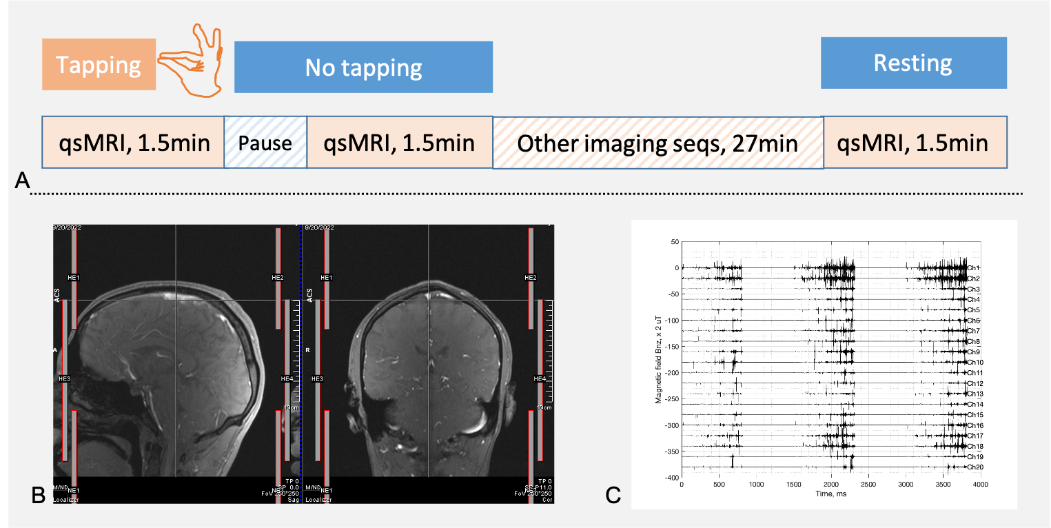
Fig. 1. Experiment set-up of the finger-tapping task and qsMRI. A) The finger-tapping at a rate of ~1.0Hz starts by the instruction of MRI operator and is performed by the index finger of subject dominant hand. The qsMRI data acquisition starts right after the tapping and is followed by a pause (~15 s) and a repeated qsMRI without finger-tapping. B) Coil element (channel) locations in a Head/Neck 20Ch array: HE1-2, HE3-4, and NE1-2 from top to bottom. C) Representative neuronal firings (magnetic field) detected by the qsMRI at the 20 individual channels for 3-TR recording at resting-state.
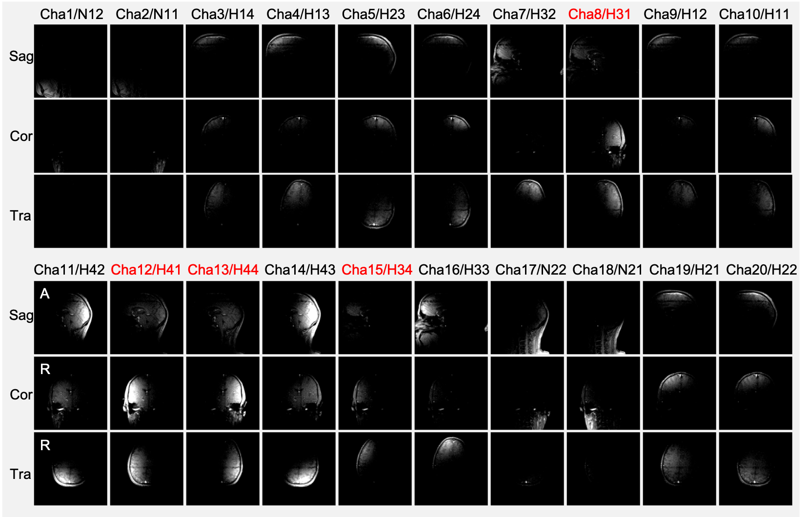
Fig. 2. Individual channel images (sensing volumes) of the qsMRI from the Head/Neck 20Ch array shown in Fig. 1. Three orthogonal slices (Sagittal, Coronal, and Transversal) cross the center of a subject’s head. The images are displayed in the same window/level for visual comparison in signal intensity among channels. The motor cortex involved in the finger-tapping task is detected in the channels (red): Cha8/H31, Cha12/H41, Cha13/H44, and Cha15/H34.
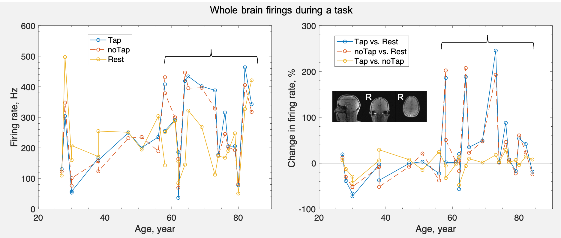
Fig. 3. Whole brain neuronal firing rates (averaged over all the 16 head channels HE1-4) and their changes in response to the tasks: finger-tapping, non-tapping, and resting state. Note: a clear variation appears at age 58 years, showing a large increase in the older ages for the tasks: tap and non-tap, compared with the resting state.
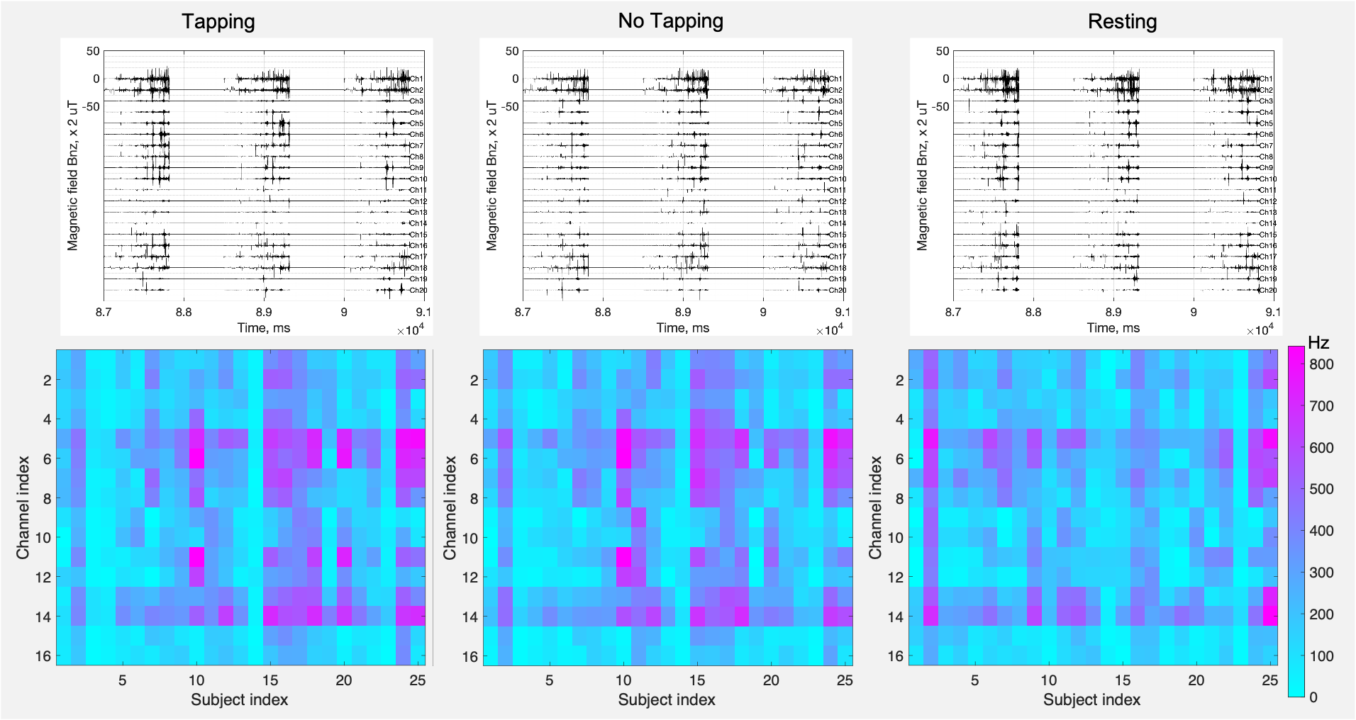
Fig. 4. Individual channel neuronal firings and rates of the subjects under three states: tapping, no-tapping, and resting. Top: Representative firings from a subject (index #8, 51-year-old healthy male) at the 20 channels. Bottom: Mean firing rate over a 1.5-min-long task at the 16 head channels. These firing rates present a big picture about the brain locations where firings were active during a task. Overall, the tapping activated more firings than the resting. Channels covering the motor cortex (index #6, 10, 11, and 13) have an increased firing rate (in pink) during the tapping.
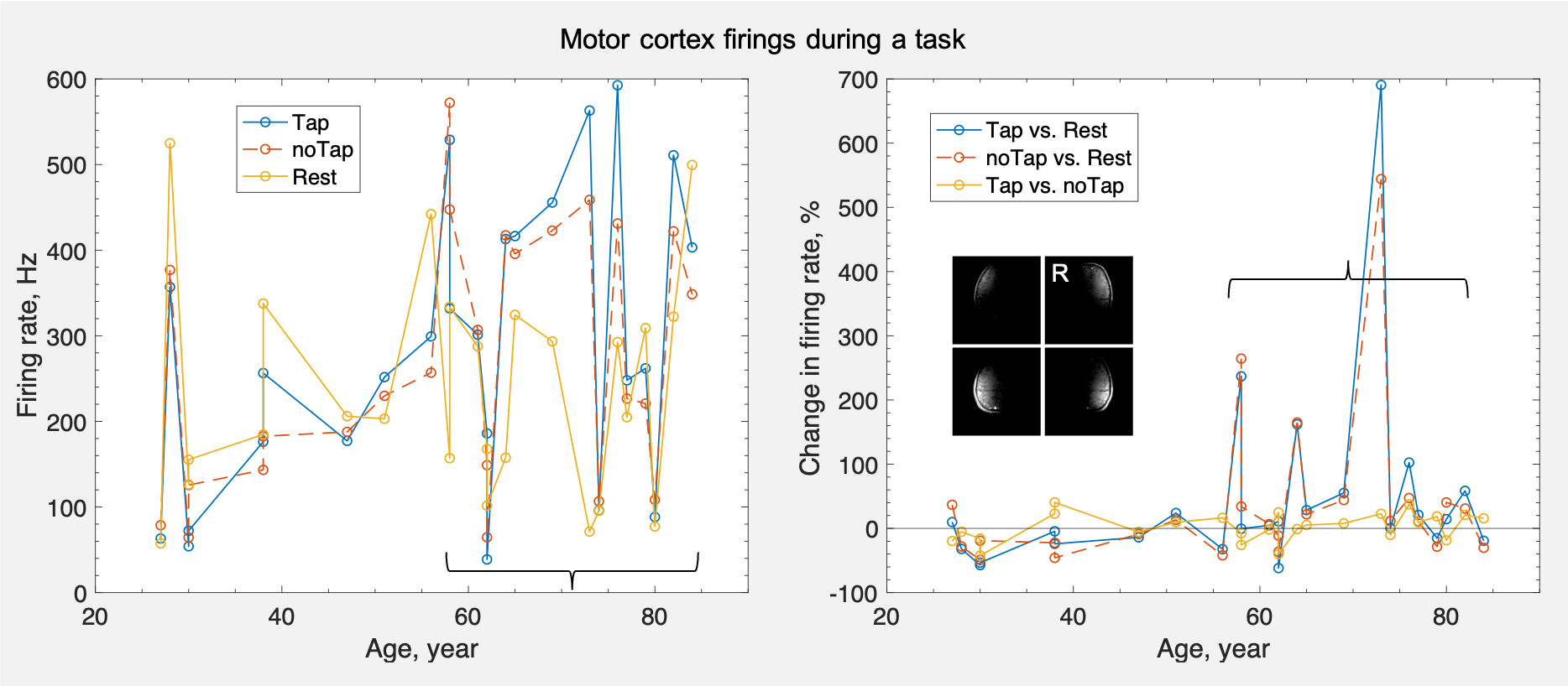
Fig. 5. Motor cortex neuronal firing rates (sum over the 4 cortex-involved channels: Cha8/H31, Cha12/H41, Cha13/H44, and Cha15/H34) and their changes in response to the tasks: tapping, non-tapping, and resting state. Note: a clear increase in firing rate also appears at age 58 years and older, with a change more than 200%.