0130
Direct Neuronal Activity-related (DIANA) fMRI in Awake Mice1Guangdong-Hong Kong-Macao University Joint Laboratory of Interventional Medicine, the Fifth Affiliated Hospital, Sun Yat-sen University, Zhuhai, China, 2Shenzhen Institute of Advanced Technology,Chinese Academy of Sciences, Zhuhai, China, 3Shenzhen Institute of Advanced Technology,Chinese Academy of Sciences, Shenzhen, China
Synopsis
Keywords: Task/Intervention Based fMRI, fMRI (task based), DIANA
Motivation: Toi et al. reported a revolutionary approach of direct imaging of neuronal activity (DIANA) by fMRI in anesthetized mice at 9.4 T. However, anesthesia has a profound impact on the central nervous system, leading to modifications in physiological parameters.
Goal(s): Our goal is to investigate Direct Neuronal Activity-related (DIANA) fMRI in awake mice.
Approach: We performed the event-related cerebral functional magnetic resonance imaging and DIANA experiment in habit-trained awake mice.
Results: In response to the electrical stimulation, a statistically significant increase in the DIANA signal was observed in the contralateral S1FL compared with the prestimulus signal (p < 0.005, n = 6 mice).
Impact: Direct detection of neural activity allows us to better understand the rapid dynamics of neural activity, which will help improve the understanding and diagnosis of neurological diseases.
Introduction
Detecting neuronal activity at high temporal and spatial resolution is essential for the development of neuroscience. A study of direct neuronal imaging published last year by Toi et al. caused a huge stir in the neuroscience community[1]. They claimed DIANA that enables direct imaging of neuronal activity with millisecond precision while retaining the high spatial resolution of MRI. Here, we used line-scanning sequence to explore DIANA fMRI in awake mice recently[2, 3].Methods
Animal preparation: Six mice were used for awake fMRI and DIANA. To minimize head motion during scanning, we built an MRI compatible glass fiber head post (Fig.1G). After placing the mouse in a stereotactic frame, all soft tissues were removed and washed away by saline with cotton buds. All the exposed region, including the head post, was covered with a smooth layer of dental cement, dental cement was coated with kwik-sil (Fig. 1G) prior to imaging to reduce distortion in the MRI images caused by air[4]. After the surgery, the mice were allowed to recover from anesthesia and then returned to their cages. Habituation for the fMRI scans was started after three days of recovery as shown in Fig. 2A. A mouse cradle (Fig.1F) that could be attached to the head post was designed to minimize motion during the fMRI scans.Behavior training setup: An awake mouse custom training box (Fig. 1A) was designed to simulates the internal magnetic resonance environment. A cradle (Fig. 1F) that could be fitted to the head post was designed to minimize motion during the fMRI scans. The same cradles were used for habituation and the fMRI studies. In all habituation and scanning sessions, the mice were initially anesthetized with 1% isoflurane then positioned in the cradle and fixed by the headpost. After isoflurane anesthesia was terminated, the animals woke up before starting habituation. All habituation session was started at 2 pm every day to match circadian rhythms. Six mice were habituated for seven consecutive days from an initial 30 min session to 180 min sessions, as described in (Fig. 2B). Weight (Fig. 2C) was monitored continuously and feces (Fig. 2D) collected during training.
fMRI image acquisition: The overall setup is shown in Fig3A, an MRI compatible infrared cameras used to monitor mice in the whole process. A 10-mm surface coil was positioned on top of the bregma of the mouse’s head to cover S1FL and S1HL. After a Localizer scanning for animal positioning, T2 RARE anatomical images (Fig3B) were acquired (TE 10ms, TR 3000 ms, field of view 19.2×14.4 mm, matrix size 192×144, slice thickness 0.4 mm, number of slices 32). High resolution (0.2×0.2×0.4 mm³) gradient-echo EPI images (Fig.3C) were acquired with minimal image distortion after optimizing setup and surgical procedures. Parameter settings and stimulus paradigm of BOLD-EPI and DIANA are shown in Fig3D, 4D, respectively.
Results
Six awake mice were used for fMRI studies of electric plantar stimulation at 9.4T. For electric plantar stimulation, a custom animal cradle was linked with headpost, which were wrapped in copper with wires attached to it. Head motion was assessed for six individual animals. Stimulation parameters were at a pulse width of 1 ms and current intensity of 1.25 mA in the six mice used for the awake fMRI, For DIANA acquisition, stimulation parameters were same as fMRI, 50 successful DIANA trails were acquired per mouse at least, only successful trials were included for further analysis. During the imaging session utilized 180-s runs with 6 blocks of plantar stimulation. One animal’s fMRI data are presented in Figure 3C. The BOLD map responding to plantar stimulation at 4 Hz (Fig.3C for one mouse) shows reliable activation in the S1FL(Fig.3E). We select the layer according to the BOLD-EPI result, and select S1FL as ROI, got the result shown as Fig.4C. The peak DIANA signal occurred with a latency of 40±0.64 ms after the plantar shock onset.Discussion
We successfully replicated DIANA in awake mice, but did not proceed to the next step of calcium imaging simultaneous direct neuroimaging. Further, we still don't know the underlying mechanism of DIANA and we will continue to verify the method. However, we can't deny is that DIANA does provide a new opportunity for direct neuroimaging with high spatiotemporal resolution.Conclusion
DIANA is an advanced noninvasive neuroimaging methods provide valuable information on the brain’s functional organization, If more and more laboratories can replicate this, it will have great clinical application value.Acknowledgements
We thank the Analysis of Functional NeuroImages (AFNI) team for software support. This work was supported by grants from the National Natural Science Foundation of China (No. RLZY20231001-01, and No. 82201447), the Fundamental Research Funds for the Central Universities, Sun Yat-sen University (No. 23hytd009), the Hundred Talents Program of Sun Yat-sen University (The Fifth Affiliated Hospital, 202101), the Guangdong-Hong Kong-Macao University Joint Laboratory of Interventional Medicine Foundation of Guangdong Province (2023LSYS001)References
1. Toi, P.T., et al., In vivo direct imaging of neuronal activity at high temporospatial resolution. Science, 2022. 378(6616): p. 160-168.
2. Yu, X., et al., Sensory and optogenetically driven single-vessel fMRI. Nat Methods, 2016. 13(4): p. 337-40.
3. Villette, V., et al., Ultrafast Two-Photon Imaging of a High-Gain Voltage Indicator in Awake Behaving Mice. Cell, 2019. 179(7): p. 1590-1608.e23.
4. Han, Z., et al., Awake and behaving mouse fMRI during Go/No-Go task. Neuroimage, 2019. 188: p. 733-742.
Figures
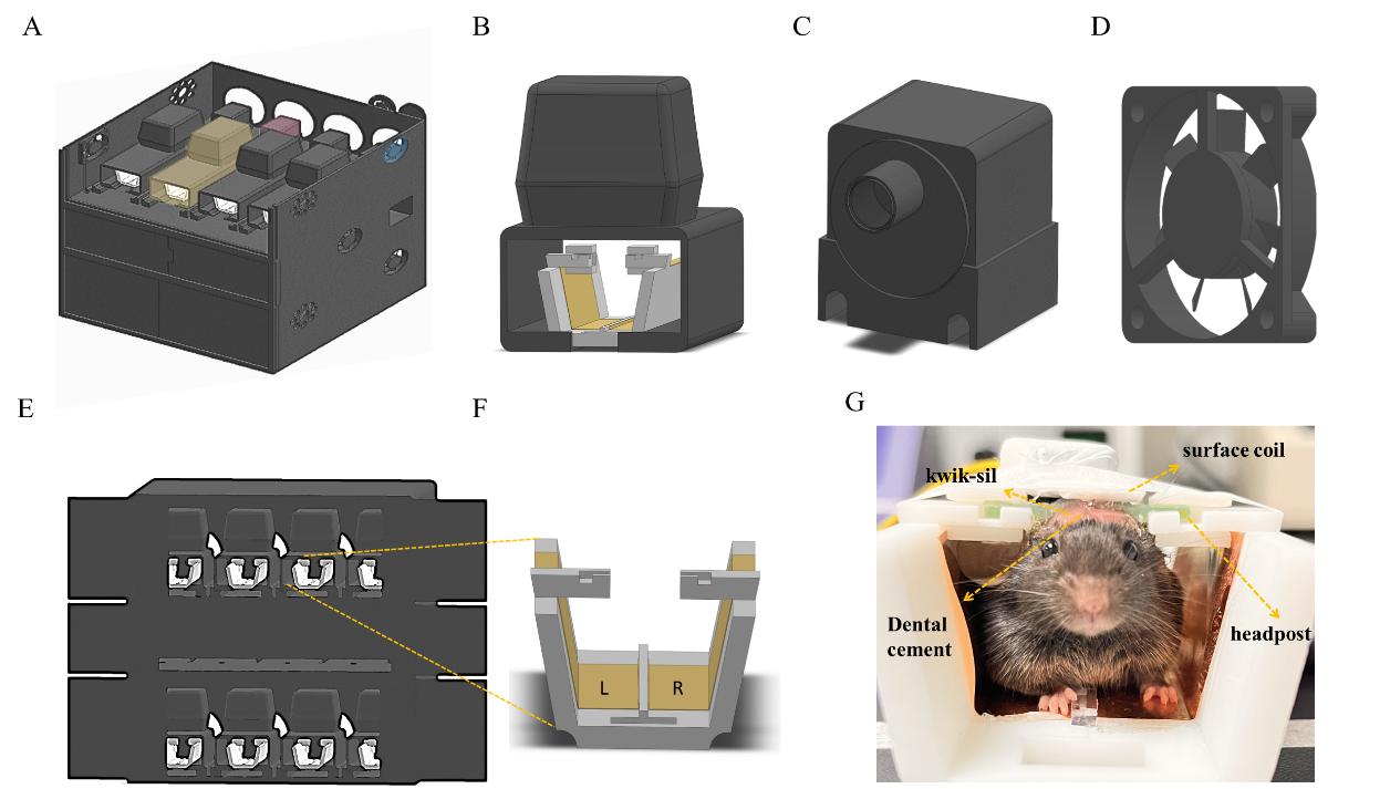
Fig1. Training device for awake mouse fMRI&DIANA:
A. Three-dimensional model of the custom mouse training box and cradle.
B. An MRI environment simulation box , assembled with a animal cradle (The yellow shaded area in (A));
C. Infrared cameras used to monitor mice during training(The pink shaded area in (A));
D. Exhaust fans for circulating air(The blue shaded area in (A));
E. Overview of the inside of the habituation box
F. Copper attached to a training bed for electrical stimulation of mice;
G. Head-fixed mice; Glass fiber headpost used for head fixing
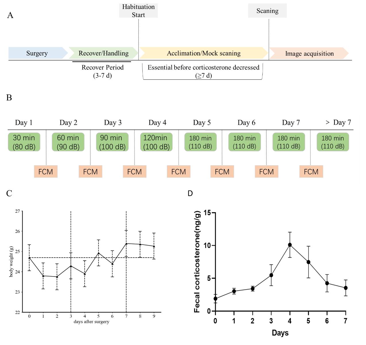
Fig2.Habituation protocol
A. Experimental timeline for habituation protocol;
B. Noise decibel Settings in awake mice during pre-imaging training;
C. Effects of surgical procedures and awake MRI habituation on mouse body weight fluctuations.Note that the left vertical line indicates the start of awake MRI aclimatization,the right vertical line indicate when awake MRI habituation length reached the maximum (180 min).
D. Timeline representation of fecal corticosterone metabolite (FCM) concentration levels at the sampling timepoints during the experimental period.
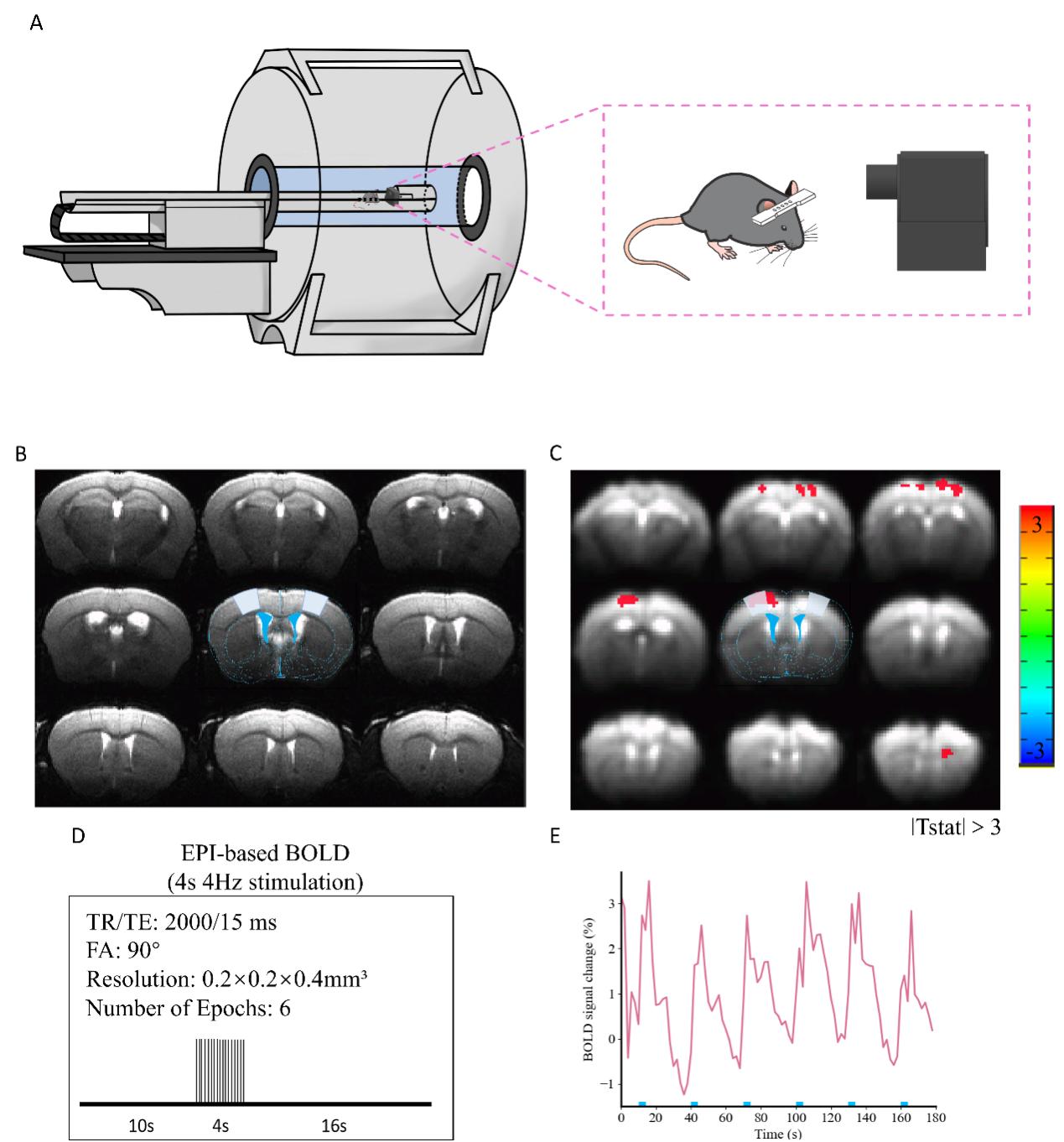
Fig3.BOLD-EPI results.
A. Overall setting within MRI (Mice were anchored to the animal bed via a head post, and the magnetic resonance compatible infrared camera was placed directly in front of the mice);
B. T2 RARE anatomical images;
C. BOLD-EPI(the red part responds to electrical stimulation)
D. Scan parameters and stimulation paradigm design of BOLD-EPI;E. Timecourse of BOLD-EPI signal(The blue block on the horizontal coordinate is for electrical stimulation)
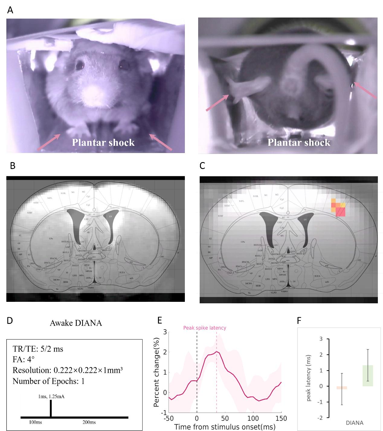
Fig4. DIANA results.
A. The left part is the front view of a mouse being scanned in an MRI, the right part is divided into the rear view of the mice undergoing electrical stimulation;
B. T1 FLASH anatomical image corresponding to DIANA;
C. The red part is the responding DIANA signal, covered ROI in the mouse brain atlas;
D. Scan parameters and stimulation paradigm design of DIANA;