0129
Neurovascular & experimental confounds when probing neuronal activity with fast fMRI: exploring evasive DIANA & DW-fMRI.1School of Psychology, University of Nottingham, Nottingham, United Kingdom, 2School of Physics, Engineering, and Technology, University of York, York, United Kingdom, 3Department of Chemistry, University of York, York, United Kingdom, 4Department of Sports and Exercise Sciences, Manchester Metropolitan University, Manchester, United Kingdom
Synopsis
Keywords: fMRI Acquisition, Neuro, Neuronal Activity, Line Scan Imaging
Motivation: Exploring the cerebrum's functional organisation and processing is challenging. Functional Magnetic Resonance Imaging (fMRI) measures neuronal activity (NA) noninvasively, but relies on indirect signals related to cerebral haemodynamics.
Goal(s): We rigorously investigate if NA in the human brain can be measured using diffusion-weighted fMRI and Direct Imaging of Neuronal Activity (DIANA).
Approach: We utilise DW-fMRI and DIANA at 3 Tesla to record the responses in the somatosensory cortex following electric stimulation of the digits.
Results: We confirm BOLD responses in somatosensory cortex. Both DW-fMRI and DIANA also show stimulus-locked responses. However, we express concerns regarding electrical stimulation noise artefacts and neuronal inhibition.
Impact: This study advances our understanding of neuronal activity measurement using innovative fMRI techniques. It sheds light on the challenges, potential artefacts, and optimal strategies for precise human brain mapping, which is crucial for both basic research and clinical applications.
Introduction
Functional Magnetic Resonance Imaging (fMRI) provides noninvasive brain physiology information at relatively high spatiotemporal resolution (~1mm3 and 1s respectively). These signals are indirect, pseudo measures of neuronal activity (NA) with interpretation hindered by complex neurophysiology and associated vascular weightings1,2. Thus, researchers keep developing novel acquisitions, moving away from haemodynamic surrogates3: diffusion weighted (DW) fMRI is used to investigate cellular-swelling as a marker of NA4, & direct imaging of neuronal activity (DIANA) was demonstrated (in rodents) using fast 2D-line scanning (TR=5ms) at high field5. Here, MR signal correlated with evoked neuronal spiking in the somatosensory cortex after stimulation of the whisker pad. Replicating these findings in humans and understanding the signal sources has proven difficult, raising concerns about these methods6-9.We investigate DW-fMRI and DIANA for measuring NA in the human somatosensory cortex (at 3T), in response to electrical stimulation of the digits. We describe stimulation design/delivery implementation, explore haemodynamic confounds and suggest optimisations of DIANA to mitigate neuronal inhibition.
Methods
Data from 6 participants (aged=21-35) was acquired on a Siemens 3T MAGNETOM Vida system (gradients 45mT/m @ 200 T/m/s), using a 64-channel head/neck coil. Electric stimulation (200ms pulse) across the thumb and ring fingers was controlled using a Digitimer (fig.1a). Stimulus synchronisation (to a 1ms 5V trigger) was handled via a MICRO1401 (CED) programmed with Spike2 software (v9.02). Stimuli were presented at 5Hz (200ms). The amplitude of stimulation (~4-12mA) was tailored for perception & comfort upon repetition. Somatosensory cortex was targeted to better replicate whisker stimulation10 in Toi et al (2022) and its localisation through BOLD fMRI measures to 200ms 40Hz stimulation (20s On/Off block design11) aided placement of DIANA & DW-fMRI acquisition planes (fig.1b-c).BOLD responses used a whole-brain 2D-GRE-EPI sequence (voxel size 3mm*3mm*3mm); base resolution 96 pixels, TR/TE=1000/30ms, FOV 290*290mm, flip angle 90o, multiband acceleration factor 2 and phase under-sampling. DW-fMRI followed a SE-EPI approach with monopolar diffusion gradients (b=1800/0 s/mm2). TE was extended to 85ms to maintain TR=1000ms. BOLD & DW-fMRI 5Hz stimulation duration varied between 16s and 60s (ISI 70s & 60s respectively). DIANA used a FLASH sequence (TR/TE=5/1.4ms, bandwidth=1444 Hz/pixel, slice thickness=8mm and 128 points, cartesian/radial readout). A line average loop (x40=~200ms) was placed inside the phase-line loop. Images were acquired every 12.8s and repeated across 45 dynamics, with total acquisition time of 576s and continuous 5Hz stimulation. At every new phase line, a 1ms trigger pulse allowed synchronisation of 50ms stimulus-delay into each 200ms line data capture period (after 10-line repeats, through Spike2).
Results & Discussion
Our optimised experimental setup (fig.1a) shows BOLD responses in the somatosensory cortex to 40Hz, 20s (fig.1b-c) and 5Hz, 16s (fig.2a) contralateral electrical finger-stimulation. DW-fMRI measures result in a rapid response onset for b=1800s/mm2 relative to BOLD (fig.2a) potentially reflecting cell swelling (as a marker of NA)4. DW-fMRI showed poorer sensitivity than BOLD, and spatial mapping proved challenging. Stimulus design affected response shape; a short inter-stimulus-interval amplified the rapid onset in the BOLD data. The response may contain haemodynamic components, and the shape be driven, in part, by arterial-weighting.DIANA analysis followed Toi et.al. (2022), comparing each image voxel-wise with reference to the baseline (average across 50ms) using a paired t-test and only displaying positive values within predefined brain areas. This appears to show a large signal peak in the somatosensory cortex ~15-20ms post-stimulus (fig.2b). Our setup achieved stimulation timing accuracies of ~1-5μs, minimising temporal blurring across the 2,880 DIANA averages (phase-lines*dynamics). Stimulation devices generally use RS232 to USB conversion, resulting in delays on the order of 10ms, negligible for standard BOLD imaging, but critical for fast imaging.
Conclusion & Summary
While our findings replicate the Toi et.al. (2022) results in human brain at 3T, with observed delayed responses possibly reflecting nerve conduction times12, examination of coil elements reveals noise banding from the electrical stimulus (fig.3a). Observed signals could therefore be artefactual in origin. By eliminating coils demonstrating banding (on PCA analysis) DIANA peaks disappear (fig.3b). We note that BOLD response to 5Hz constant stimulation is not insignificant over long experiments (fig.4) and potentially demonstrates neuronal inhibition. Our findings, however, do not explain observation of the DIANA signal in the original optogenetic experiments5. Cartesian based DIANA sequences measure the centre of k-space for 40/2880 repeats of the stimulus. Constant stimulation can result in habituation of neural responses over time13,14 with repercussions for the DIANA signal: simply averaging across multiple repeats to obtain the ‘200ms time course’ window of NA will not guarantee high sensitivity if habituation is high during these repeats. Radial based readouts15 recording the k-space centre every repeat will improve DIANA statistical power.Acknowledgements
JMS/AJK acknowledge provision of MR scanning time and development from the Manchester Metropolitan University.References
1. Drew, P. J. Vascular and neural basis of the BOLD signal. Curr. Opin. Neurobiol. 58, 61–69 (2019).
2. Kennerley, A. J., Mayhew, J. E., Redgrave, P. & Berwick, J. Vascular origins of BOLD and CBV fMRI signals: Statistical mapping and histological sections compared. Open Neuroimag. J. 4, 1–8 (2010).
3. Huber, L., Uludağ, K. & Möller, H. E. Non-BOLD contrast for laminar fMRI in humans: CBF, CBV, and CMRO2. Neuroimage 197, 742–760 (2019).
4. Le Bihan, D., Urayama, S.-I., Aso, T., Hanakawa, T. & Fukuyama, H. Direct and fast detection of neuronal activation in the human brain with diffusion MRI. Proc. Natl. Acad. Sci. U. S. A. 103, 8263–8268 (2006).
5. Toi, P. T. et al. In vivo direct imaging of neuronal activity at high temporospatial resolution. Science 378, 160–168 (2022).
6. Tsurugizawa, T., Ciobanu, L. & Le Bihan, D. Water diffusion in brain cortex closely tracks underlying neuronal activity. Proc. Natl. Acad. Sci. U. S. A. 110, 11636–11641 (2013).
7. Editorial expression of concern. Science 381, 1058–1058 (2023).
8. Choi, S.-H. et al. No replication of direct neuronal activity-related (DIANA) fMRI in anesthetized mice. bioRxiv 2023.05.26.542419 (2023) doi:10.1101/2023.05.26.542419.
9. Hodono, S., Rideaux, R., van Kerkoerle, T. & Cloos, M. A. Initial experiences with Direct Imaging of Neuronal Activity (DIANA) in humans. Imaging Neuroscience 1, 1–11 (2023).
10. Arkley, K., Grant, R. A., Mitchinson, B. & Prescott, T. J. Strategy change in vibrissal active sensing during rat locomotion. Curr. Biol. 24, 1507–1512 (2014).
11. Sanchez Panchuelo, R. M., Besle, J., Schluppeck, D., Humberstone, M. & Francis, S. Somatotopy in the Human Somatosensory System. Front. Hum. Neurosci. 12, (2018).
12. Chouhan, S. Normal motor and sensory nerve conduction velocity of radial nerve in young adult medical students. J. Clin. Diagn. Res. (2016) doi:10.7860/jcdr/2016/14618.7037.
13. Grissom, N. & Bhatnagar, S. Habituation to repeated stress: Get used to it. Neurobiol. Learn. Mem. 92, 215–224 (2009).
14. Klingner, C. M., Hasler, C., Brodoehl, S. & Witte, O. W. Excitatory and inhibitory mechanisms underlying somatosensory habituation. Hum. Brain Mapp. 35, 152–160 (2014).
15. Kennerley, A. J., Mitchell, D. A., Sebald, A. & Watson, I. Real-time magnetic resonance imaging: mechanics of oral and facial function. Br. J. Oral Maxillofac. Surg. 60, 596–603 (2022).
Figures
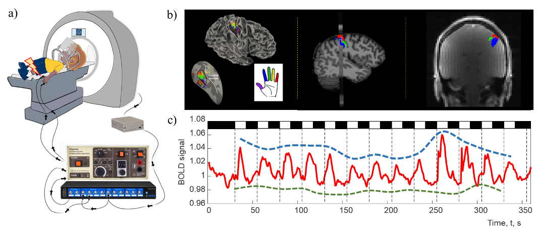
Figure 1 – Experimental setup; a) optical signals from MR trigger (per phase line) are recorded (10kHz sf) and trigger a Micro1401 CED (running Spike2) to pulse a Digitimer (DS7A: 200ms, 4-12mA) across the thumb and ring finger (@50ms). Timing is confirmed to an accuracy of 1-5μs. b) BOLD based digit mapping shows orderly representation (digit-topography by colour). Data used to guide positioning of DIANA & DW-fMRI; c) Resultant BOLD time series shows variance in the magnitude of response linking to neuronal inhibition/vascular interplay.
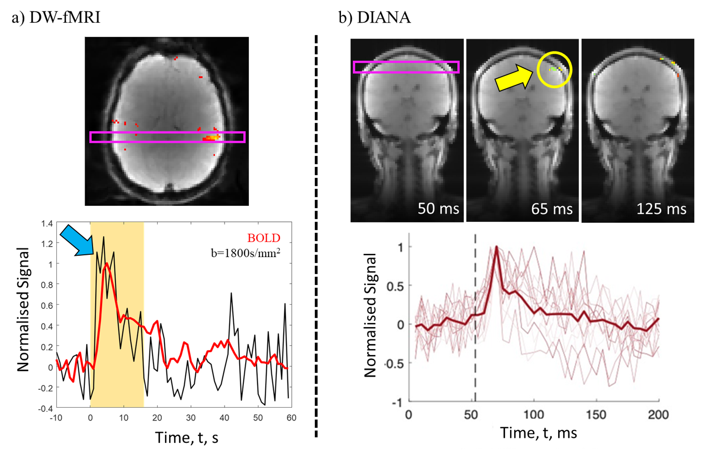
Figure 2 – ‘Direct’ measures of NA in human somatosensory cortex with a) DW-fMRI, with b=1800s/mm2 showing rapid onset to 16s/5Hz stimulation (ISI 70s) Vs. equivalent BOLD signal. Data across stimulus duration suggests vascular confounds; & b) DIANA. Following published analysis methods we observe a robust peak in MRI signal ~15-20ms after 200ms stimulation (at the 50ms time point) ‘localised’ to somatosensory cortex. Mean & individual trials shown. Note – data confounded by the stimulation presentation (see fig.3).
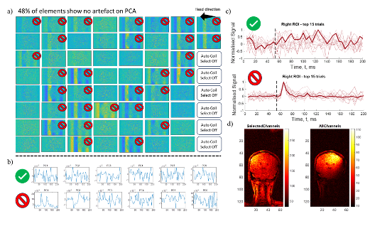
Figure 3 – PCA of a) individual coil elements reveal noise banding, across the brain, in ~50% of channels. This is linked to electrical stimulus of the digits (no artifact seen when the stimulus is off). b) PCA data from single ‘good’ & ‘bad’ coils demonstrates time-based peaks confounding the regional based analysis (see fig.2). c) upon manual elimination of ‘noisy’ coil elements, the DIANA peaks vanish. Suggestive of an artefactual origin for DIANA d) tSNR maps from images reco’ed from selected Vs. all channels.
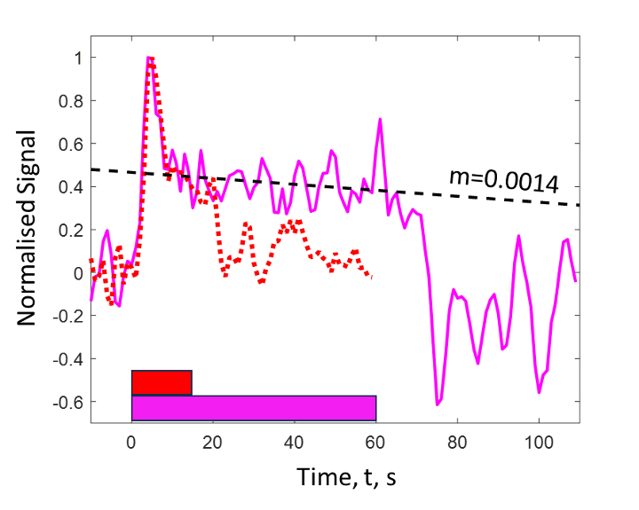
Figure 4 – Haemodynamic effects of 5Hz constant stimulation over the DIANA imaging period are not insignificant. BOLD response 1min 5Hz stimulation shows the same arterial peak seen for shorter 16s stims. This a fast arterial dynamic that would affect the first DIANA image (@12.8s). Furthermore, response does not plateau & slowly decreases in amplitude. We estimate ~335s (5mins 35s) for the haemodynamic response to drop to zero across the 9-min DIANA experiments. Cartesian sampling of the response will be heavily influenced by these changes.