0127
Investigating neural responses using fast, non-selective MRI and simultaneous EEG1NIMH, Bethesda, MD, United States
Synopsis
Keywords: fMRI Acquisition, fMRI, DIANA, neural
Motivation: Recent reports of DIANA responses open the possibility of non-invasively recording neural activity using fMRI.
Goal(s): We aim to test fast, non-selective MRI to better evaluate the feasibility of capturing DIANA responses in the human brain.
Approach: We develop a fast bSSFP sequence without gradient encoding to record the center of k-space during neural activation with a temporal resolution of 3ms.
Results: We observe MRI response dynamics on the order of tens to hundreds of milliseconds, and compare to simultaneously acquired EEG measurements.
Impact: We tested a fast, non-selective MRI sequence to provide preliminary evidence for direct measurement of neural responses in the human brain, and compare these responses to simultaneously acquired EEG measurements.
Purpose
Several groups have recently reported on attempts to directly measure neural responses using MRI sequences (DIANA responses) in mice1,2 and humans3,4,5. This presents great potential for impact on the field of functional MRI (fMRI), as it would circumvent the necessity for indirect inference of neural activity based on vascular signals1,6. However, the feasibility of measuring DIANA responses is still an open question, as these responses are reportedly very small1, and require many trials to average away noise2. Furthermore, the structure of 2D line-scanning sequences used to image DIANA responses necessitates trials be repeated for every acquired k-space line5, inflating the required number of experimental trials by (at least) an order of magnitude. Here, we measured DIANA responses from the center of k-space using a fast (3 ms), non-selective MRI sequence without phase or frequency encoding, negating the need to repeat trials for multiple k-space lines. For comparison to a robust form of direct neuronal recording, we simultaneously acquired electroencephalography (EEG).Methods
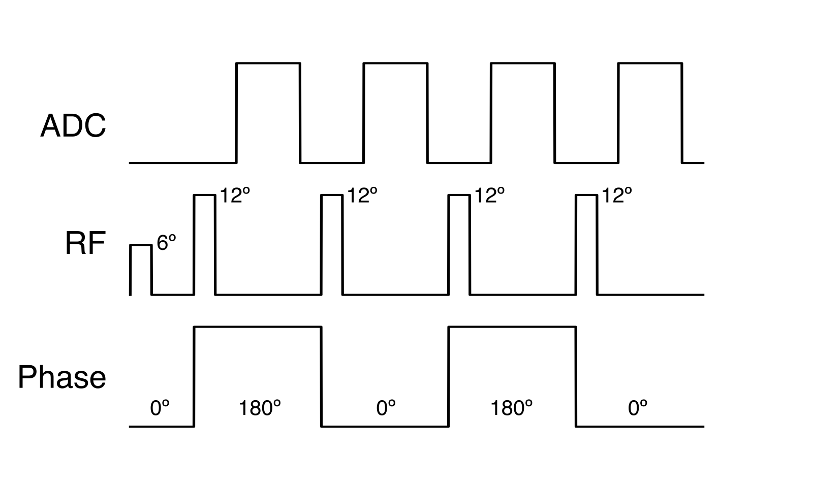
We tested a fast, non-selective, balanced steady-state free precession (bSSFP) sequence7 without gradient encoding to record the center of k-space (Fig. 1). MRI scan parameters: TR=3 ms; TEs=0.75-2.25 ms; RF: hard pulse length=0.5 ms, FA=12º; ADC length=1.5 ms, BW=21370 Hz, vector size=64; RF and ADC phase progressed by 180º with every TR; an SSFP preparation preceded the first TR (FA=6º; TR=1.5 ms).Two participants underwent testing on a Siemens 3T Skyra with a Siemens 64-receiver coil. The first participant viewed a flashing strobe light (10 us; Grass Instruments PS33) via a fiber optic cable with an interstimulus interval (ISI) of 834 ms. The participant completed 10 runs (acquisition time=6:40), each consisting of 10 blocks (20 s rest, 20 s stimulation; 2300 total trials). The second participant heard two auditory tones (1000 and 2000 Hz; duration=300 ms), with random presentation order and pseudo-random ISIs (800-2000 ms). Each run had 72 trials (acquisition time=2:00), and the participant completed 12 runs (864 total trials). The participant tracked the occurrence of the two tones, but did not respond.
MRI time series were formed by Fourier transforming every ADC and taking the magnitude of the spectral peak response, after which each data set size was [64 receiver channels x 40000 time points]. The first 5 s before the scan reached steady state were discarded, and data were bandpass filtered between 0.5 and 50 Hz. MRI receiver coil locations were estimated by computing the center of mass of sensitivity maps within a skull ROI. We cut epochs between -100 and 800 ms from stimulus time and averaged within conditions.
EEG data were recorded using a MR-conditional 256-channel EGI GES 400 (Magstim EGI; Eugene, OR) with Geodesic Hydrocel Sensor Nets. To reduce the impact of the ballistocardiogram (BCG), QSR was estimated from the EEG channels, and BCG was modeled and subtracted using optimal basis sets8 (OBS), bandpass filtered 0.1-30Hz, segmented -100 and 700 ms from stimulus onset, baseline corrected, average referenced, and averaged.
Results
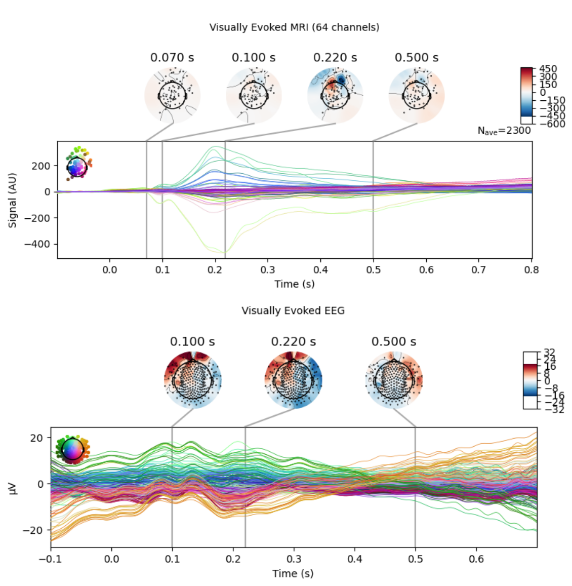
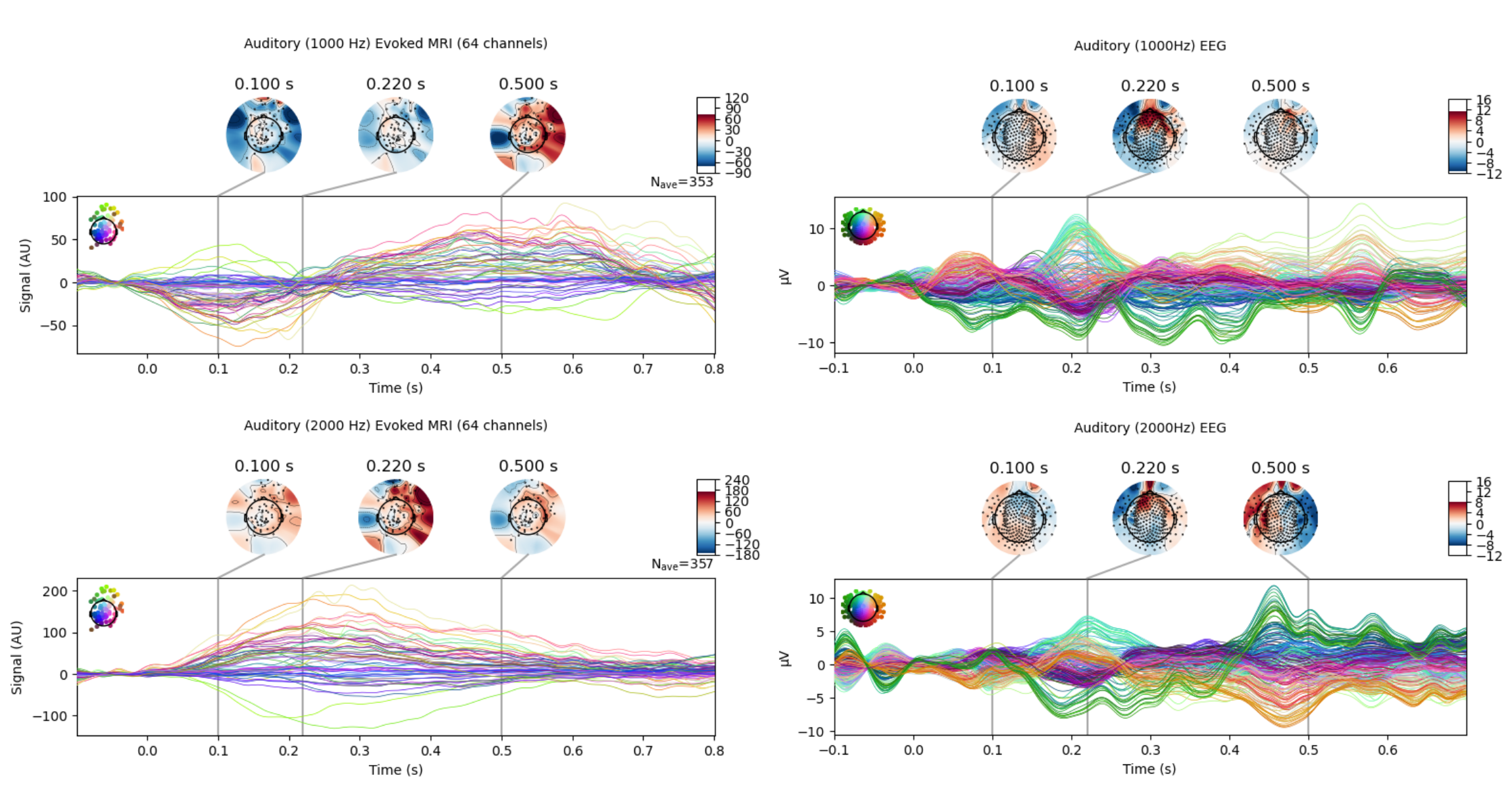
Evoked responses (MRI) and potentials (EEG) to visual flash stimulation are shown in Figure 2. Impressively, we observe response dynamics at 100 ms and observe correspondence between MRI and EEG topographical features. However, a prominent feature of the MRI time series is a substantial positive and negative peak at 200 ms. This feature is likely caused by a blink artifact, and is less visible in the EEG potentials due to automatic blink removal.Responses and potentials to auditory stimulation are shown in Figure 3. As expected, these responses are more lateralized than visual responses, and we see increased dynamics between conditions in the MRI signal, with an initial negative response for the 1000 Hz tone (100 ms) and a delayed positive response (500 ms). There is some correspondence between MRI and EEG topographies, though MRI responses are comparatively delayed. The initial negative response was not visible with the 2000 Hz tone and the response peak occurred earlier (250 ms).
Conclusion
In the current study, we tested a fast, non-selective MRI sequence to better evaluate the feasibility of capturing DIANA responses in the human brain. We provide preliminary evidence for MRI response dynamics on the order of hundreds of milliseconds, and compare these responses to simultaneously acquired EEG measurements. However, it remains an important avenue for future work to better model what relationships exist between fast MRI signals and EEG recordings to understand the sources and mechanisms of DIANA responses.Acknowledgements
The research was conducted within the NIMH Intramural Research Program (#ZIAMH002783). We thank Isabel Gephart for kind assistance with participant recruiting.References
1. Toi, Phan Tan, Hyun Jae Jang, Kyeongseon Min, Sung-Phil Kim, Seung-Kyun Lee, Jongho Lee, Jeehyun Kwag, and Jang-Yeon Park. “In Vivo Direct Imaging of Neuronal Activity at High Temporospatial Resolution.” Science 378, no. 6616 (October 14, 2022): 160–68. https://doi.org/10.1126/science.abh4340.
2. Choi, Sang-Han, Geun Ho Im, Sangcheon Choi, Xin Yu, Peter A. Bandettini, Ravi S. Menon, and Seong-Gi Kim. “No Replication of Direct Neuronal Activity-Related (DIANA) fMRI in Anesthetized Mice.” bioRxiv, May 29, 2023. https://doi.org/10.1101/2023.05.26.542419.
3. Zhang, Yifei, Kaibao Sun, Jianxun Ren, Qingyu Hu, Yezhe Wang, Shiyi Li, Tienzheng Chen, et al. “High-Resolution Dynamic Human Brain Neural Activity Recording Using 3T MRI.” bioRxiv, June 4, 2023. https://doi.org/10.1101/2023.05.31.542967.
4. Hodono S, Rideaux R, van Kerkoerle T, Cloos MA. Initial experiences with Direct Imaging of Neuronal Activity (DIANA) in humans. 2023. https://arxiv.org/abs/2303.00161
5. Wilson, Joshua M., Hua Wu, Adam B. Kerr, Brian A. Wandell, and Justin L. Gardner. “Limitations of Line-Scan MRI for Directly Measuring Neural Activity.” bioRxiv, November 5, 2023. https://doi.org/10.1101/2023.11.03.565394.
6. O’Herron, Philip, Pratik Y. Chhatbar, Manuel Levy, Zhiming Shen, Adrien E. Schramm, Zhongyang Lu, and Prakash Kara. “Neural Correlates of Single-Vessel Haemodynamic Responses in Vivo.” Nature 534, no. 7607 (June 2016): 378–82. https://doi.org/10.1038/nature17965.
7. Scheffler, Klaus, and Stefan Lehnhardt. “Principles and Applications of Balanced SSFP Techniques.” European Radiology 13, no. 11 (November 1, 2003): 2409–18. https://doi.org/10.1007/s00330-003-1957-x.
8. Niazy, R. K., C. F. Beckmann, G. D. Iannetti, J. M. Brady, and S. M. Smith. “Removal of FMRI Environment Artifacts from EEG Data Using Optimal Basis Sets.” NeuroImage 28, no. 3 (November 15, 2005): 720–37. https://doi.org/10.1016/j.neuroimage.2005.06.067.
Figures