0124
A Monte Carlo simulation framework for histology-informed diffusion MRI parameter estimation in cancer1Radiomics Group, Vall d’Hebron Institute of Oncology, Vall d’Hebron Barcelona Hospital Campus, Barcelona, Spain, 2Department of Biomedicine, Faculty of Medicine and Health Sciences, University of Barcelona, Barcelona, Spain, 3Molecular Oncology Group, Vall d’Hebron Institute of Oncology, Barcelona, Spain, 4Prostate Cancer Translational Research Group, Vall d’Hebron Institute of Oncology, Vall d’Hebron Barcelona Hospital Campus, Barcelona, Spain, 5Institut de Diagnòstic per la Imatge (IDI), Barcelona, Spain, 6Department of Radiology, Hospital Universitari Vall d’Hebron, Barcelona, Spain, 7GU, Sarcoma and Neuroncology Unit, Hospital Universitari Vall d’Hebron, Barcelona, Spain, 8Drug Development Unit, Vall d’Hebron Institute of Oncology, Vall d’Hebron Barcelona Hospital Campus, Barcelona, Spain, 9Biomarkers and Clonal dynamics group, Vall d’Hebron Institute of Oncology, Vall d’Hebron Barcelona Hospital Campus, Barcelona, Spain, 10Early Clinical Drug Development Group, Vall d’Hebron Institute of Oncology, Vall d’Hebron Barcelona Hospital Campus, Barcelona, Spain, 11Department of Biomedicine, Faculty of Medicine, Institute of Neurosciences, Institut d’Investigacions Biomèdiques August Pi i Sunyer (IDIBAPS), Barcelona, Spain, 12Centro de Investigación Biomédica en Red de Bioingenierı́a, Biomateriales y Nanomedicina (CIBER-BBN), Barcelona, Spain, 13Cardiff University Brain Research Imaging Centre (CUBRIC), School of Psychology, Cardiff University, Cardiff, United Kingdom, 14School of Computer Science and Informatics, Cardiff University, Cardiff, United Kingdom
Synopsis
Keywords: Simulation/Validation, Microstructure, Monte-Carlo, Histology
Motivation: Analytical biophysical diffusion MRI (dMRI) models fail to capture the full complexity
of diffusion processes.
Goal(s): We propose a Monte Carlo (MC) simulation framework enabling the numerical implementation
of biophysical models with unprecedented fidelity to histology.
Approach: Our framework enables simulating diffusion within cancer environments reconstructed
from histology. It provides paired examples of dMRI signals and histological properties, which can be
used to build numerical microstructure parameter estimators.
Results: Our approach enables more accurate estimation of key properties such as cell size compared
to fitting of classical multi-compartment analytical models.
Impact: We propose a Monte Carlo (MC) simulation framework enabling the implementation of biophysicalmodels with unprecedented fidelity to histology. The framework improves microstructure inference compared to standard analytical fitting, and may provide more robust biomarkers in diseases such ascancer.
Introduction
Multi-compartment biophysical diffusion MRI (dMRI) models enable the estimation of properties such as cell size (CS), promising biomarkers in oncology [1, 2, 3, 4]. However, they often rely on idealisations of microstructural components (e.g., spheres for cells [5]), that fail to capture the full complexity of diffusion processes. Conversely, Monte Carlo (MC) approaches enable realistic diffusion simulations [6, 7, 8] that may be used to develop more accurate signal models [9, 10]. Here we present a MC simulation framework tailored for cancer applications, demonstrating its utility by improving microstructural parameter estimation.Methods
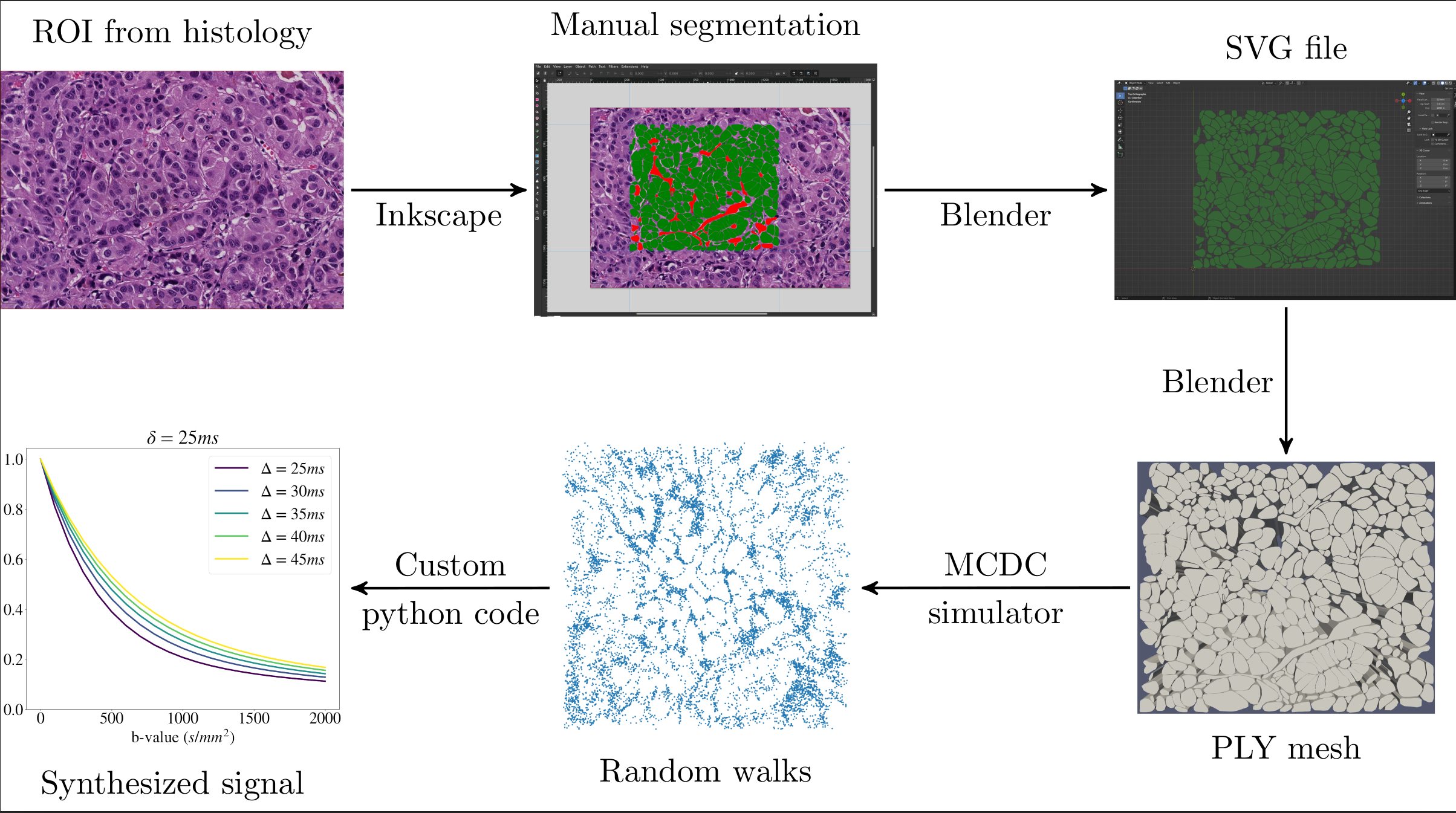
2.1 MC frameworkUsing freely-available software (Inkscape, Blender, MCDC simulator [11]), our framework synthesises random walks within meshes generated from manual segmentations of histological images (Figure 1).The signal sn for the n-th structure is computed with in-house code as [12]
\begin{equation}
s_{n}\,\,=\,\,\left | \frac{1}{W_n} \sum_{w=1}^{W_n} e^{ -j \, \gamma \, \Delta t \, \sum_{t=0}^{TE} \mathbf{g}(t)^{\mathrm{T}}\, \mathbf{r}_{n,w}(t) } \right |.
\label{eq:spinphase}
\end{equation}
Above, $$$\mathbf{r}_{n,w}(t)$$$ is the w-th walker within the n-th structure; ∆t/TE are temporal resolution and echo time; g(t) is the diffusion-encoding gradient. IC/EC signals $$$s_{in}$$$/$$$s_{ex}$$$ are obtained as
\begin{equation}
s_{in}=\sum_{n \in IC}\frac{V_n}{\sum_{k \in IC}V_k}s_n
\end{equation}
and
\begin{equation}
s_{ex}=\sum_{n \in EC}\frac{V_n}{\sum_{k \in EC}V_k}s_n,
\end{equation}
Vn is the volume of the n-th cell in Eq. 2, or of the n-th lumen and of EC space in Eq. 3. No spins are placed inside vessels, fat and debris, but their presence affects EC diffusion. The total signal is
\begin{equation}
s \,\,=\,\, f_{in}\,s_{in}+(1 - f_{in})\,s_{ex},
\label{sEC}
\end{equation}
with $$$f_{in}$$$ being the intra-cellular volume fraction (ICVF).
2.2 Substrates and simulations
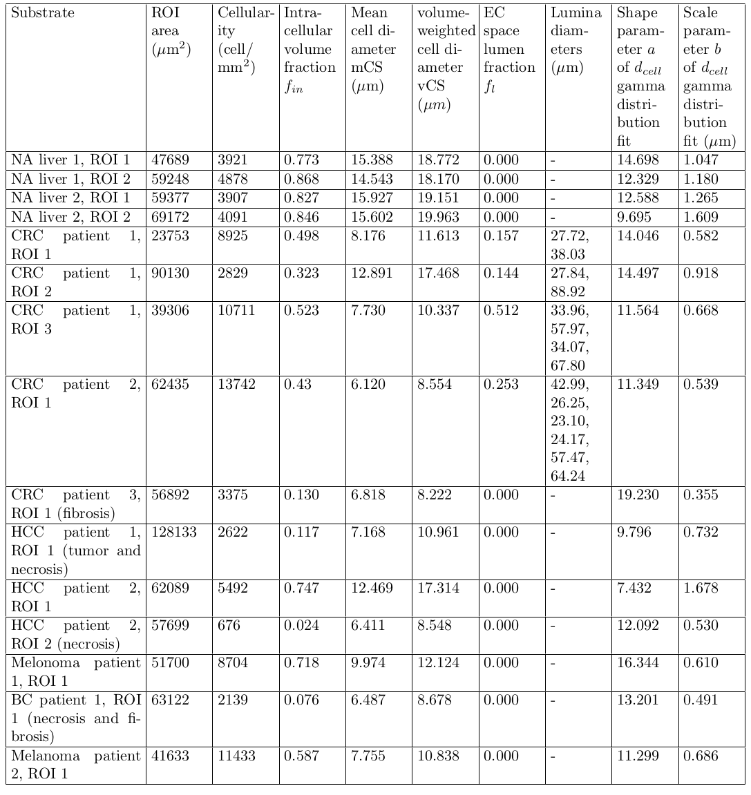
We synthesised random walks within 15 2D substrates, reconstructed from digitised hematoxylin-eosin(HE) liver tumour biopsies (resolution: 0.454 µm) of colorectal cancer (CRC), breast cancer (BC),melanoma and hepatocellular carcinoma (HCC) patients. For each substrate we computed:
- ROI area and cellularity (cell/mm$$$^2$$$);
- ICVF $$$f_{in}$$$;
- lumen fraction of EC space $$$f_l$$$;
- a list of lumen diameters $$$d_{lumen}=2\sqrt{A_{lumen}/\pi}$$$, with $$$A_{lumen}$$$ being the lumen area;
- mean CS index $$$\text{mCS}=<d_{cell}>$$$, where $$$d_{cell}=2\sqrt{A_{cell}/\pi}$$$ with $$$A_{cell}$$$ being the cell area;
- volume-weighted [13] CS index $$$\text{vCS} = \Bigg(\frac{<d_{cell}^7>}{<d_{cell}^3>} \Bigg)^{1/4}$$$;
- shape (a) and scale (b) parameters of a a $$$d_{cell}$$$ gamma-distribution [14] fit
Random walks were simulated for 10$$$\times$$$10 linearly-spaced IC/EC intrinsic diffusivities ($$$D_{0|in}$$$/$$$D_{0|ex}$$$, each varying in [0.8, 3] $$$\mu$$$m$$$^2$$$/ms; $$$\Delta t$$$ = 21.43 $$$\mu$$$s; 1000 walkers per cell, 10000 for EC space).
2.3 In silico parameter estimation
We informed parameter estimation with synthetic signals. We simulated a pulsed-gradient spin-echo (PGSE) protocol matching that of available in vivo scans (see below). It included 3 b-values = 0 and 18 diffusion-weighted (DW) measurements (6 non-zero b-values; minimum/maximum gradient duration $$$\delta$$$ and separation $$$\Delta$$$: 3.9/21.0 ms, 27.8/42.3 ms).Signals were corrupted with Rician noise (signal-to-noise ratio = 50 at b-value = 0) and tissue parameters $$$\mathbf{p}$$$ were estimated according to:
- MC-informed fitting (proposed): a radial-basis function regressor, implementing the forward model $$$\mathbf{p} \mapsto s(\mathbf{p})$$$ was trained and deployed in maximum-likelihood [15] fitting. Estimated parameters: $$$f_{in}$$$, $$$D_{0|in}$$$, $$$vCS$$$, $$$D_{0|ex}$$$.
- Classical analytical model fitting: benchmark maximum-likelihood fitting of a two-compartment analytical model [15,2] (restrictions within spheres [14]; hindered EC diffusion). Estimated parameters: $$$f_{in}$$$, $$$D_{0|in}$$$, $$$vCS$$$, EC apparent diffusion coefficient ($$$ADC_{ex}$$$)
2.4 In-vivo parameter estimation
The two approaches were tested on in-vivo images of BC liver metastases (48 y.o. female patient), acquired on a 3T GE SIGNA-Pioneer system (resolution 2.4 $$$\times$$$ 2.4 $$$\times$$$ 6 mm$$$^3$$$; $$$TR$$$ $$$\sim$$$ 6000 ms, respiratory-gated; effective NEX = 6; ASSET = 2; same dMRI protocol as in simulations; $$$TE = \{75, 90, 105\}$$$ ms). Descriptive statistics were calculated within regions-of-interest (ROIs; normal-appearing (NA) liver, active/necrotic tumour).
Results and discussion
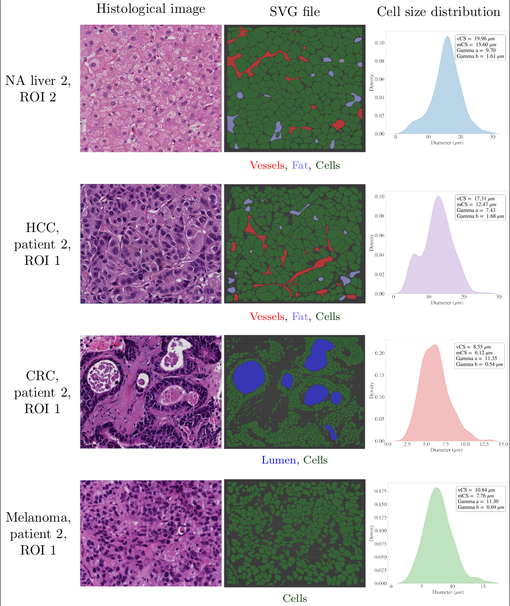
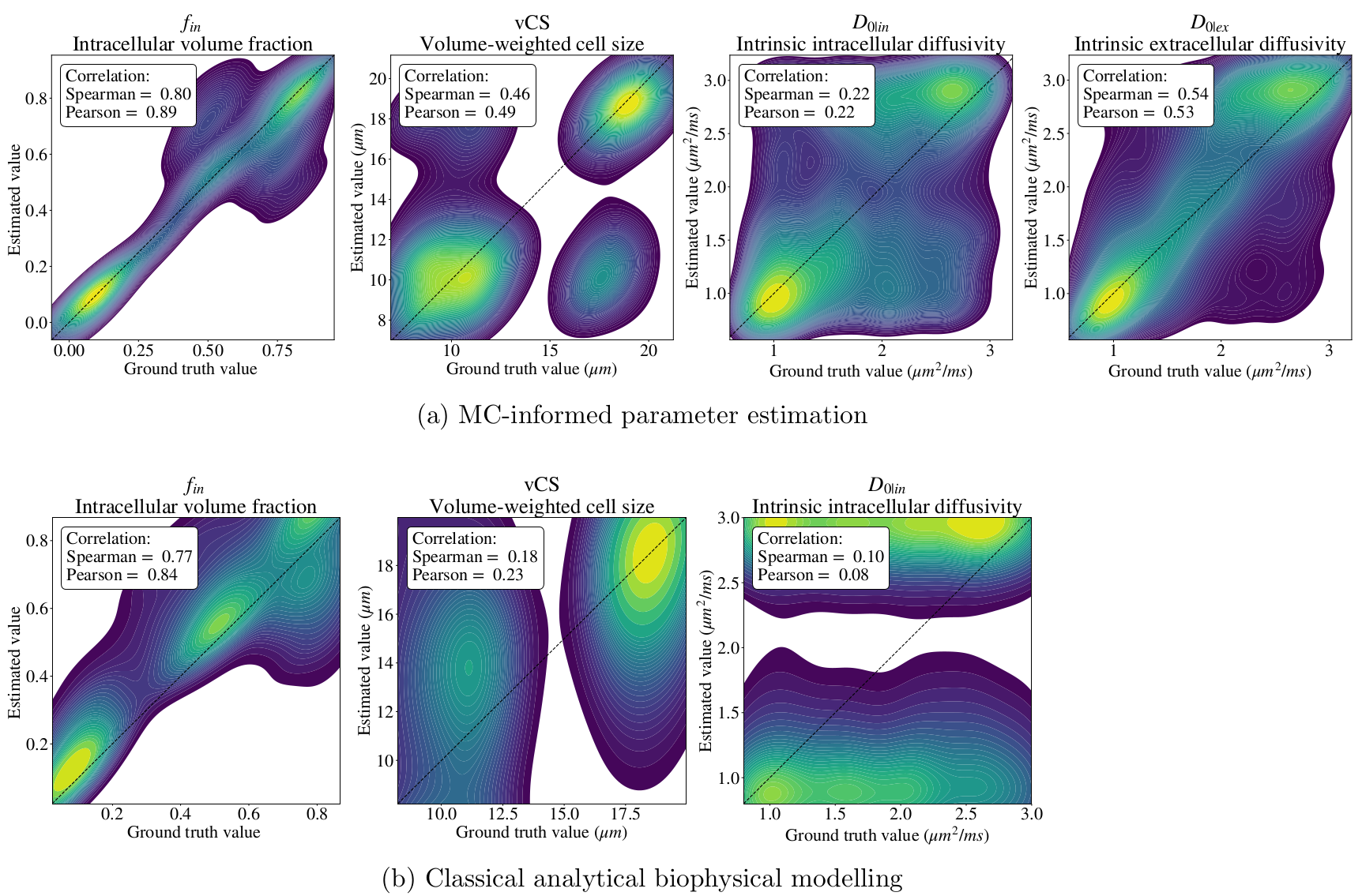
Table 1 describes all substrates, including NA liver, found in the biopsies. Figure 2 shows four substrates, highlighting the variety of microstructures that can be seen in cancer, e.g., smaller cells compared to NA liver, or luminal spaces in CRC metastases.Figure 3 reports on in-silico experiments. MC-informed fitting improves $$$f_{in}$$$, $$$D_{0|in}$$$ and $$$vCS$$$ estimation compared to classical fitting, and enables intrinsic EC diffusivity ($$$D_{0|ex}$$$) quantification.
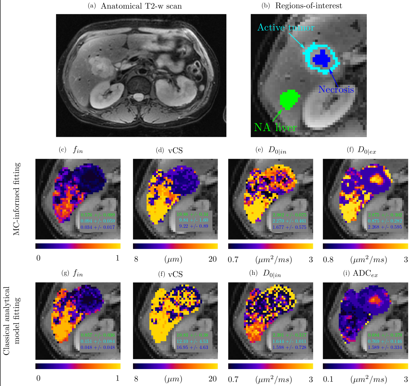
Figure 4 shows in-vivo maps, with ROI-wise means/standard deviations. Larger $$$f_{in}$$$ is seen in the NA liver compared to active/necrotic tumour with both strategies. $$$vCS$$$ shows smaller cells in the tumour compared to the liver for MC-informed fitting; trends are less clear for analytical model fitting. Between-tissue contrasts are consistent between the two strategies, although systematic differences are seen. MC-informed fitting provides visually smoother maps.
Conclusions
Our MC framework enables the implementation of realistic biophysical dMRI models with fewer assumptions than analytical models. Future work will include vascular signals, exchange and relaxation in our simulations
Acknowledgements
RPL and FG are joint last (senior) and corresponding authors. The authors are thankful to Prof.Dmitry S. Novikov and Prof. Els Fieremans for useful discussion, and to the whole MRI radiology team and GE HealthCare for their support with in vivo diffusion imaging. This project received support from AstraZeneca (AZ); AZ was not involved in the acquisition and analysis of the data,interpretation of the results, or the decision to submit this abstract. RPL is supported by ”la Caixa”Foundation, a CRIS Foundation Talent Award (TALENT19-05), the FERO Foundation, the Instituto de Salud Carlos III-Investigación en Salud (PI18/01395 and PI21/01019) and the Prostate Cancer Foundation (18YOUN19). FG receives the support of a fellowship from ”la Caixa” Foundation (ID100010434). The fellowship code is “LCF/BQ/PR22/11920010”, and the fellowship also supports AV.AG is supported by a Severo Ochoa PhD fellowship (PRE2022-102586). KB is funded by a Generalitat de Catalunya Beatriu de Pinós post-doctoral grant (2019 BP 00182). MP is supported by the UKRI Future Leaders Fellowship MR/T020296/2.References
[1] E. Hoffmann, M. Gerwing, S. Niland, R. Niehoff, M. Masthoff, C. Geyer, L. Wachsmuth,E. Wilken, C. Höltke, W. L. Heindel, V. Hoerr, R. Schinner, P. Berger, T. Vogl, J. A. Eble,B. Maus, A. Helfen, M. Wildgruber, and C. Faber, “Profiling specific cell populations within the inflammatory tumor microenvironment by oscillating-gradient diffusion-weighted MRI,” Journal for Immuno Therapy of Cancer, vol. 11, p. e006092, Mar. 2023.
[2] X. Jiang, J. Xu, and J. C. Gore, “Mapping hepatocyte size in vivo using temporal diffusion spectroscopy MRI,” Magnetic Resonance in Medicine, vol. 84, pp. 2671–2683, Nov. 2020.
[3] M. Palombo, V. Valindria, S. Singh, E. Chiou, F. Giganti, H. Pye, H. C. Whitaker, D. Atkinson,S. Punwani, D. C. Alexander, and E. Panagiotaki, “Joint estimation of relaxation and diffusion tissue parameters for prostate cancer with relaxation-VERDICT MRI,” Scientific Reports, vol. 13,p. 2999, Feb. 2023.
[4] G. Lemberskiy, E. Fieremans, J. Veraart, F.-M. Deng, A. B. Rosenkrantz, and D. S. Novikov,“Characterization of Prostate Microstructure Using Water Diffusion and NMR Relaxation,” Frontiers in Physics, vol. 6, p. 91, Sept. 2018.
[5] E. Panagiotaki, R. W. Chan, N. Dikaios, H. U. Ahmed, J. O’Callaghan, A. Freeman, D. Atkinson,S. Punwani, D. J. Hawkes, and D. C. Alexander, “Microstructural Characterization of Normaland Malignant Human Prostate Tissue With Vascular, Extracellular, and Restricted Diffusion for Cytometry in Tumours Magnetic Resonance Imaging:,” Investigative Radiology, vol. 50, pp. 218–227, Apr. 2015.
[6] M. Palombo, C. Ligneul, C. Najac, J. Le Douce, J. Flament, C. Escartin, P. Hantraye, E. Brouillet,G. Bonvento, and J. Valette, “New paradigm to assess brain cell morphology by diffusion-weighted MR spectroscopy in vivo,” Proceedings of the National Academy of Sciences, vol. 113, pp. 6671–6676, June 2016.
[7] J. Xu, J. Xie, N. B. Semmineh, S. P. Devan, X. Jiang, and J. C. Gore, “Diffusion time dependency of extracellular diffusion,” Magnetic Resonance in Medicine, p. mrm.29594, Feb. 2023.
[8] H.-H. Lee, E. Fieremans, and D. S. Novikov, “Realistic Microstructure Simulator (RMS): Monte Carlo simulations of diffusion in three-dimensional cell segmentations of microscopy images,”Journal of Neuroscience Methods, vol. 350, p. 109018, Feb. 2021.
[9] M. Nilsson, E. Alerstam, R. Wirestam, F. Sta, S. Brockstedt, J. Lätt, et al., “Evaluating the accuracy and precision of a two-compartment kärger model using monte carlo simulations,” Journal of Magnetic Resonance, vol. 206, no. 1, pp. 59–67, 2010.4
[10] L. Morelli, M. Palombo, G. Buizza, G. Riva, A. Pella, G. Fontana, S. Imparato, A. Iannalfi,E. Orlandi, C. Paganelli, et al., “Microstructural parameters from dw-mri for tumour characterization and local recurrence prediction in particle therapy of skull-base chordoma,” MedicalPhysics, 2023.
[11] J. Rafael-Patino, D. Romascano, A. Ramirez-Manzanares, E. J. Canales-Rodrı́guez, G. Girard,and J.-P. Thiran, “Robust Monte-Carlo Simulations in Diffusion-MRI: Effect of the Substrate Complexity and Parameter Choice on the Reproducibility of Results,” Frontiers in Neuroinformatics, vol. 14, p. 8, Mar. 2020.
[12] E. Fieremans and H.-H. Lee, “Physical and numerical phantoms for the validation of brain microstructural MRI: A cookbook,” NeuroImage, vol. 182, pp. 39–61, Nov. 2018.
[13] F. Grussu, K. Bernatowicz, I. Casanova-Salas, N. Castro, P. Nuciforo, J. Mateo, I. Barba, andR. Perez-Lopez, “Diffusion MRI signal cumulants and hepatocyte microstructure at fixed diffusion time: Insights from simulations, 9.4T imaging, and histology,” Magnetic Resonance in Medicine,vol. 88, pp. 365–379, July 2022.
[14] Y. Assaf, T. Blumenfeld-Katzir, Y. Yovel, and P. J. Basser, “Axcaliber: A method for measuring axon diameter distribution from diffusion MRI,” Magnetic Resonance in Medicine, vol. 59,pp. 1347–1354, June 2008.
[15] E. Panagiotaki, T. Schneider, B. Siow, M. G. Hall, M. F. Lythgoe, and D. C. Alexander, “Compartment models of the diffusion MR signal in brain white matter: A taxonomy and comparison,”NeuroImage, vol. 59, pp. 2241–2254, Feb. 2012.
[16] B. Balinov, B. Jonsson, P. Linse, and O. Soderman, “The NMR self-diffusion method applied to restricted diffusion. Simulation of echo attenuation from molecules in spheres and between planes,” Journal of Magnetic Resonance, Series A, vol. 104, no. 1, pp. 17–25, 1993.
Figures