0123
White matter changes across the migraine cycle evaluated with Diffusion Tensor Imaging and the impact of Free Water1Universidad Autónoma de Madrid, Madrid, Spain, 2Institute for Systems and Robotics - Lisboa and Department of Bioengineering, Instituto Superior Técnico, Universidade de Lisboa, Lisbon, Portugal, 3Laboratorio de Procesado de Imagen, Universidad de Valladolid, Valladolid, Spain, 4Learning Health, Hospital da Luz, Lisbon, Portugal, 5Imaging Department, Hospital da Luz, Lisbon, Portugal, 6Neurology Department, Hospital da Luz, Lisbon, Portugal, 7Center for Interdisciplinary Research in Health, Universidade Católica Portuguesa, Lisbon, Portugal
Synopsis
Keywords: Diffusion Modeling, Diffusion Tensor Imaging, Migraine
Motivation: About 25% of female migraine patients suffer from menstrual-related migraine, which has been poorly studied.
Goal(s): To identify white matter alterations across the migraine cycle in patients with episodic menstrual-related migraine without aura.
Approach: Diffusion MRI allows to assess alterations in the brain tissue microenvironment. Moreover, including the free-water contribution in the diffusion signal can give information about biological mechanisms, such as inflammation, and more directly expose the tissue alterations by removing free water contamination.
Results: Significant differences were found in the diffusion parameters of the white matter tracts of the menstrual-related migraine patients.
Impact: We found significant alterations in the diffusion parameters of the white matter tracts of episodic menstrual-related migraine patients across migraine cycle using standard diffusion tensor imaging (DTI) and Free-Water corrected DTI.
Introduction
Migraine is a neurological disorder affecting 15% of the global population, with a higher incidence in women1. One type of episodic migraine related to menstruation affects almost 25% of female migraine patients, with regular attacks within two days of menstruation and the first three days of bleeding. The current view is that menstrual-related migraine attacks are related with estrogen withdrawal2. Brain alterations caused by migraine have been investigated with MRI, including diffusion MRI (dMRI). dMRI can detect changes in the tissue microenvironment, assessing alterations in white matter. Previous migraine studies with diffusion tensor imaging (DTI) revealed lower mean diffusivity (MD) in migraineurs, suffering from attacks not specifically related to menstruation3,4. Recently, the application of a diffusion signal model including two compartments, tissue plus isotropic free water diffusion5, allowed more direct characterization of tissue properties and to obtain a biologically relevant parameter, the free water (FW) partial volume fraction. This work investigates white matter alterations in menstrual-related migraine without aura patients along the migraine cycle using DTI parameters calculated with and without free water correction (FW-DTI).Methods
dMRI datasets were acquired in a 3T Siemens Vida scanner with a 64-channel receive RF coil from healthy and menstrual-related migraine without aura female subjects. The migraine patients (n=14, age 35 ± 8 years) were studied in four sessions: preictal (n=9, before menstruation), ictal (n=8, during a migraine episode), postictal (n=10, after the migraine attack) and interictal (n=14, between migraine attacks). The healthy control group (n=15, age 31 ± 7 years) was evaluated in two sessions in corresponding phases of the menstrual cycle: peri-menstrual and midcycle (after ovulation), respectively. The diffusion sequence used 2 shells (b = 400, 1000s/mm2) along 64 gradient directions (32 for each b value) and eight non-diffusion weighted volumes. Pre-processing followed the DESIGNER pipeline6. The Spherical Means method was used to calculate the FW maps with the dMRI-Lab toolbox7. The FW contribution was subtracted from the diffusion signal, and the DTI parameters estimated with DIPY’s TensorModel tool8. The DTI parameters were also estimated directly from the original signal, using the same tool. All the parametric maps were skeletonised using FSL’s Tract-based spatial statistics (TBSS)9. Mean diffusion parameter values in the white matter (WM) regions identified in the Johns Hopkins University ICBM-DTI-81 White-Matter Labels (JHU-WM) Atlas10 were calculated for each subject. Then, the mean values for the control sessions (midcycle and peri-menstrual) for each parameter were subtracted from the migraine sessions: midcycle from interictal and peri-menstrual from preictal, ictal and postictal. To test for significant differences across the migraine cycle, ANOVA was performed in MatLab. P-values were corrected for multiple comparisons by false discovery rate (FDR) correction.Results
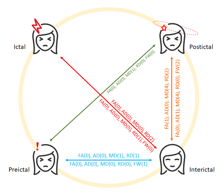
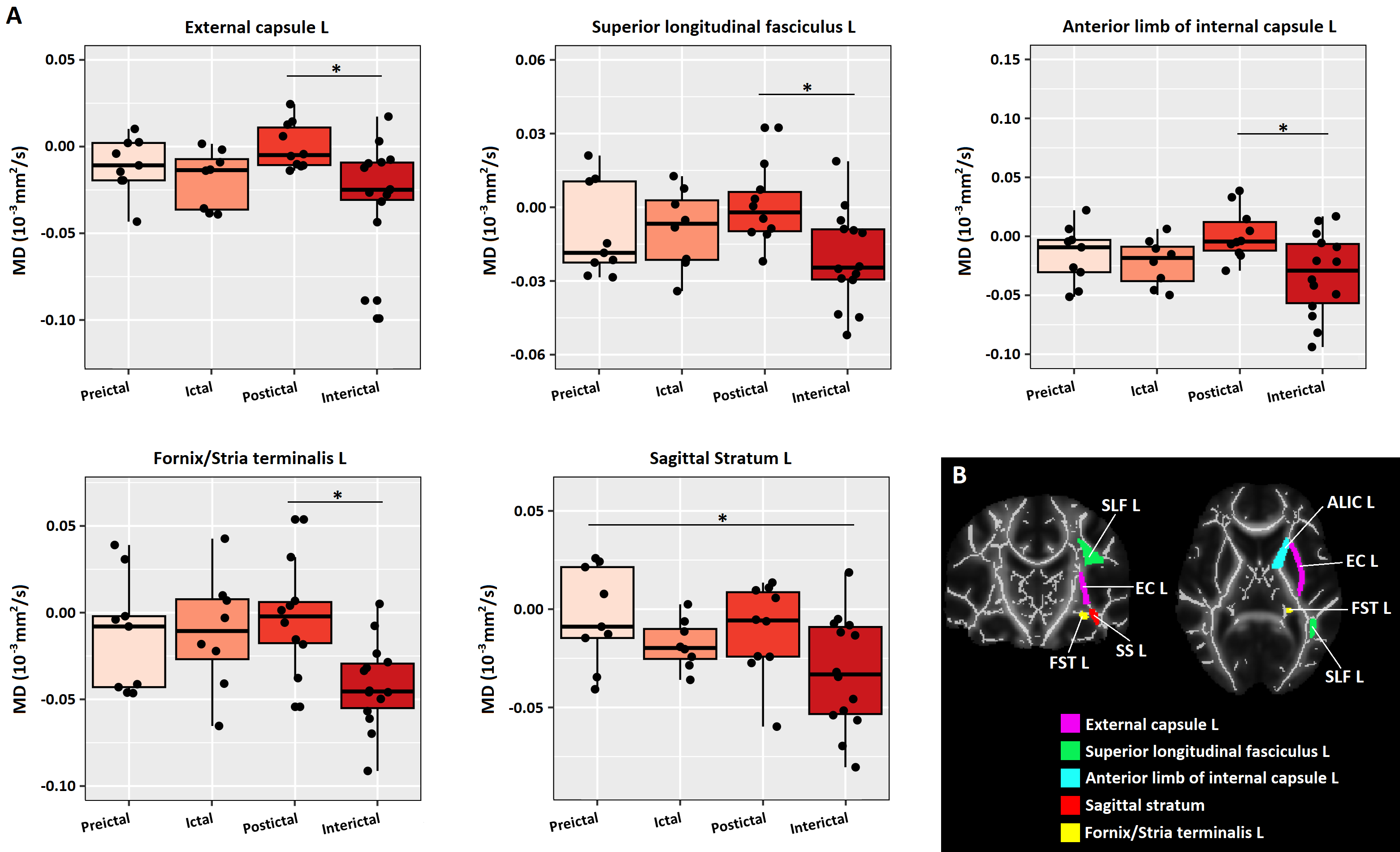
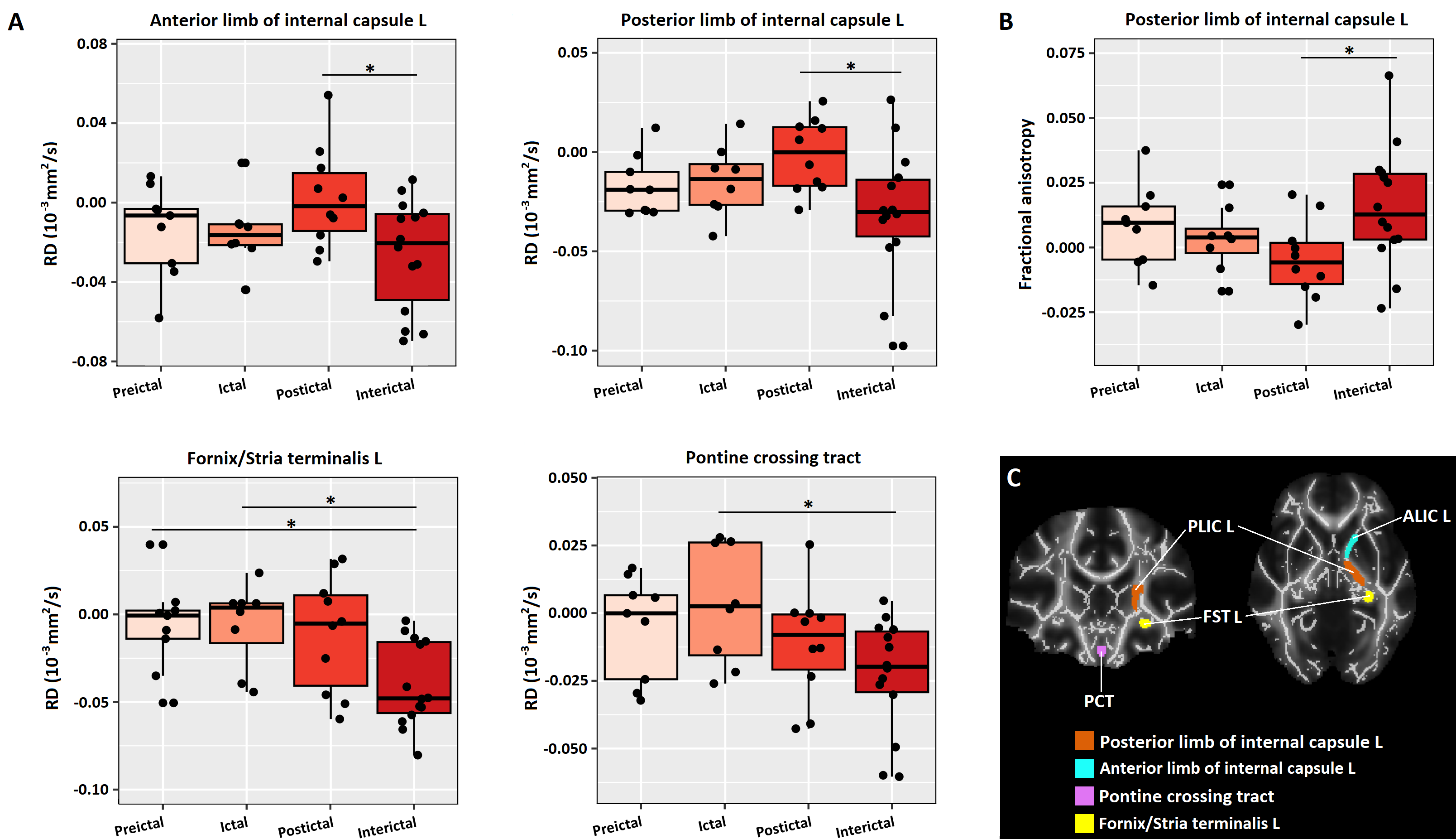
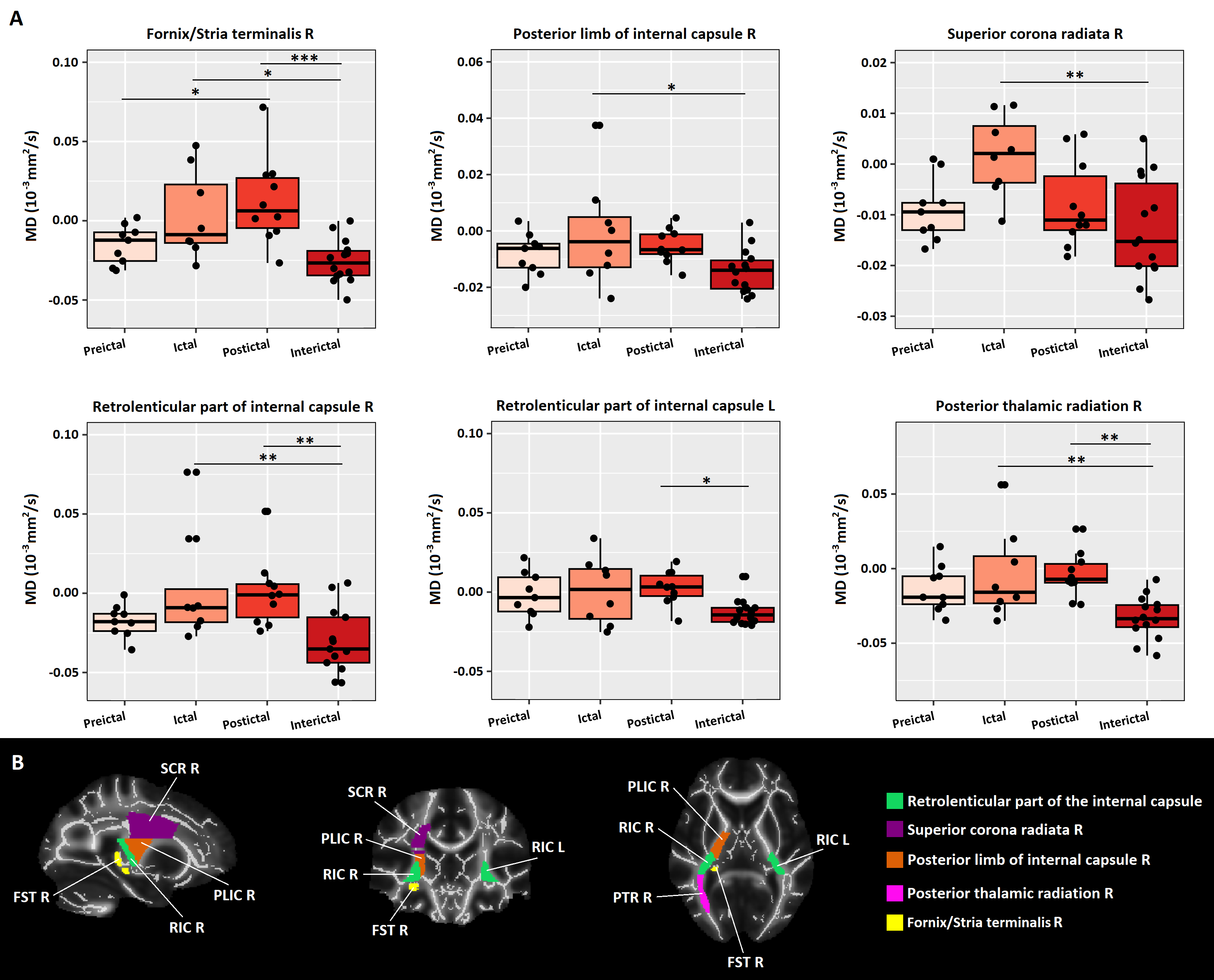
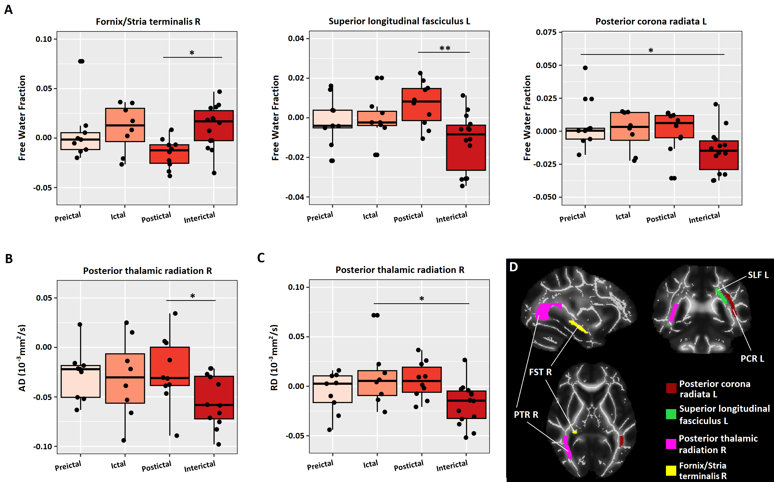
The statistical analyses revealed several significant differences in the white matter skeleton regions for the DTI parameters and the FW values across sessions (Figure 1), although differences did not survive FDR correction. Differences were found between the interictal and remaining sessions, mostly with the postictal session, for both diffusion signal descriptions. In general, the interictal session axial (AD), radial (RD) and mean (MD) diffusivity values tended to be lower compared to other phases. For the standard DTI parameters, differences were found in the left hemisphere of some WM tracts for MD (Figure 2) and RD (Figure 3A), while only one region displayed differences in FA (Figure 3B) and no regions presented differences in AD. The FW signal correction led to differences being found mainly in the WM tracts of the right hemisphere. Most differences were obtained in the MD parameter (Figure 4). However, one WM tract, right posterior thalamic radiation presented changes in AD, RD (Figure 5, B and C respectively) and MD (Figure 4A, bottom right). FW fraction differences were detected in some left hemisphere WM tracts (Figure 5A) while none were detected for FA.Discussion/Conclusions
dMRI allowed to detect alterations along the migraine cycle. Using both DTI and FW-DTI, differences were mainly in the MD values, with FW correction revealing a higher number of MD alterations. No significant differences were detected in FA. Further analyses are needed to determine the possible processes that could explain these results.Acknowledgements
This work was supported by Ministerio de Ciencia e Innovación PID2021-124407NB-I00 and TED2021-130758B-I00, funded by MCIN/AEI/10.13039/501100011033 and the European Union NextGenerationEU/PRTR, and Margarita Salas grants CA1/RSUE/2021-00801 from Universidad Autónoma de Madrid, Spain. We acknowledge the Portuguese Science Foundation through grants PTDC/EMD-EMD/29675/2017, LISBOA-01-0145-FEDER-029675 and UIDB/50009/2020.References
1. Hoffmann J, Baca SM, Akerman S. Neurovascular mechanisms of migraine and cluster headache. Journal of Cerebral Blood Flow & Metabolism. 2019;39(4):573-594.
2. Vetvik K, MacGregor E. Menstrual migraine: a distinct disorder needing greater recognition. The Lancet Neurology, 2021, 20(4):304-315.
3. Yu D, Yuan K, Qin W, et al. Axonal loss of white matter in migraine without aura: A tract-based spatial statistics study. Cephalalgia. 2013;33(1):34-42.
4. Rocca MA, Colombo B, Inglese M, Comi G, Filippi M. A diffusion tensor magnetic resonance imaging study of brain tissue from patients with migraine. Journal of Neurology, Neurosurgery & Psychiatry 2003;74:501-503.
5. Pierpaoli C, Jones DK. Removing CSF contamination in brain DT-MRIs by using a two-compartment tensor model. In: Proc 12th Annual Meeting ISMRM, Kyoto; 2004: 1215.
6. Ades-Aron B, Veraart J, Kochunov P, McGuire S, Sherman P, Kellner E, Novikov D, Fieremans E. Evaluation of the accuracy and precision of the diffusion parameter EStImation with Gibbs and NoisE removal pipeline. NeuroImage 2018; 183:532-543.
7. Tristán-Vega A, París G, de Luis-García R, Aja-Fernández S. Accurate free-water estimation in white matter from fast diffusion MRI acquisitions using the spherical means technique. Magn Reson Med. 2021; 87: 1028–1035.
8. Chang LC, Jones DK, Pierpaoli C. (2005), RESTORE: Robust estimation of tensors by outlier rejection. Magn. Reson. Med., 53: 1088-1095.
9. Smith S, Jenkinson M, Johansen-Berg H, Rueckert D, Nichols T, Mackay C, Watkins K, Ciccarelli O, Zaheer Cader M, Matthews P, Behrens T. Tract-based spatial statistics: voxelwise analysis of multi-subject diffusion data. NeuroImage 2006, 31(4):1487-1505.
10. Oishi K, Zilles K, Amunts K, Faria A, Jiang H, Li X, Akhter K, Hua K, Woods R, Toga A, Pike G, Rosa-Neto P, Evans A, Zhang J, Huang H, Miller M, van Zijl P, Mazziotta J, Mori S. Human brain white matter atlas: identification and assignment of common anatomical structures in superficial white matter. Neuroimage, 2008, 43(3):447–457.
Figures