0121
In vivo tensor-valued diffusion MRI evaluates isotropic and anisotropic kurtosis mismatch in a middle cerebral artery occlusion stroke model1the Fifth Affiliated Hospital, Sun Yat-sen University, Zhuhai, China, 2Guangdong-Hong Kong-Macao University Joint Laboratory of Interventional Medicine, the Fifth Affiliated Hospital, Sun Yat-sen University, Zhuhai, China, 3Department of Biomedical Engineering, Shanghai jiaotong university, Shanghai, China
Synopsis
Keywords: Microstructure, Ischemia, MKI MKA mismatch isotropic kurtosis anisotropic kurtosis
Motivation: Diffusion-weighted imaging (DWI) is widely used in the early detection of stroke, providing valuable information on the infarct core and ischemic penumbra. The mismatch between DWI and more advanced dMRI enhances the accuracy of stroke lesion characterization.
Goal(s): Our goal is to explore whether advanced tensor-valued diffusion MRI (dMRI) can yield sensitive microstructural readouts and evaluate the mismatch between anisotropic and isotropic kurtosis as a potential biomarker for stroke.
Approach: We performed tensor-valued dMRI in a middle cerebral artery occlusion (MCAO) rodent model.
Results: The tensor-valued diffusion MRI demonstrated significant mean diffusivity, mean kurtosis, anisotropic kurtosis, and isotropic kurtosis lesion mismatch.
Impact: Tensor-valued diffusion MRI reveals the isotropic and anisotropic in kurtosis/diffusion lesion mismatch in an animal model of acute stroke, the tensor-valued dMRI may help characterize different microstructural features of acute stroke lesions for precision medicine.
Introduction
Stroke is a leading cause of death and disability, characterized by a significant rate of impairment 1, 2 . the development of advanced MRI has the potential to not only understand the acute ischemia but also transform the management of acute stroke patients3. In this study, we used a tensor-valued diffusion MRI to successfully separated anisotropic and isotropic in the ischemic lesions4.Methods
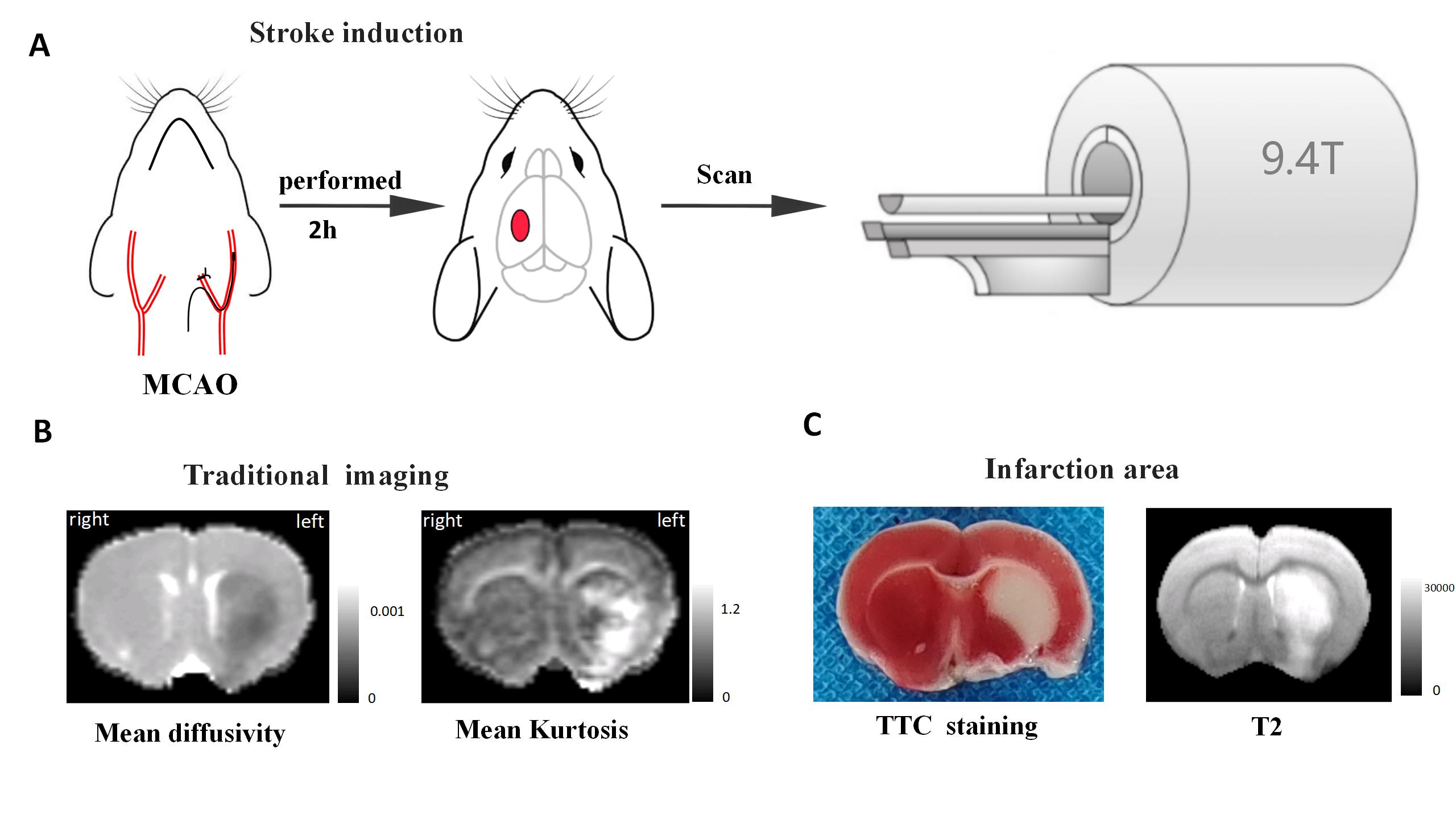
We used a middle cerebral artery occlusion (MCAO) rodent model of Ischemia-reperfusion stroke and images were acquired at a 9.4 T MRI (Bruker).A middle cerebral artery occlusion (MCAO) rodent model: the thread plug enters the internal carotid artery through the external carotid artery to block the origin of the middle cerebral artery. After 2 hours, the thread plug of the external carotid artery is removed to establish a model of ischemia-reperfusion in rats.
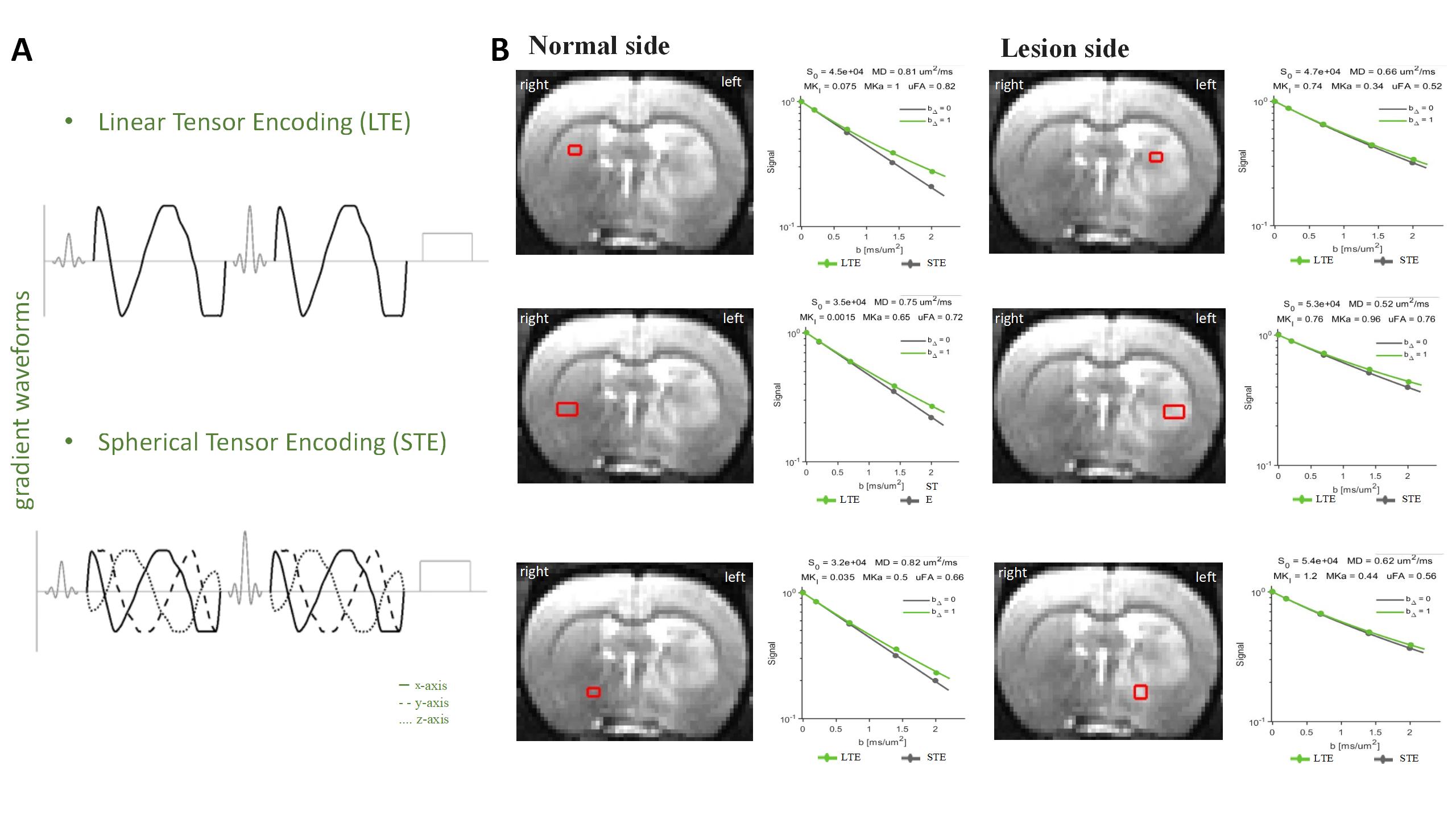
Diffusion MRI (dMRI) images: The tensor-valued dMR included a Linear Tensor Encoding (LTE) protocol and a Spherical Tensor Encoding (STE) protocol: b=200, 700, 1400, and 2000 s/mm2.; TR = 6500 ms, TE = 45ms, matrix = 128×128; FOV = 32 × 32 mm2 ; in plane resolution = 0.25 x 0.25 mm2; number of segments = 8; slice thickness = 800 µm.
Results
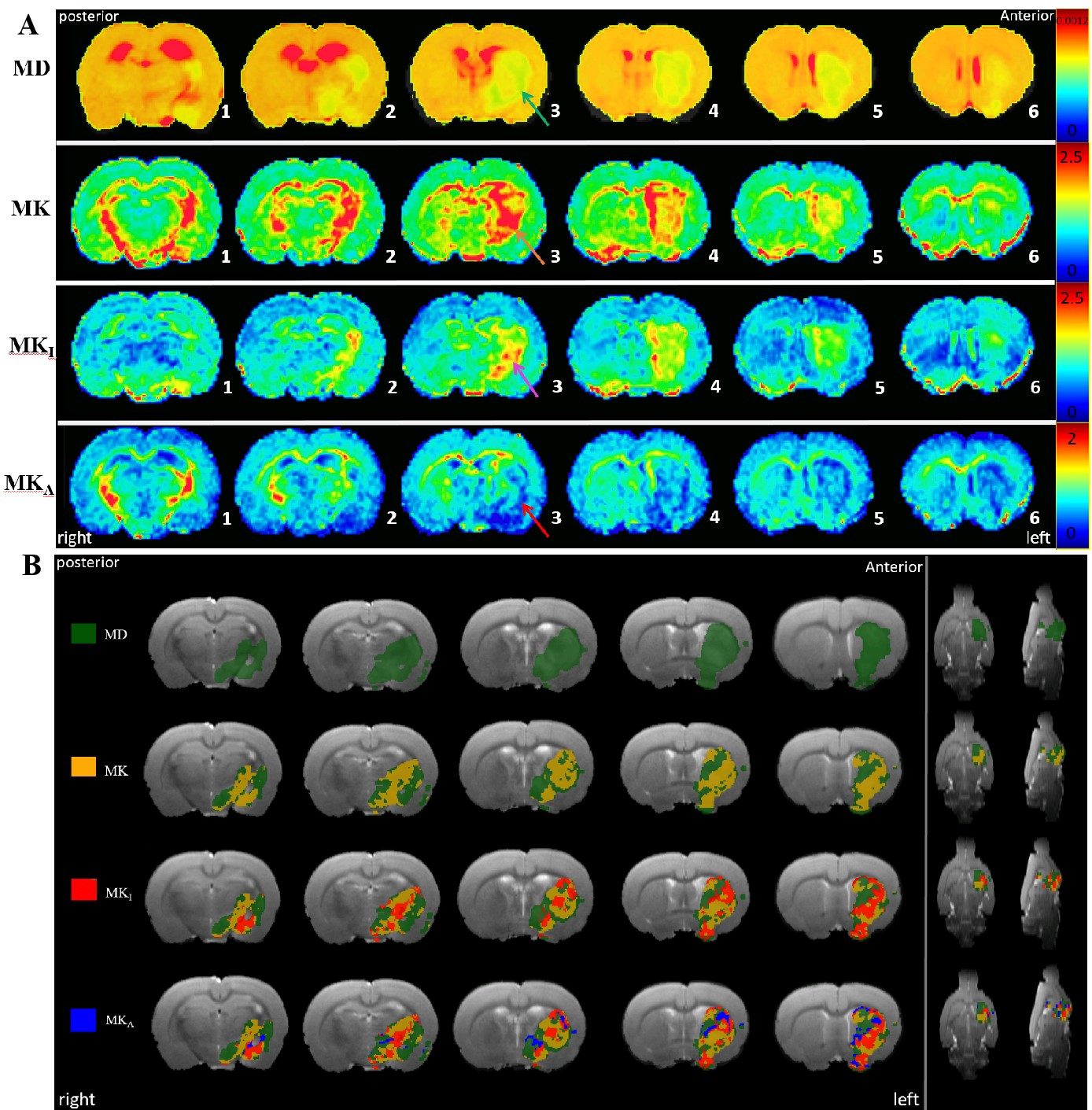
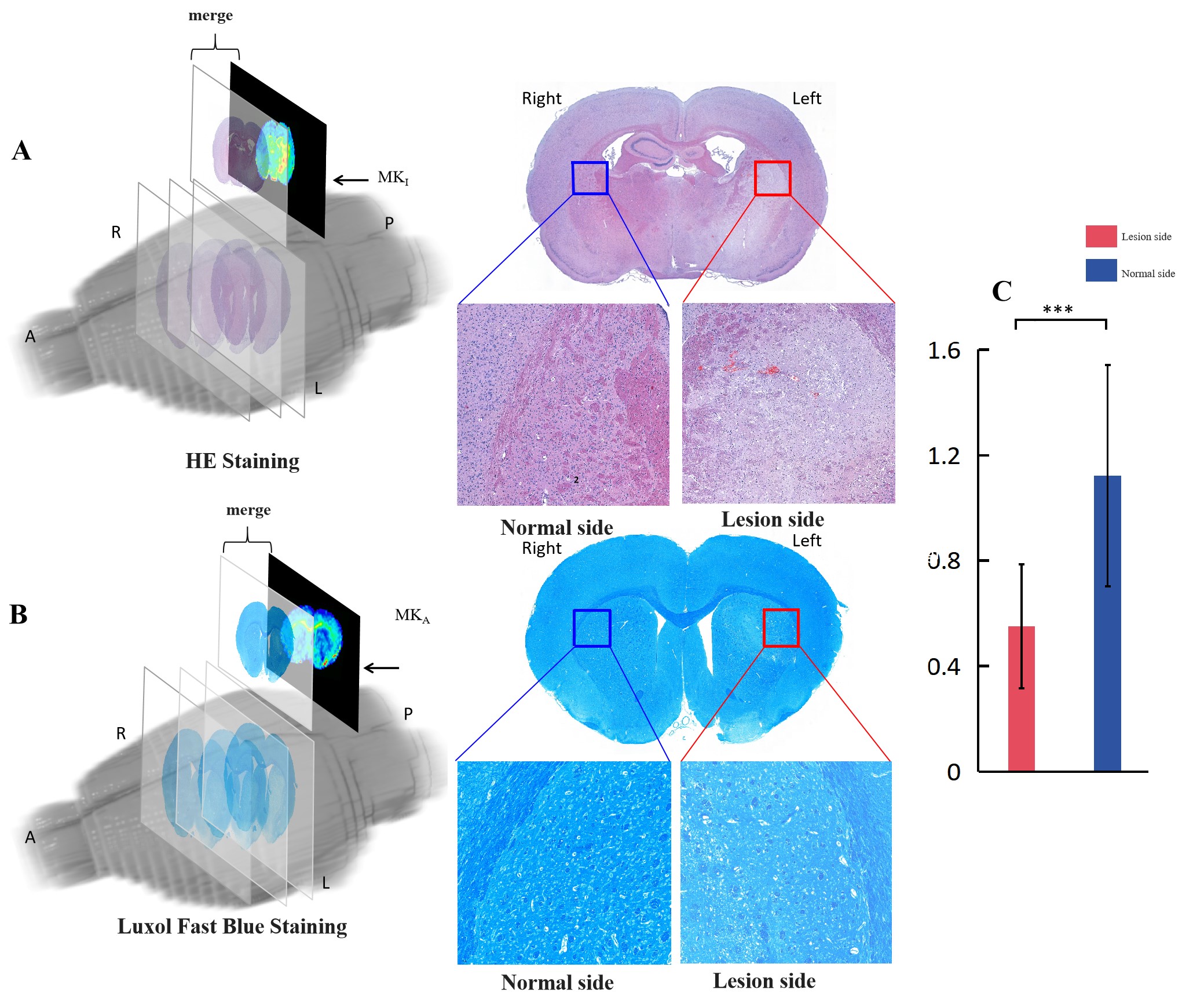
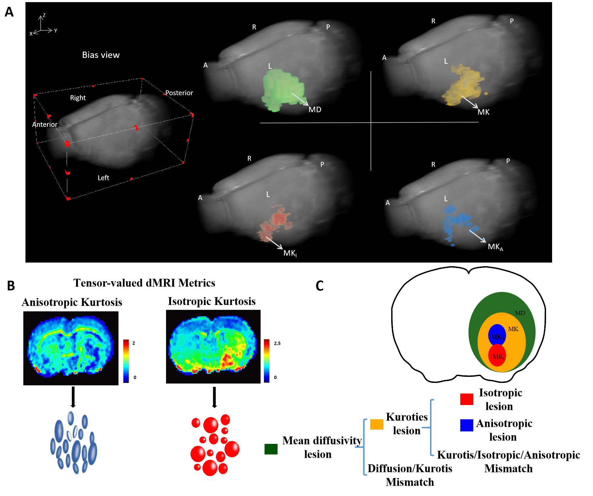
We use tensor-valued diffusion MRI evaluates isotropic and anisotropic kurtosis mismatch in MCAO. Firstly, we validated that ischemic lesions and stroke location are highly repeatable (Fig. 1 C). Using the raw powder averaged to more accurately detect and identify the characteristics and anomalies of normal and Ischemia Lesion data (Fig.2 B). Through MRI, we defined the regions of MKA and MKI and overlaid them with different regions affected by each indicator to evaluate the distribution of stroke infarcts (Fig. 3 A-B), both evidencing the stroked area very clearly. The 3D reconstruction clearly showed the mismatch in rat brain. The histological evaluation in the stroke area clearly showed abnormalities in HE staining and Luxol Fast Blue staining, with cell density quantifying MKI and axonal density quantifying MKA. When quantifying (Fig 4. A), when comparing the ipsilateral hemisphere with the contralateral hemisphere, most of these trends showed statistical significance (Fig 4. B), The 3D reconstruction clearly showed the mismatch in the rat brain. Finally, Tensor value diffusion MRI interprets kurtosis as isotropic and anisotropic kurtosis, while tensor-value diffusion MRI shows significant mean diffusion rate, mean kurtosis, anisotropic kurtosis, and isotropic kurtosis lesions that do not match (Fig.5 A-B).Discussion
Our findings suggest that the isotropic and anisotropic kurtosis mismatch may serve as an advanced imaging biomarker for identifying the ischemic penumbra, potentially leading to improved patient stratification for intervention and a better understanding of stroke pathology. Further research should focus on the longitudinal assessment of the isotropic and anisotropic kurtosis mismatch and its correlation with long-term functional outcomes.Conclusion
Tensor-valued diffusion MRI deciphers kurtosis to be isotropic and anisotropic kurtosis, thereby potentially enhancing sensitive microstructural readouts in stroke. This mismatch may also serve as a predictive tool for clinical outcomes, offering valuable insights for personalized therapeutic strategies. We expect that this study could be used as a steppingstone for developing tensor-valued dMRI in stroke imaging and for assessing novel therapies. Furthermore, this technology can be used not only in a middle cerebral artery occlusion stroke model, but also in other neural injury model, and that tensor-valued dMRI can be generalized towards the interrelationships and change mechanisms between the structures of biological from the perspective of microstructure, and develops related processes such as disease occurrence and development from them. This work shows great promise of tensor-valued dMRI in basic and applied research in the future.Acknowledgements
We thank the Analysis of Functional NeuroImages (AFNI) team for software support. This work was supported by grants from the National Natural Science Foundation of China (No. RLZY20231001-01, and No. 82201447), the Fundamental Research Funds for the Central Universities, Sun Yat-sen University (No. 23hytd009), the Hundred Talents Program of Sun Yat-sen University (The Fifth Affiliated Hospital, 202101), the Guangdong-Hong Kong-Macao University Joint Laboratory of Interventional Medicine Foundation of Guangdong Province (2023LSYS001).References
1. He Y, Aznar S, Siebner HR, Dyrby TB. In vivo tensor-valued diffusion MRI of focal demyelination in white and deep grey matter of rodents. Neuroimage Clin. 2021;30:102675.
2. Global, regional, and national burden of stroke and its risk factors, 1990-2019: a systematic analysis for the Global Burden of Disease Study 2019. Lancet Neurol. 2021;20(10):795-820.
3. Campbell BCV, De Silva DA, Macleod MR, Coutts SB, Schwamm LH, Davis SM, Donnan GA. Ischaemic stroke. Nat Rev Dis Primers. 2019;5(1):70.
4. Cheung J, Doerr M, Hu R, Sun PZ. Refined Ischemic Penumbra Imaging with Tissue pH and Diffusion Kurtosis Magnetic Resonance Imaging. Transl Stroke Res. 2021;12(5):742-753.
Figures