0112
Dichotomizing Motor and Non-motor Correlates of Cholinergic Network Denervation in Parkinson’s Disease using Correlational Tractography1Diagnostic Radiology, Singapore General Hospital, Singapore, Singapore, 2Graduate Institute of Medicine, Yuan-Ze University, Taipei, Taiwan, 3Department of Neurology, National Neuroscience Institute, Singapore, Singapore, 4Radiography Department, Singapore General Hospital, Singapore, Singapore, 5National Neuroscience Institute, Singapore, Singapore, 6Singapore General Hospital, Singapore, Singapore
Synopsis
Keywords: Tractography, Parkinson's Disease
Motivation: Cholinergic denervation underlying clinical manifestations in Parkinson’s disease (PD) is complicated.
Goal(s): To clarify motor vs non-motor correlates of cholinergic denervation in PD and identify potential novel therapeutic targets.
Approach: We evaluated for significant associations between projections of the nucleus basalis of Meynert and pedunculopontine nucleus and motor/non-motor scores using correlational tractography in a case-control PD cohort.
Results: Intracellular and extracellular diffusivity demonstrated significant correlations with motor, cognitive and sleep assessment scores in patients. Significantly reduced intra- and extracellular diffusivity of the PPN-cholinergic-motor projection were seen. The cholinergic projections were dichotomized where the most correlated segments innervated ventral posterolateral thalamic nuclei.
Impact: The motor and non-motor correlates of cholinergic denervation show potential as objective clinical markers to characterize the PD spectrum while mapping of the cholinergic projection with highest correlation could identify substructure areas as novel stimulation target.
Introduction
Motor impairment is the typical classic presentation in Parkinson’s disease (PD), preceded by subclinical non-motor symptomatologies of mood and behavioral changes, cognitive impairment, sleep disorders, and olfactory dysfunction [1-4]. Cholinergic degeneration has often been linked to impaired cognition, falling, slower gait speed, rapid eye movement sleep behaviour disorder (RBD) and impaired olfaction, while motor dysfunction has been ascribed to the dopaminergic system [1,6]. Non-responsiveness of gait deficits and sleep disturbances to dopaminergic treatment or deep brain stimulation surgery has lent impetus to exploring the heterogeneity of cholinergic system degeneration in PD. The major cholinergic neurotransmitter systems comprises of the basal fore-brain complex [projections via nucleus basalis of Meynert (NBM)] and the brainstem [pedunculopontine nucleus (PPN)-thalamic projections] [7,8]. Cholinergic denervation of the NBM and PPN have deep implications in the pathophysiology of motor symptoms, cognitive impairment and sleep disturbances in PD [9,10].Methods
Eighty PD patients and 110 healthy controls (HC) underwent brain MRI using diffusion spectrum imaging (DSI) on a 3T scanner (Skyra, Siemens Healthcare, Erlangen, Germany). The DSI imaging parameters were as follows: TR/TE=4100/110 ms, in-plane resolution=2x2x2 mm3, diffusion sampling=129 directions, and maximum b-value=3000 s/mm2. Clinical motor [Hoehn and Yahr Scale (H&Y) and Unified Parkinson’s Disease Rating Scale-Part III (UPDRS-III)] and non-motor assessments were performed for all participants. The latter characterized cognition [Montreal Cognitive Assessment (MOCA) and Mini Mental State Examination (MMSE)) and sleep disturbances [REM Sleep Behavior Disorder Screening Questionnaire (RBD), Pittsburgh Sleep Quality Index (PSQI) and Epworth Sleepiness Scale (ESS)]. Whole brain correlational tractography [11-14] was performed using connectometry analysis in DSI studio (http://dsi-studio.labsolver.org) with the NBM and PPN as seeds to map cholinergic projections exhibiting significant correlations with motor and non-motor scores. The false discovery rate (FDR) threshold was 0.05 and age and gender were included as covariates. The restricted (RDI) and non-restricted (NRDI) diffusion metrics were sampled along the cholinergic projections, which significantly correlated with the variable of interests, to quantify the intracellular and extracellular diffusivity respectively [15]. The Mann-Whitney test was carried out to establish group differences on diffusion metrics of the cholinergic projections between PD and HC. The cholinergic segments were further clustered using FSL (FMRIB Software Library) to investigate the mapping of tracts with highest correlation to the clinical variable of interest.Results
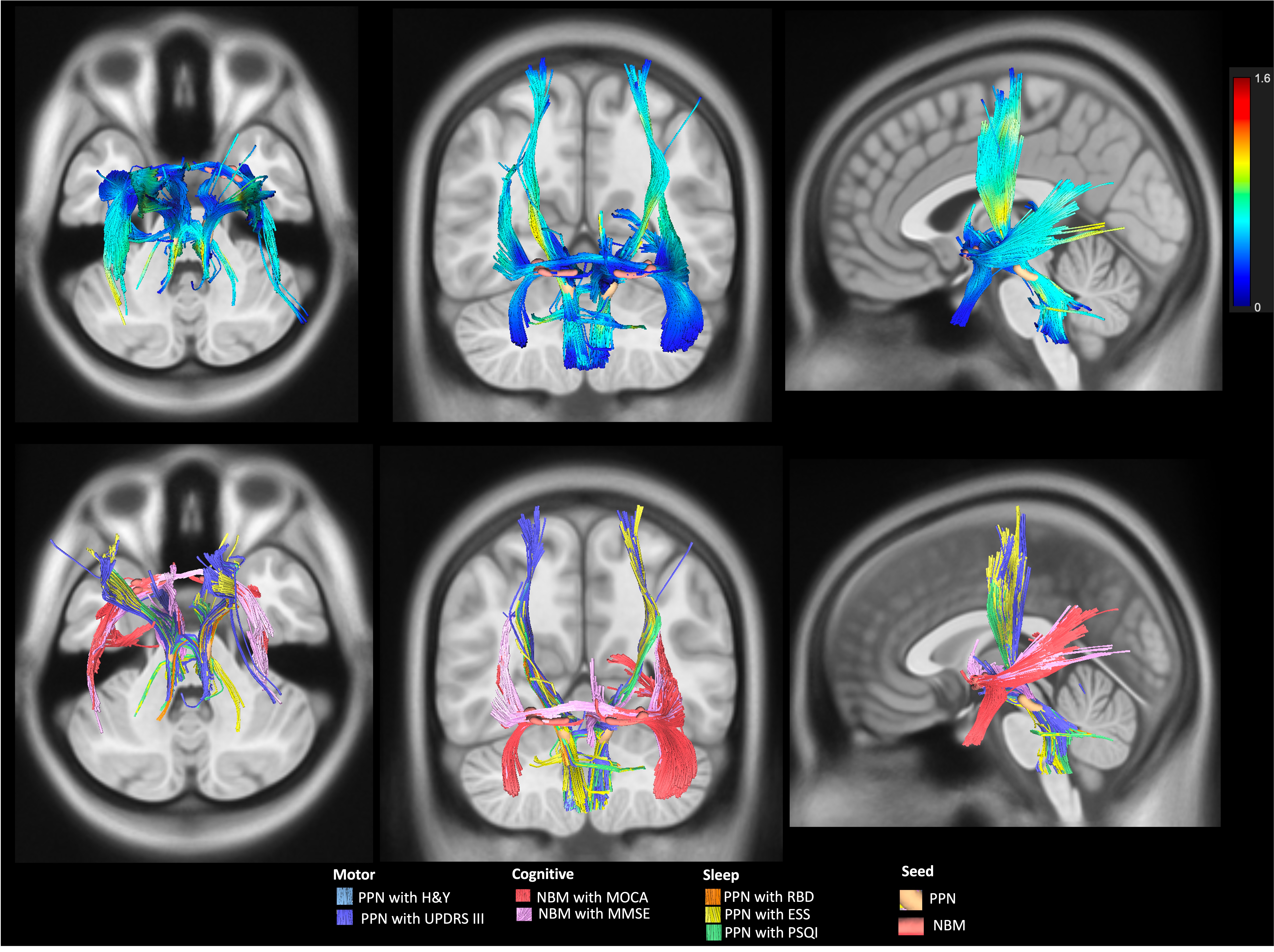
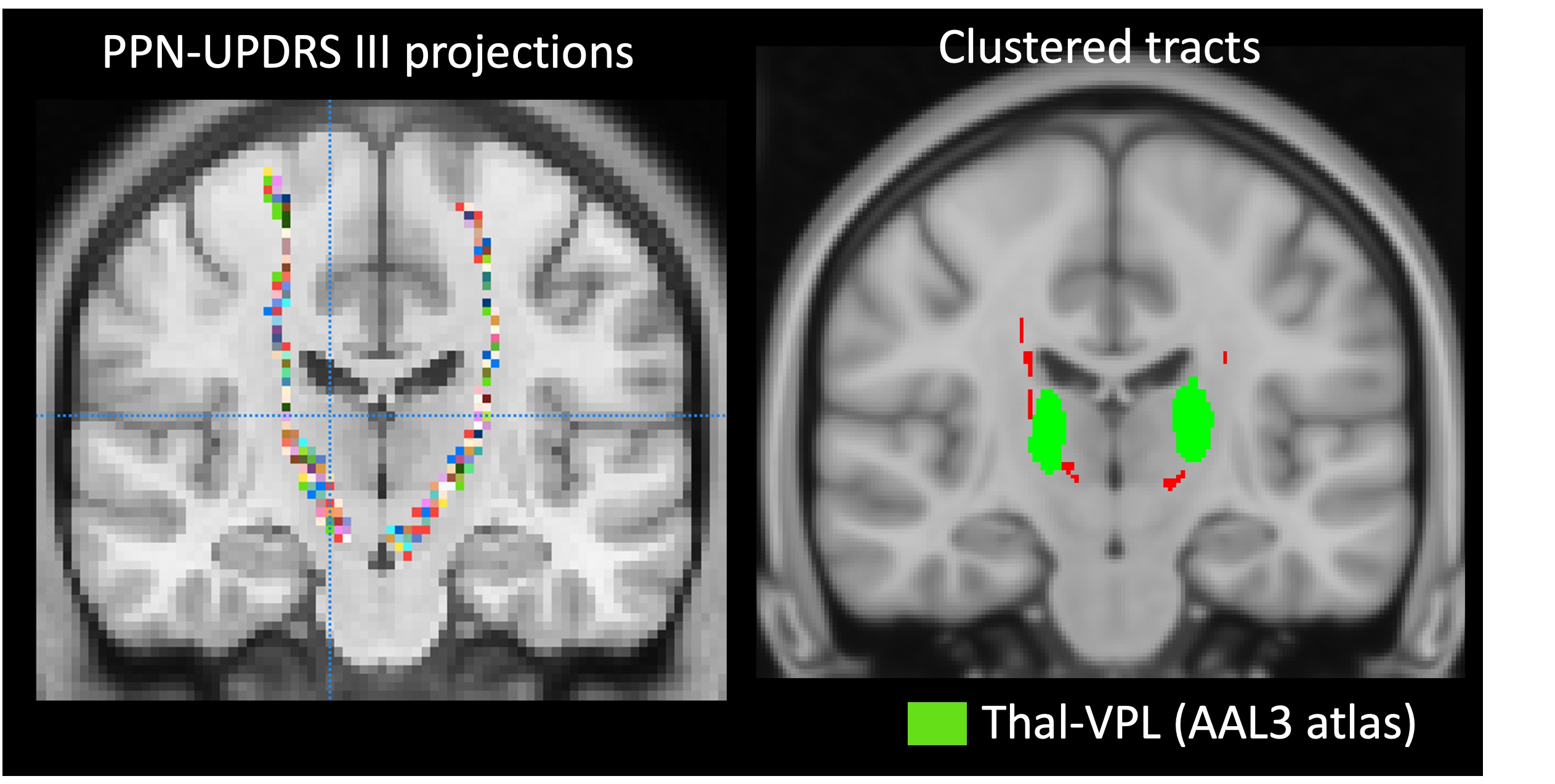
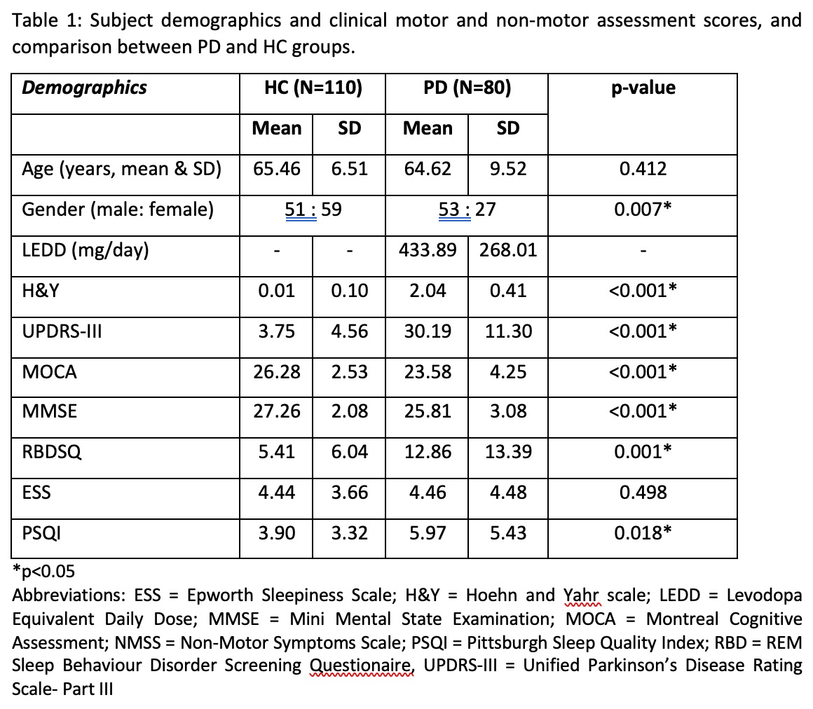
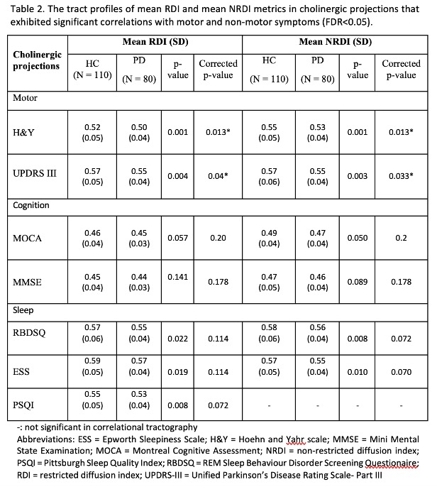
Table 1 describes the subject clinico-demographics. Correlational tractography showed projections that exhibited significant correlations between RDI/NRDI (r=0.06-0.26, FDR<0.05) and motor, cognitive and sleep scores (Figure 1). Tract profiles of the cholinergic segments showed significant associations only between reduced RDI and NRDI, and H&Y (mean RDI_HC, mean RDI_PD, p-value: 0.52, 0.50, 0.013; and mean NRDI_HC, mean NRDI_PD, p-value: 0.55, 0.53, 0.013) and UPDRS-III (mean RDI_HC, mean RDI_PD, p-value: 0.52, 0.50, 0.04; and mean NRDI_HC, mean NRDI_PD, p-value: 0.57, 0.55, 0.033) in PD and HC after correction for multiple comparisons (Table 2). PPN-cholinergic projections that significantly correlated with motor and sleep scores overlapped, while NBM-cholinergic projections that correlated with cognitive scores were well isolated (Figure 1). The PPN-projections that correlated to motor assessments were further clustered to identify segments with the highest correlation with UPDRS-III scores. This demonstrated notable connections passing through the ventral posterolateral (Thal-VPL) thalamic nuclei (Figure 2).Discussion
We found cholinergic projections with significant correlations between diffusivity and motor, cognitive and sleep assessment metrics. The PPN-cholinergic projections showed overlapping projections that correlated with motor and sleep score; these were well isolated from the NBM-cholinergic projections that correlated with cognitive scores. However, only the tract profiles that correlated with motor dysfunction survived multiple comparison correction. Significantly decreased intracellular and extracellular diffusivity correlating with motor dysfunction in cholinergic neuronal connectivity in PD may be related to reduced axonal density and myelination.Cholinergic system degeneration has been increasingly identified as a major pathophysiologic component in motor and non-motor manifestations across the PD spectrum [15-17] despite heterogeneous early reports in neurodegenerative disorders. PPN cholinergic deficits have been associated with dopamine non-responsive gait and balance impairment, [18,19] which lends support to our findings that reduced RDI reflects lower axonal density in the PPN-cholinergic projections. The thalamopetal cholinergic projection has roles in producing fast thalamocortical activation associated with arousal, wakefulness, and REM sleep [20,21]. The clustered PPN-motor-cholinergic segments showed connections passing through the Thal-VPL, further supporting the fact that the ventral thalamic nuclei are deeply involved in motor functions [22].
Conclusion
Correlational tractography shows promise as a sensitive and robust method for mapping cholinergic connectivity in relation to clinical dysfunction. Understanding cholinergic neurotransmission system changes may provide novel therapeutic targets, and mapping of clustered cholinergic projections may allow precise substructure targeting in deep brain stimulation surgery.Acknowledgements
We thank the National Medical Research Council, Singapore, our funding agency, and excellent teams of MR radiographers and research coordinators at the Department of Diagnostic Radiology, Singapore General Hospital, for their kind assistance and support in this study.References
Bergamino M, Keeling EG, Mishra VR, Stokes AM, Walsh RR. Assessing White Matter Pathology in Early-Stage Parkinson Disease Using Diffusion MRI: A Systematic Review. Front Neurol. 2020;11:314.
2. Postuma RB, Bertrand JA, Montplaisir J, et al. Rapid eye movement sleep behavior disorder and risk of dementia in Parkinson's disease: a prospective study. Movement disorders. 2012;27(6):720-726.
3. Gama RL, Távora DG, Bomfim RC, Silva CE, de Bruin VM, de Bruin PF. Sleep disturbances and brain MRI morphometry in Parkinson's disease, multiple system atrophy and progressive supranuclear palsy–a comparative study. Parkinsonism & related disorders. 2010;16(4):275-279.
4. Ashraf-Ganjouei A, Kheiri G, Masoudi M, et al. White matter tract alterations in drug-naive Parkinson's disease patients with excessive daytime sleepiness. Frontiers in neurology. 2019;10:378.
5. Morita H, Hass CJ, Moro E, Sudhyadhom A, Kumar R, Okun MS. Pedunculopontine Nucleus Stimulation: Where are We Now and What Needs to be Done to Move the Field Forward? Frontiers in Neurology. 2014;5(243).
6. Müller ML, Bohnen NI, Kotagal V, et al. Clinical markers for identifying cholinergic deficits in Parkinson's disease. Movement Disorders. 2015;30(2):269-273.
7. Bohnen NI, Müller ML, Dauer WT, Albin RL. Parkinson’s disease: what role do pedunculopontine cholinergic neurons play? Future neurology. 2014;9(1):5-8.
8. Bohnen NI, Müller ML, Kotagal V, et al. Heterogeneity of cholinergic denervation in Parkinson's disease without dementia. Journal of Cerebral Blood Flow & Metabolism. 2012;32(8):1609-1617.
9. Benarroch EE. Pedunculopontine nucleus: functional organization and clinical implications. Neurology. 2013;80(12):1148-1155.10. Wilson H, de Natale ER, Politis M. Nucleus basalis of Meynert degeneration predicts cognitive impairment in Parkinson's disease. Handb Clin Neurol. 2021;179:189-205. doi: 10.1016/B978-0-12-819975-6.00010-8. PMID: 34225962.11. Yeh FC, Badre D, Verstynen T. Connectometry: A statistical approach harnessing the analytical potential of the local connectome. NeuroImage. 2016;125:162-171.12. Yeh FC, Panesar S, Barrios J, et al. Automatic Removal of False Connections in Diffusion MRI Tractography Using Topology-Informed Pruning (TIP). Neurotherapeutics : the journal of the American Society for Experimental NeuroTherapeutics. 2019;16(1):52-58.13. Yeh FC, Tseng WY. NTU-90: a high angular resolution brain atlas constructed by q-space diffeomorphic reconstruction. NeuroImage. 2011;58(1):91-99.14. Yeh FC, Wedeen VJ, Tseng WY. Generalized q-sampling imaging. IEEE Trans Med Imaging. 2010;29(9):1626-1635.15. Yeh F-C, Liu L, Hitchens TK, Wu YL. Mapping immune cell infiltration using restricted diffusion MRI. Magnetic resonance in medicine. 2017;77(2):603-612.15. Kotagal V, Albin RL, Müller MLTM, et al. Symptoms of rapid eye movement sleep behavior disorder are associated with cholinergic denervation in Parkinson disease. Ann Neurol. 2012;71(4):560-568.16. Bohnen, Nicolaas I., et al. "Parkinson’s disease: what role do pedunculopontine cholinergic neurons play?." Future neurology 9.1 (2014): 5-8.17. Müller, Martijn LTM, and Nicolaas I. Bohnen. "Cholinergic dysfunction in Parkinson’s disease." Current neurology and neuroscience reports 13 (2013): 1-9.18. Yarnall A, Rochester L, Burn DJ. The interplay of cholinergic function, attention, and falls in Parkinson's disease. Movement disorders : official journal of the Movement Disorder Society. 2011;26(14):2496-2503.19. French IT, Muthusamy KA. A Review of the Pedunculopontine Nucleus in Parkinson's Disease. Frontiers in aging neuroscience. 2018;10:99.20. Benarroch EE. Pedunculopontine nucleus: functional organization and clinical implications. Neurology. 2013;80(12):1148-1155.21. Jenkinson N, Nandi D, Muthusamy K, et al. Anatomy, physiology, and pathophysiology of the pedunculopontine nucleus. Movement Disorders. 2009;24(3):319-328.
Figures