0108
White matter neurite alterations in dementia with Lewy body bodies: influence of amyloid-β and tau1Department of Radiology, Mayo Clinic, Rochester, MN, United States, 2Department of Psychiatry, University of Cambridge, Cambridge, United Kingdom, 3Department of Quantitative Health Sciences, Mayo Clinic, Rochester, MN, United States, 4Department of Radiology, Mayo Clinic, Jacksonville, FL, United States, 5Department of Neurology, Mayo Clinic, Rochester, MN, United States, 6Department of Psychiatry and Psychology, Mayo Clinic, Rochester, MN, United States, 7Department of Psychiatry and Psychology, Mayo Clinic, Jacksonville, FL, United States, 8Laboratory of Medicine and Pathology, Mayo Clinic, Jacksonville, FL, United States, 9Department of Neurology, Mayo Clinic, Jacksonville, FL, United States
Synopsis
Keywords: Microstructure, Dementia, Lewy bodies, NODDI, DTI, Amyloid, Tau
Motivation: The influence of Alzheimer’s disease (AD) copathologies on white matter neurite changes in dementia with Lewy bodies (DLB) remains unclear.
Goal(s): To delineate the severity of neurite abnormalities and their associations with amyloid and tau PET imaging in DLB.
Approach: We compared Neurite Orientation Dispersion and Density Imaging metrics in the DLB spectrum (DLBs, n=45) against controls (n=45), and evaluated their correlations with amyloid-β ([11C]-PiB) and tau ([18F]-Flortaucipir) PET.
Results: The DLBs exhibited widespread white matter injury relative to controls. Elevated tau deposition, but not amyloid-β burden, was significantly associated with neurite abnormalities, predominantly involving the temporal and limbic white matter tracts.
Impact: These findings demonstrate the impact of AD copathologies on widespread neurite abnormalities in people with DLB, underscoring the importance of further elucidating the mechanisms underlying amyloid-β and tau deposition, and evaluating anti-AD disease-modifying interventions for DLB.
INTRODUCTION
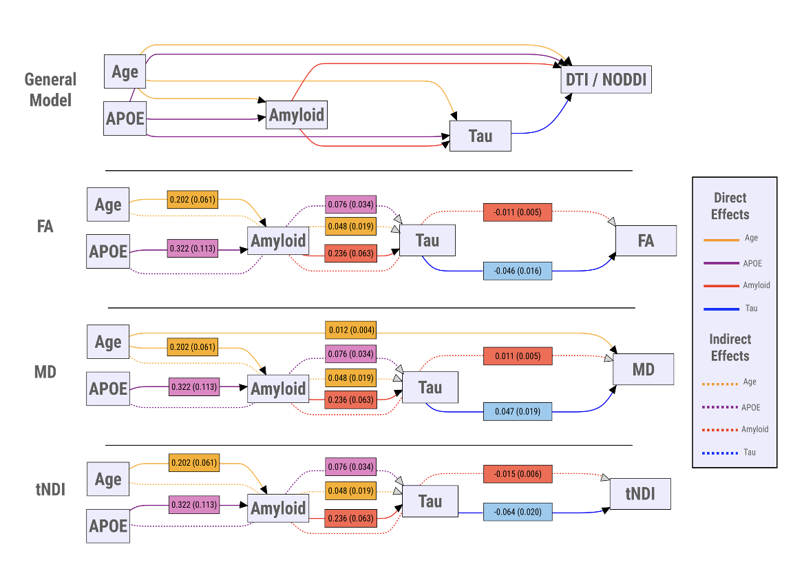
Dementia with Lewy bodies (DLB) is a neurodegenerative disorder often comorbid with Alzheimer’s disease (AD) pathology. While diffusion tensor imaging (DTI) has been widely used in numerous studies to delineate white matter pathology in DLB [1, 2], there is a lack of data on the contribution of AD pathologies to DTI changes, and ambiguous interpretations of changes in DTI metrics remains unresolved. Emerging biophysical models of diffusion weighted data such as Neurite Orientation Dispersion and Density Imaging (NODDI) may help delineate the biological underpinnings of white matter injury in DLB [3].To date, no study has characterized the regional distributions of NODDI parameters in DLB or delineated the in vivo topographical correlations of white matter microstructural changes with PET biomarkers of amyloid-β and tau. We addressed these gaps by: (1) Comparing the regional distributions of DTI and NODDI parameters between individuals on the spectrum of DLB (DLBs) and clinically unimpaired (CU) controls; (2) investigating the topographical associations of white matter integrity with cortical [11C]-PiB and [18F]-Flortaucipir uptake in DLBs to identify the white matter tracts most closely associated with AD pathologies, (3) assessing the degree to which AD-related white matter injury is associated with clinical disease severity; (4) and evaluating the multivariable associations of age, APOE genotype, cortical [11C]-PiB and [18F]-Flortaucipir uptake with DTI and NODDI parameters using Structural Equation Models (SEM).
METHODS
The DLBs (mild cognitive impairment with Lewy bodies (n=13) [4] and probable DLB (n=32) [5] ) and 45 controls underwent diffusion-weighted imaging and PET imaging of [11C]-Pittsburgh compound-B and [18F]-Flortaucipir. Group differences in DTI and NODDI metrics were compared using conditional logistic models.To investigate the influence of amyloid-β and tau on regional DTI and NODDI parameters, separate multivariable regression models were constructed in the DLBs and CU groups, while adjusting for age and APOE genotype and correcting for multiple comparisons across white matter regions-of-interst (ROIs) using False Discovery Rate (FDR). Next, we derived composite ROIs based on the significant FDR results representing white matter changes associated with tau and evaluated age-adjusted partial Pearson’s correlations between these composite ROIs and the Clinical Dementia Rating (CDR) sum of boxes in the DLBs. SEMs were used to explore relationships among age, APOE ε4, amyloid-β, tau, and white matter injury.
RESULTS
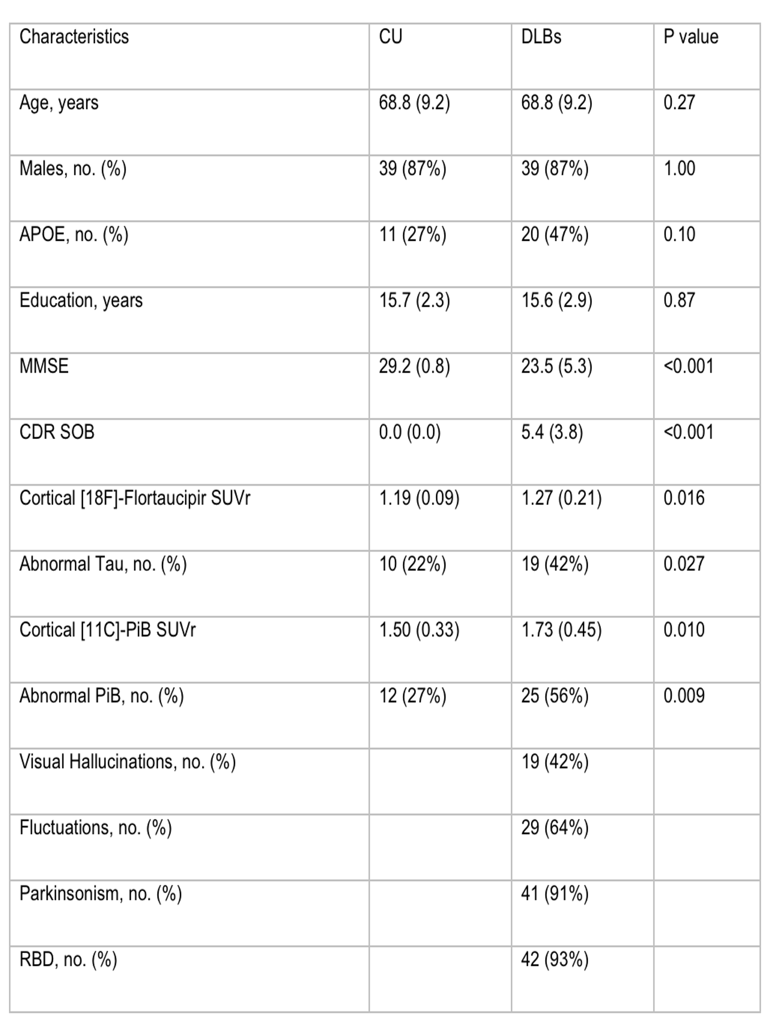
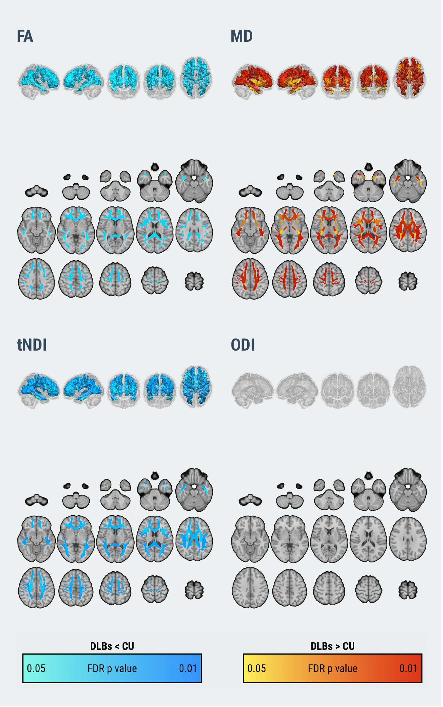
Participant characteristics are summarized in Figure 1.The DLBs group exhibited widespread white matter abnormalities relative to controls, including lower fractional anisotropy (FA), elevated mean diffusivity (MD), and lower tissue-weighted neurite density (tNDI) in topographically similar regions (Figure 2, FDR p < 0.05). Dice coefficients indicated that the topography of group differences between FA and MD were moderately similar (Dice index = 0.62), while tNDI changes were highly similar to both FA (Dice index = 0.71) and MD (Dice index = 0.89).
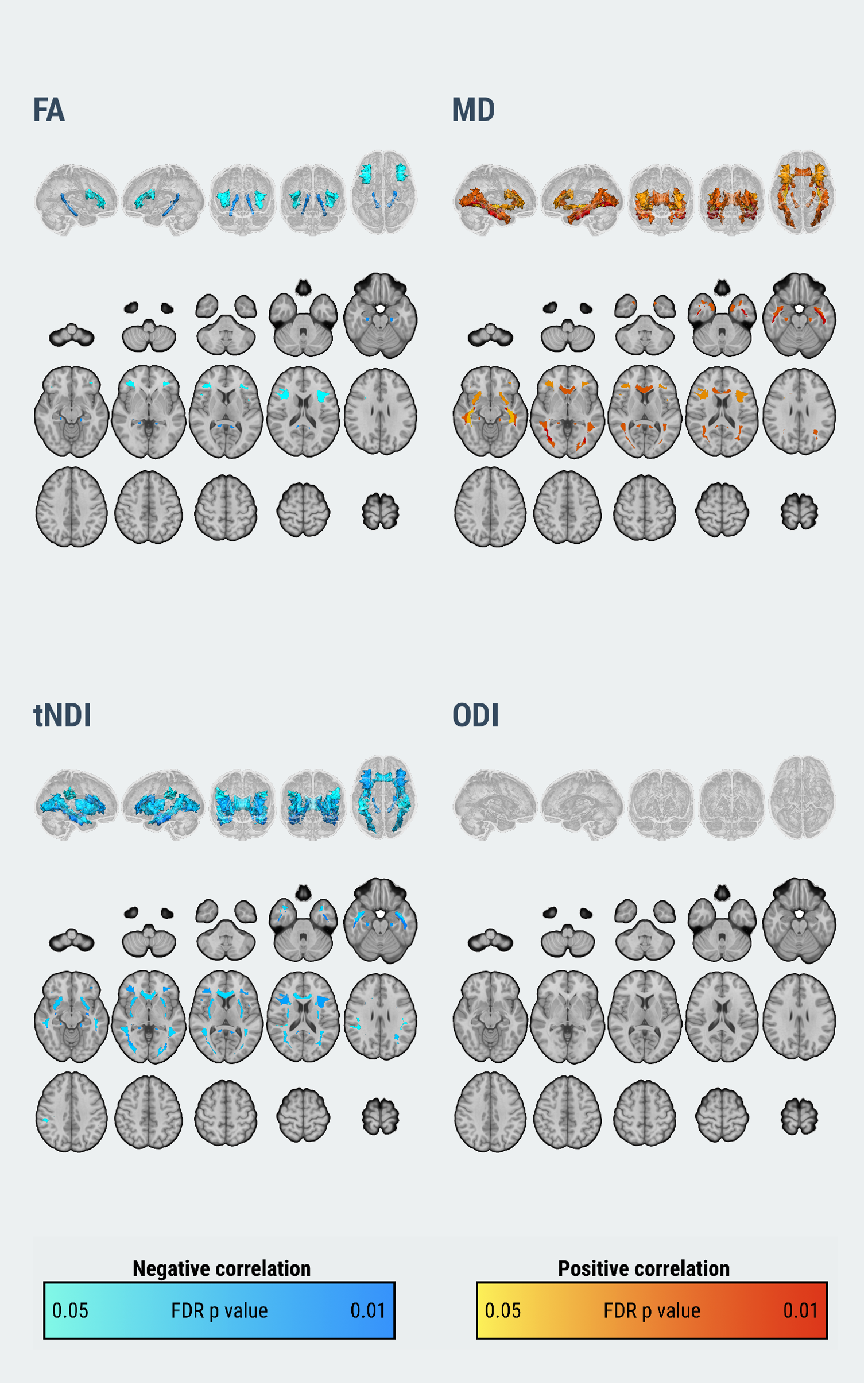
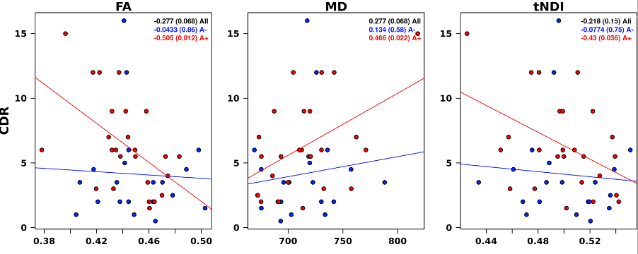
Among the DLBs, multivariable regression models adjusting for age, APOE and [11C]-PiB uptake, demonstrated that increased cortical [18F]-Flortaucipir correlated with lower FA, higher MD and lower tNDI values, predominantly in the temporal and limbic white matter tracts (Figure 3, FDR p < 0.05). There were no significant associations in the CU group. These tau-related neurite abnormalities were, in turn, associated with worse CDR, particularly in the subgroup of DLBs who were amyloid-positive (FA: r = -0.51, p = 0.01; MD: r = 0.47, p = 0.02; tNDI: r = -0.43, p = 0.036; Figure 4).Structural equation modeling revealed that amyloid-β did not directly affect white matter; instead, it exerted indirect effects through its influence on tau (Figure 5).
DISCUSSION
In the first study to combine biophysical NODDI parameters with multi-modal PET imaging of amyloid-β and tau in DLBs, our findings implicated tau-related neurite loss as a pathological substrate of widespread microstructural deficits in DLB.Greater cortical [18F]-Flortaucipir uptake was associated with a stereotypical AD pattern of white matter abnormalities that were in turn associated with worsened clinical disease severity, especially in the presence of elevated amyloid-β deposition. Nevertheless, amyloid-β may influence white matter microstructure through its mediating effect on tau. Finally, the topographic overlap between neurite density loss and diffusion tensor alterations emphasized the value of NODDI in conferring biological specificity to interpret the pathological underpinnings of white matter changes in DLBs.
CONCLUSION
Considered together with previous evidence of tau-associated cognitive decline6 and grey matter atrophy [7, 8] , these results support the need for further research into the underlying mechanisms of AD pathology and may have potential implications for disease-modifying trials that aim to target not only Lewy body pathology but also AD in individuals with DLB.Acknowledgements
The authors thank AVID Radiopharmaceuticals, Inc, for provision of the AV-1451 precursor, chemistry production, advice and oversight, and Food and Drug Administration regulatory cross-filing permission and documentation needed for this work. They thank the patients and their family members for participating in this research.References
1. Kantarci, K. et al. Dementia with Lewy bodies and Alzheimer disease: Neurodegenerative patterns characterized by DTI. Neurology 74, 1814–1821 (2010).
2. Nedelska, Z. et al. White matter integrity in dementia with Lewy bodies: A voxel-based analysis of diffusion tensor imaging. Neurobiology of Aging 36, 2010–2017 (2015).
3. Zhang, H., Schneider, T., Wheeler-Kingshott, C. A. & Alexander, D. C. NODDI: Practical in vivo neurite orientation dispersion and density imaging of the human brain. NeuroImage 61, 1000–1016 (2012).
4. McKeith, I. G. et al. Research criteria for the diagnosis of prodromal dementia with Lewy bodies. Neurology 94, 743–755 (2020).
5. McKeith, I. G. et al. Diagnosis and management of dementia with Lewy bodies. Neurology 89, 88–100 (2017).
6. Mak, E. et al. Imaging tau burden in dementia with Lewy bodies using [18F]-AV1451 positron emission tomography. Neurobiology of Aging 101, 172–180 (2021).
7. Ferreira, D. et al. Cross-sectional associations of -Amyloid, tau, and cerebrovascular biomarkers with neurodegeneration in probable dementia with lewy bodies. Neurology 10.1212/WNL.0000000000201579.
8. Chen, Q. et al. Longitudinal Tau Positron Emission Tomography in Dementia with Lewy Bodies. Movement Disorders 37, 1256–1264 (2022).
Figures